Abstract
Context:
The use of mineral trioxide aggregate (MTA) and Biodentine as a root-end filling materials used in the root-end cavities prepared by laser or ultrasonic technique is a current topic in the branch of dentistry and push-out bond strength is used to measure the adhesiveness provided by the root-end filling materials.
Aim:
The aim of this study is to evaluate the push-out bond strength of MTA and Biodentine in root-end cavities prepared by erbium:yttrium-aluminium-garne (Er:YAG) laser and ultrasonic retrotip.
Materials and Methods:
A total of 40 extracted maxillary central incisors and canines were selected. Chemomechanical preparation and obturation were done. Root-end resections were performed followed by the root-end cavity preparation and root-end filling. Specimens were divided into four groups. Root-end cavities prepared by Er:YAG laser and filled with MTA, root-end cavities prepared by Er:YAG laser and filled with Biodentine, root-end cavities prepared by ultrasonic retrotip and filled with MTA and root-end cavities prepared by ultrasonic retrotip and filled with Biodentine, respectively. The apical end was again sectioned perpendicular to the long axis. The push-out bond strength was evaluated using a universal testing machine.
Statistical Analysis Used:
The data were analyzed using the analysis of variance and post hoc Tukey test.
Results:
Difference between push-out bond strength of root-end filling materials to root-end cavity walls prepared by laser and ultrasonic retrotips was statistically nonsignificant. Push-out bond strength of MTA and Biodentine did not differ significantly.
Conclusion:
Difference between push-out bond strength of MTA and Biodentine to root-end cavity walls prepared by Er:YAG Laser or ultrasonic retrotip were statistically nonsignificant.
Keywords: Biodentine, laser, mineral trioxide aggregate, push-out bond strength, root end cavity, ultrasonic retrotips
INTRODUCTION
The presence of accessory canals and apical ramifications may hamper chemomechanical preparation and leads to failure of nonsurgical root canal therapy. Surgical approach then, is the treatment of choice which involves resecting the apical portion of the root, preparing a root-end cavity and filling it with root-end filling material.[1] The rationale behind this is to remove 98% of apical ramifications and 93% of lateral canals.[2]
While doing root-end cavity preparation, many instruments and techniques have been used. Latest among them are the use of laser[3] and ultrasonic retrotips.[4]
Erbium:yttrium-aluminium-garne (Er:YAG) laser has a high potential for ablating hard tissues with less heat generation because of their high water absorption characteristics.[5] The ultrasonic retrotips allows cleaner and deeper root-end cavity preparations. They are considered to be faster and more effective in producing quality root-end preparations.[6]
Mineral trioxide aggregate (MTA), which is biocompatible and has the capacity to induce osteointegration[7] and Biodentin which has the ability to create a micromechanical bond formation with dentin have been used as root-end filling materials.[8]
Marginal adaptation and bond strength of root-end filling materials are crucial factors for endodontic success because most endodontic failures arise from leakage at the root-end.[9]
The push-out test is used to measure the interfacial shear strength developed between different surfaces. It provides information about the adhesiveness of the tested material to the surface.[10]
Therefore, in the present study, push-out bond strength of MTA and Biodentine were tested in root-end cavities prepared by Er:YAG laser and ultrasonic retrotip.
MATERIALS AND METHODS
Forty freshly extracted maxillary permanent anteriors and canines devoid of any caries were selected and stored in distilled water until use.
A conventional endodontic access opening was done in all the samples, and working length for each tooth was recorded with the help of the K file (Mani Inc., Japan). Chemomechanical preparation was done with hand K files using step-back technique. Apical preparation was done till ISO tip size 70. Irrigation was done using 5 ml of 3% sodium hypochlorite (Prevest Denpro Limited, India) and 5 ml of 0.9% normal saline (Beryl, MP, India) solution alternatively after each instrumentation. Ethylenediaminetetra aceticacid (MetaBiomed® Co. Ltd, Korea) was used as a lubricant with each instrument. For the final flush, normal saline was used after which the canals were dried with sterile paper points (MetaBiomed® Co. Ltd., Korea) and obturation was done using 2% gutta-percha points (DiaDent Group International Inc., Canada) and resin-based sealer using cold lateral compaction technique. All teeth were stored in normal saline at room temperature for 24 h.
Root-end resections were performed by removing 3 mm of the apical portion of the root perpendicular to the long axis of root with a low-speed diamond disc using a straight handpiece (Marathon 3 Plus, India). Apical gutta-percha was removed from the apical end of resected root using a heat carrying instrument.
The specimens were randomly divided into four groups of 10 samples each, based on the method of root-end cavity preparation and root-end filling material used.
Method of cavity preparation
3-mm deep root-end cavities were prepared either using Er:YAG laser (Lite Touch™, Syneron dental lasers, Israel) with energy of 100 mJ and frequency 10 Hz or diamond coated ultrasonic retro tips ([ED 11, Wood Pecker, China] on a piezoelectric unit at a frequency of 32 KHz and medium power setting with distilled water irrigation according to the division of groups.
Group I: Root-end cavities were prepared using Er:YAG laser and later filled with MTA (Dentsply Tulsa, USA) which was prepared according to the manufacturer's instructions with a powder-to-liquid ratio of 3:1
Group II: Root-end cavities were prepared using Er:YAG laser and later filled with Biodentine (Septodont, France). It was prepared by adding 5 drops of its liquid into the capsule containing powder and then mixed in a triturator for 30 s, at 4300 oscillations per minute
Group III: Root-end cavities were prepared with diamond coated ultrasonic retrotrip. After that, root-end cavities were filled with MTA following the same procedure as in Group I
Group IV: Root-end cavities were prepared with the ultrasonic retrotip and filled with Biodentine following the same procedure as in Group II.
All samples were stored in 100% humidity for 21 days in a humidifier at 37°C.
The apical portion of each resected root was sectioned perpendicular to the long axis into 2-mm thick slice with a low-speed diamond disc using a straight handpiece.
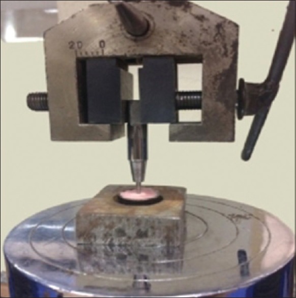
The push-out bond strength was evaluated using a universal testing machine (Instron India Pvt., Ltd.) at a speed of 1 mm/min by keeping specimens in an apico-coronal direction. The specimen were kept in the prefabricated jig and the bond failure was checked by the extrusion of the root-end filling material into the mounting jig [Figure 1]. The maximum load applied to the filling materials was recorded in Newtons (N) and that values were used to calculate the push-out bond strength in Megapascals (MPa) as per the surface area of specimens. The formula used to calculate the bond strength was:
Figure 1.

Ring placed in jig and plunger mounted in universal testing machine
MPa = N/2 πrh
Where N = the maximum load for each specimen,
r = root canal radius in mm,
h = the thickness of the root dentin disc in millimeters and
π =3.14.
Statistical analysis
The results are presented in mean ± standard deviation. The two-way analysis of variance was used to compare the push-out bond strength of MTA and Biodentine in the root-end cavity preparation. The post hoc Tukey test was used to compare the push-out bond strength of two root-end filling materials (MTA and Biodentine) in root-end cavities prepared by laser or ultrasonic technique. The P > 0.05 was considered to be statistically nonsignificant. All the analysis was carried out using the SPSS 16.0 version (Chicago, Inc., USA).
RESULTS
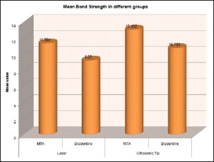
Graph 1 shows the mean values and standard deviation among all the four groups.
Graph 1.

Descriptive values of all the groups
It shows that the Group-III has the maximum push-out bond strength and Group-II has least push-out bond strength among all the four groups.
Table 1 shows the difference between push-out bond strength of root-end filling materials used was statistically nonsignificant.
Table 1.
Comparison between mineral trioxide aggregate and Biodentine
| Comparison between the type of material used | P | Significance |
|---|---|---|
| MTA* and Biodentine | 0.318 | Nonsignificant |
*MTA: Mineral trioxide aggregate
Table 2 shows the effect of different methods of root-end cavity preparation (laser and ultrasonic retro tip) on the push-out bond strength of root-end filling materials to prepare cavity walls. Results were found to be statistically nonsignificant.
Table 2.
Comparison between laser and ultrasonic RetroTip
| Comparison between the methods used to prepare a cavity | P | Significance |
|---|---|---|
| Laser and ultrasonic retrotip | 0.464 | Nonsignificant |
DISCUSSION
If nonsurgical root canal treatment followed by retreatment attempts prove unsuccessful, then surgical periapical surgery followed by root-end resection and the periapical seal may be needed to save the tooth. While doing root-end resection, at least 3 mm of the root-end must be cut to reduce 98% of the apical ramifications and 93% of the lateral canals.[11]
The presence of obturating material effects the efficacy of both laser and ultrasonic tips.[12] Therefore, apical gutta-percha was removed using a heat carrying instrument before starting with the root-end cavity preparation procedure in this study.
According to Arens, Class I root-end cavity prepared parallel to the long axis of the root, 3 mm deep in the center of the root exhibits the least leakage.[13] Therefore, in this study, Class I root-end cavities were prepared.
The bond strength of root-end filling materials to root canal walls is an important factor because it is beneficial in maintaining the integrity of the cement-dentin interface.[14] In push-out strength test, there is uniform stress distribution at the dentin-cement interface.[15] Hence, push-out bond strength test may be more efficient, practical and reliable method to check the bond strength of cement to dentin. Hence, this test was used in this study.
In the present study, the push-out bond strength values of root-end filling materials to dentin walls of root-end cavities prepared with Er: YAG laser was lower than that of cavities prepared with diamond coated ultrasonic retrotip. The reason behind this may be, the irradiation with laser causes an irregular and rough surface of dentin which might affect the bond strength of root-end filling materials to dentinal walls of root-end cavities because of their incomplete penetration into the irregularities due to their high viscosity.[16] Moreover, gap was observed by between root-end filling materials and lased dentin surface, which indicated inappropriate adaptation of root-end filling material.[17]
Smear layer is a moist layer which might act as a coupling agent, which in turn improves the adaptation of hydrophilic materials such as MTA or Biodentine to the dentinal walls.[18] However, laser results in the removal of the smear layer. El-Ma'aita et al. examined the effects of smear layer on push-out bond strength of Biodentine and pro-root MTA. They found that push-out bond strength decreases with the removal of the smear layer.[19]
Based on the findings of this study, MTA had greater push-out bond strength than Biodentine. This is in accordance with in vitro study conducted by Uzunoglu et al. who also showed the same results. They also compared the effect of mixing techniques on the push-out bond strength of Biodentine and found decreased values when Biodentine was mixed mechanically.[20] This was in accordance with our study as we mixed Biodentine mechanically and found less bond strength values of Biodentine.
In the present study, the analysis of the mean push-out bond strength values of each group revealed that there were nonsignificant differences among all the groups (P > 0.05). In this study, the ultrasonic tip group, using MTA as a root-end filling material showed the maximum push-out bond strength and the Laser group, using Biodentine as a root-end filling material showed the least push-out bond strength.
According to Vallés et al., Biodentine has higher push-out bond strength than MTA after 24 h of setting time.[21] After 7 days, MTA and Biodentine have similar push-out bond strength.[22] Gancedo-Caravia and Garcia-Barbero showed that if MTA is kept under wet conditions for 3–21 days, its push-out bond strength increases significantly, indicating prolonged maturation process of the material.[23] According to Torabinejad et al. push-out bond strength of MTA increases under wet conditions after being immersed in 21 days.[1] Therefore, in the present study, samples were kept under wet conditions for 21 days to allow optimal hydration, solidification and to achieve optimal strength before testing.
CONCLUSION
The difference between push-out bond strength of root-end filling materials used was statistically nonsignificant. The root-end cavities prepared by ultrasonic retrotip and filled with MTA had maximum push-out bond strength and root-end cavities prepared by laser and filled with Biodentine had least push-out bond strength. The effect of different methods of root-end cavity preparation (laser and ultrasonic retro tip) on the push-out bond strength of root-end filling materials to prepare cavity walls was statistically nonsignificant.
Financial support and sponsorship
Nil.
Conflicts of interest
There are no conflicts of interest.
Acknowledgment
The authors would like to thank Genesis Institute of Dental Sciences and Research, Ferozepur, Punjab, India, for its support.
REFERENCES
- 1.Torabinejad M, Hong CU, McDonald F, Pitt Ford TR. Physical and chemical properties of a new root-end filling material. J Endod. 1995;21:349–53. doi: 10.1016/S0099-2399(06)80967-2. [DOI] [PubMed] [Google Scholar]
- 2.Kim S, Kratchman S. Modern endodontic surgery concepts and practice: A review. J Endod. 2006;32:601–23. doi: 10.1016/j.joen.2005.12.010. [DOI] [PubMed] [Google Scholar]
- 3.Esen E, Yoldas O, Kürkçü M, Doǧan MC, Seydaoǧlu G. Apical microleakage of root-end cavities prepared by CO2 laser. J Endod. 2004;30:662–4. doi: 10.1097/01.don.0000125316.89703.e2. [DOI] [PubMed] [Google Scholar]
- 4.Sumi Y, Hattori H, Hayashi K, Ueda M. Ultrasonic root-end preparation: Clinical and radiographic evaluation of results. J Oral Maxillofac Surg. 1996;54:590–3. doi: 10.1016/s0278-2391(96)90639-4. [DOI] [PubMed] [Google Scholar]
- 5.Yu LL. Multiple applications with Er: YAG laser in apicoectomy – A clinical case report. Dentistry. 2017;7:1–5. [Google Scholar]
- 6.Liu Z, Zhang D, Li Q, Xu Q. Evaluation of root-end preparation with a new ultrasonic tip. J Endod. 2013;39:820–3. doi: 10.1016/j.joen.2013.03.004. [DOI] [PubMed] [Google Scholar]
- 7.Vidal K, Martin G, Lozano O, Salas M, Trigueros J, Aguilar G. Apical closure in apexification: A review and case report of apexification treatment of an immature permanent tooth with biodentine. J Endod. 2016;42:730–4. doi: 10.1016/j.joen.2016.02.007. [DOI] [PubMed] [Google Scholar]
- 8.Jose J, Shoba K, Tomy N, Aman S. One-step apexification with two bioactive materials – A CBCT evaluation. Int J Oral Health Med Res. 2015;2:46–9. [Google Scholar]
- 9.Ertas H, Kucukyilmaz E, Ok E, Uysal B. Push-out bond strength of different mineral trioxide aggregates. Eur J Dent. 2014;8:348–52. doi: 10.4103/1305-7456.137646. [DOI] [PMC free article] [PubMed] [Google Scholar]
- 10.Thompson JI, Gregson PJ, Revell PA. Analysis of push-out test data based on interfacial fracture energy. J Mater Sci Mater Med. 1999;10:863–8. doi: 10.1023/a:1008929201918. [DOI] [PubMed] [Google Scholar]
- 11.Soundappan S, Sundaramurthy JL, Raghu S, Natanasabapathy V. Biodentine versus mineral trioxide aggregate versus intermediate restorative material for retrograde root end filling: An in vitro study. J Dent (Tehran) 2014;11:143–9. [PMC free article] [PubMed] [Google Scholar]
- 12.Batista de Faria-Junior N, Tanomaru-Filho M, Guerreiro-Tanomaru JM, de Toledo Leonardo R, Camargo Villela Berbert FL. Evaluation of ultrasonic and ErCr: YSGG laser retrograde cavity preparation. J Endod. 2009;35:741–4. doi: 10.1016/j.joen.2009.02.007. [DOI] [PubMed] [Google Scholar]
- 13.Agarwal V, Nayak D, Sharma M, Reddy YG, Singla M, Nanda Z. Comparative evaluation of different retrograde cavity designs of amalgam for assessment of microleakage by dye penetration method – An in vitro study. Endodontology. 2013;6:91–9. [Google Scholar]
- 14.Akcay H, Arslan H, Akcay M, Mese M, Sahin NN. Evaluation of the bond strength of root-end placed mineral trioxide aggregate and biodentine in the absence/presence of blood contamination. Eur J Dent. 2016;10:370–5. doi: 10.4103/1305-7456.184150. [DOI] [PMC free article] [PubMed] [Google Scholar]
- 15.Mathew S, Raju IR, Sreedev CP, Karthick K, Boopathi T, Deepa NT. Evaluation of push out bond strength of fiber post after treating the intra radicular post space with different post space treatment techniques: A randomized controlled In vitro trial. J Pharm Bioallied Sci. 2017;9:S197–S200. doi: 10.4103/jpbs.JPBS_156_17. [DOI] [PMC free article] [PubMed] [Google Scholar]
- 16.Shokouhinejad N, Nekoofar MH, Iravani A, Kharrazifard MJ, Dummer PM. Effect of acidic environment on the push-out bond strength of mineral trioxide aggregate. J Endod. 2010;36:871–4. doi: 10.1016/j.joen.2009.12.025. [DOI] [PubMed] [Google Scholar]
- 17.Winik R, Araki AT, Negrão JA, Bello-Silva MS, Lage-Marques JL. Sealer penetration and marginal permeability after apicoectomy varying retrocavity preparation and retrofilling material. Braz Dent J. 2006;17:323–7. doi: 10.1590/s0103-64402006000400011. [DOI] [PubMed] [Google Scholar]
- 18.Ballal NV, Ulusoy Öİ, Chhaparwal S, Ginjupalli K. Effect of novel chelating agents on the push-out bond strength of calcium silicate cements to the simulated root-end cavities. Microsc Res Tech. 2018;81:214–9. doi: 10.1002/jemt.22969. [DOI] [PubMed] [Google Scholar]
- 19.El-Ma'aita AM, Qualtrough AJ, Watts DC. The effect of smear layer on the push-out bond strength of root canal calcium silicate cements. Dent Mater. 2013;29:797–803. doi: 10.1016/j.dental.2013.04.020. [DOI] [PubMed] [Google Scholar]
- 20.Uzunoglu E, Turker SA, Uyanik MO, Nagas E. Effects of mixing techniques and dentin moisture conditions on push-out bond strength of ProRoot MTA and biodentine. J Adhes Sci Technol. 2016;30:1–7. [Google Scholar]
- 21.Vallés M, Mercadé M, Duran-Sindreu F, Bourdelande JL, Roig M. Influence of light and oxygen on the color stability of five calcium silicate-based materials. J Endod. 2013;39:525–8. doi: 10.1016/j.joen.2012.12.021. [DOI] [PubMed] [Google Scholar]
- 22.Gupta A, Makani S, Vachhani K, Sonigra H, Attur K, Nayak R. Biodentine: An effective pulp capping material. Sch J Dent Sci. 2016;3:15–9. [Google Scholar]
- 23.Gancedo-Caravia L, Garcia-Barbero E. Influence of humidity and setting time on the push-out strength of mineral trioxide aggregate obturations. J Endod. 2006;32:894–6. doi: 10.1016/j.joen.2006.03.004. [DOI] [PubMed] [Google Scholar]


