Abstract
Objective:
The objective of this report was to describe a patient with Graves acropachy, a rare manifestation of Graves disease (GD) that is clinically defined by skin tightness, digital clubbing, small-joint pain, and soft tissue edema progressing over months or years with gradual curving and enlargement of the fingers.
Methods:
The patient was evaluated regarding thyroid function (serum free T4 [FT4] and thyroid-stimulating hormone [TSH] quantifications) and autoimmunity biomarkers (thyroid receptor antibody [TRAb]) as well as radiographic investigation of the extremities.
Results:
A 52-year-old man presented with a history of thyrotoxicosis and clinical signs of Graves orbitopathy. Laboratory tests showed suppressed TSH (0.01 UI/L; normal, 0.4 to 4.5 UI/L) and elevated serum FT4 (7.77 ng/dL; normal, 0.93 to 1.7 ng/dL), with high TRAb levels (40 UI/L; normal, <1.75 UI/L). A diagnosis of thyrotoxicosis due to GD was made and the patient was treated with methimazole. After the patient complained of swelling in hands and feet, X-ray evaluation was conducted and established the thyroid acropachy.
Conclusion:
We present a case of a patient with GD associated with worsening extrathyroid manifestations during orbitopathy, dermopathy, and developed acropachy in hands and feet.
INTRODUCTION
Graves disease (GD) is an autoimmune disease that affects the thyroid and is the major cause of thyrotoxicosis. The disease results from the development of thyroid receptor antibodies (TRAbs). Following the binding of thyroid-stimulating hormone (TSH) receptors in the thyroid follicle membrane, these antibodies trigger thyroid inflammation and hyperplasia and increased thyroid hormone production, resulting in clinical thyrotoxicosis (1).
Thyroid acropachy is an extrathyroidal manifestation of the autoimmunity (2) that affects less than 1% of GD patients. The peak prevalence is among people in their 50s, and the prevalence is higher in women (3,4). The clinical characteristics of acropachy are digital clubbing, diaphyseal proliferation, and hypertrophy of the surrounding soft tissue caused by glycosaminoglycan deposition (2), which is often symmetrical and bilateral (5).
CASE REPORT
A 52-year-old male with a 1-year history of weight loss (16 kg in 4 months), tachycardia, hot and moist skin, and peripheral tremor presented to our hospital center. The patient was a smoker. The estimated thyroid volume was 45 cm3 (normal, 12 to 18 cm3) according to an ultrasound examination.
The ophthalmologic examination showed a bilateral proptosis using the Hertel exophthalmometer, (right eye 24 mm and left eye 23 mm; right eyelid 12 mm and left eyelid 11 mm). The clinical activity score (CAS) was 2 bilateral (diffuse redness of the conjunctiva and swelling of the eyelids), and there was no diplopia. The laboratory data showed suppressed serum TSH (0.01 UI/L; normal, 0.4 to 4.5 UI/L) and elevated serum free T4 (FT4; 7.77 ng/dL; normal, 0.93 to 1.7 ng/dL) and TRAb (40 UI/L; normal, <1.75 UI/L). There was no clinical manifestation of dermopathy or acropachy observed.
The patient's thyrotoxicosis was treated with methimazole and he was instructed to stop smoking. Four months later, he complained of progressive swelling of the hands and inferior limbs. The Graves orbitopathy (GO) was worse with a CAS of 4 bilateral (diffuse redness of the conjunctiva, swelling of the eyelids, swollen caruncle, and chemosis). There was hard edema and erythematous-infiltrative plaques in the feet, legs, and toes similar to so called “orange peel skin” or “peau d'orange.”
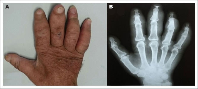
The thyroid function tests showed a FT4 of 4.16 ng/dL and a TSH of 0.005 IU/L. He had not stopped smoking. Methimazole was continued and he was again instructed to stop smoking. He was referred to dermatology for guidance and follow-up management of the skin lesions. Two months later, the patient returned and was improved as far as clinical manifestations were concerned. Thyroid function test showed an FT4 of 0.77 ng/dL and a TSH of 0.001 IU/L. However, an X-ray examination of the left hand indicated soft tissue swelling in the interphalangeal region from the 2nd to 5th fingers, interphalangeal subluxation on the 3rd finger, and anatomic loss of the distal phalange (Fig. 1). X-rays of the left foot indicated distal phalanx tapering and bone ankyloses in the hallux, with anatomic loss of phalanges in the 3rd and 4th left toes. X-rays of the right foot indicated tapering in the proximal phalanx of the 5th digit with signs of osteoarthrosis in the hallux.
Fig. 1.

A, Hand impairment. Swelling of interphalangeal region in both hands associated with finger clubbing. Dorsal. B, Left hand X-ray: presence of soft tissue swelling in the interphalangeal region from the 2nd to 5th fingers. The 3rd finger had signs of interphalangeal subluxation and anatomic loss of distal phalange.
The radiologic pattern confirmed the features of Graves acropachy. A skin biopsy was then performed, which indicated skin lesions compatible with myxedema. The treatment involved occlusive bandages of corticosteroids and cold cream formulation (10% urea, 10% salicylic acid, 5% lactic acid).
DISCUSSION
Graves acropachy was first reported in 1933 in a female patient with GD who was treated with thyroidectomy (6). It is the rarest component of the triad of GO, dermopathy, and acropachy, and occurs in only 0.8 to 1% of patients with GO (4,7). Graves acropachy is strongly associated with severe GO and with dermopathy (7). Most patients present with digital clubbing with radiologic study showing a periosteal reaction in phalangeal bones. Complaints of lower extremity pain, skin and nail changes may point to the diagnosis of acropachy (7).
The pathogenesis of acropachy is unknown, except for the anatomic location, in that it is probably similar to that of pretibial myxedema. It appears that TRAb molecules bind to the TSH receptors of fibroblasts present in the periosteum region and trigger an inflammatory response, producing cell proliferation and glycosaminoglycan deposition (7,8). The musculoskeletal manifestation is almost never seen without the remaining components of the triad of orbitopathy, dermopathy, and acropachy (9,10). Some studies suggest smoking is a predisposing factor for acropachy in GD patients (9).
In most cases, acropachy is asymptomatic, but the main clinical manifestations are digital clubbing, skin tightness with or without digital clubbing and usually with small-joint pain (in severe cases), soft tissue edema, and reactional periosteum, and skin alterations in fingers and nails may also be present (7). The disorder mostly affects the metacarpus phalangeal and proximal interphalangeal regions in the upper and lower limbs, especially the ankles and metatarsal phalangeal joints (11).
The soft tissue edema is predominantly hard, it does not have temperature elevation, and frequently is associated with bone alterations (11). Acropachy progresses over months or years, with gradual curving and enlargement of the fingers but without pain associated with initial manifestations (6,8).
Acropachy is extremely rare before the manifestation of thyrotoxicosis, with 95% of patients developing the disease during the treatment of GD (12). In the case of patients presenting digital clubbing in the hands, the diagnosis is made with only clinical findings (7). Nevertheless, X-rays of the extremities should provide a more accurate diagnosis (3). Histologic examination demonstrates nodular fibrosis in the distal periosteum (11).
In most cases, the condition affects the 1st, 2nd, and 3rd metacarpus or metatarsus in the radial face and 4th and 5th metatarsus or metacarpus in the ulnar face. The compromise of a single digit is rare and may suggest malignity (3). X-rays may also present layers of new bone formation deposited over the progression of the disease. This deposition occurs from the cortex towards the periosteum and mainly in the terminal segment of the bone (6). New bone formation depositions are restrained to the periosteum, with no signs of articular compromise and more intensity in the medial region of short bones, although it may affect long bones (6). Small bones deletion is rare and can be the result of intense inflammatory process.
Although there is no specific treatment for acropachy, immune modulators such as high potency corticosteroids have been used, and some patients were successfully treated with rituximab (12,13). Patients with joint pain can be treated with anti-inflammatory drugs such as ketoprofen (12). Correction of thyrotoxicosis may be associated with an improvement in the clinical manifestations of acropachy, but the role of thyroid function control in acropachy evolution is unclear (12,7).
CONCLUSION
Acropachy is a rare manifestation of thyroid autoimmunity, always followed by GO and dermopathy. Treatment with corticosteroids may have a good therapeutic effect. Restoration of euthyroidism is always intended; however, the clinical benefit in acropachy is not known.
Abbreviations
- FT4
free T4
- GD
Graves disease
- GO
Graves orbitopathy
- TRAb
thyroid receptor antibodies
- TSH
thyroid-stimulating hormone
Footnotes
DISCLOSURE
The authors have no multiplicity of interest to disclose.
REFERENCES
- 1.Weetman AP. Graves' Disease. N Engl J Med. 2000;343:1236–1248. doi: 10.1056/NEJM200010263431707. [DOI] [PubMed] [Google Scholar]
- 2.Anderson CK, Miller OF., 3rd. Triad of exophthalmos, pretibial myxedema, and acropachy in a patient with Graves' disease. J Am Acad Dermatol. 2003;48:970–972. doi: 10.1067/mjd.2003.323. [DOI] [PubMed] [Google Scholar]
- 3.Vanhoenacker FM, Pelckmans MC, De Beuckeleer LH, Colpaert CG, De Schepper AM. Thyroid acropachy: correlation of imaging and pathology. Eur Radiol. 2001;11:1058–1062. doi: 10.1007/s003300000735. [DOI] [PubMed] [Google Scholar]
- 4.Moule B, Grant MC, Boyle IT, May H. Thyroid acropachy. Clin. Radiol. 1970;21:329–333. doi: 10.1016/s0009-9260(70)80065-4. [DOI] [PubMed] [Google Scholar]
- 5.Neves C, Alves M, Delgado JL, Medina JL. Doença de Graves. Arq Med. 2008;22:137–146. [Google Scholar]
- 6.Henry MT., JR. Acropachy: secondary subperiosteal new bone formation. Arch Intern Med. 1933;51:571–588. [Google Scholar]
- 7.Fatourechi V, Ahmed DD, Schwartz KM. Thyroid acropachy: report of 40 patients treated at a single institution in a 26-year period. J Clin Endocrinol Metab. 2002;87:5435–5441. doi: 10.1210/jc.2002-020746. [DOI] [PubMed] [Google Scholar]
- 8.Jameson JL, De Groot L. Graves' Disease. In: Jamerson JL, De Groot L, de Krester DM, Giudice LC, Grossman AB, Melmed S, Potts JT Jr, Weir GC, editors. Endocrinology: Adult & Pediatric. 7th ed. Philadelphia, PA: Elsevier Saunders; 2015. pp. 1451–1453. [Google Scholar]
- 9.Anwar S, Gibofsky A. Musculoskeletal manifestations of thyroid disease. Rheum Dis Clin North Am. 2010;36:637–646. doi: 10.1016/j.rdc.2010.09.001. [DOI] [PubMed] [Google Scholar]
- 10.Boswell SB, Patel DB, White EA et al. Musculoskeletal manifestations of endocrine disorders. Clin Imaging. 2014;38:384–396. doi: 10.1016/j.clinimag.2014.02.014. [DOI] [PubMed] [Google Scholar]
- 11.Bland JH, Frymoyer JW, Newberg AH, Revers R, Norman RJ. Rheumatic syndromes in endocrine disease. Semin Arthritis Rheum. 1979;9:23–65. doi: 10.1016/0049-0172(79)90002-7. [DOI] [PubMed] [Google Scholar]
- 12.Bartalena L, Fatourechi V. Extrathyroidal manifestations of Graves' disease: a 2014 update. J Endocrinol Invest. 2014;37:691–700. doi: 10.1007/s40618-014-0097-2. [DOI] [PubMed] [Google Scholar]
- 13.Ferreira-Hermosillo A, Casados-V R, Paúl-Gaytán P, Mendoza-Zubieta V. Utility of rituximab treatment for exophthalmos, myxedema, and osteoarthropathy syndrome resistant to corticosteroids due to Graves' disease: a case report. J Med Case Rep. 2018;12:38. doi: 10.1186/s13256-018-1571-9. [DOI] [PMC free article] [PubMed] [Google Scholar]


