Abstract
Background
miRNAs are small noncoding RNAs that function as posttranscriptional regulators during development and disease. Aberrant expression of miRNAs has been associated with various types of malignant tumors. Decreased levels of miR-124 have been observed in human cancers. RACK1 is a scaffold protein that acts as an oncogene in various human cancers. The association between miR-124 and RACK1 in melanoma has not been characterized.
Materials and methods
Real-time quantitative PCR was used to analyze RACK1 and miR-124 expression in melanoma tissue and cell lines. Dual-Luciferase reporter assay was performed to evaluate the effect of miR-124 inhibition on RACK1 expression. The effects of miR-124 on RACK1 in melanoma cell lines were evaluated using Western blot analysis and immunocytochemical staining. Wound-healing, transwell, and MTT assays, and annexin V-fluorescein isothiocyanate/propidium iodide followed by flow cytometry were used to evaluate the effects of miR-124 on RACK1-mediated proliferation, migration, invasion, and apoptosis of melanoma cells.
Results
The expression of miR-124 in melanoma tissue was lower than that in normal skin tissue, and the expression of RACK1 was higher in melanoma tissue than that in normal skin tissue. Analysis using Dual-Luciferase reporter assay showed that RACK1 was a direct target of miR-124. Western blot and immunocytochemical staining showed that the expression of RACK1 was significantly inhibited by miR-124 in both A375 and A875 melanoma cells. Furthermore, the results of functional experiments showed that degradation of RACK1 by miR-124 inhibited proliferation, migration, and invasion of melanoma cells, and promoted melanoma cell apoptosis.
Conclusion
The results suggested that miR-124 affected melanoma cells by directly targeting RACK1. miR-124 and RACK1 may be biomarkers for clinical diagnosis, and prognostic factors of human melanoma. Furthermore, miR-124 and RACK1 may be targets for the treatment of melanoma.
Keywords: melanoma, miR-124, RACK1, proliferation, migration, invasion, apoptosis
Introduction
Melanoma, basal cell carcinoma, and squamous cell carcinoma are the three main types of skin cancer, and melanoma is the most difficult to cure due to high malignancy and few treatment options.1 Studies have shown that genetic factors play a key role in the development of melanoma. However, the molecular mechanisms of development of melanoma have not been characterized.2,3 Identification of new gene targets to determine the molecular mechanisms of onset and development of melanoma is needed to improve clinical diagnosis and treatment.4
miRNAs are small noncoding RNAs with a length of about 22 nucleotides that regulate posttranscriptional gene expression in plants and animals.5 miRNAs can regulate the expression of many target genes and are critical factors in multiple diseases and biological processes.6 Recently, a large number of miRNAs and their target genes were identified in human cancers. Studies have shown that miRNAs act as tumor suppressors or oncomiRs (miRNAs associated with the development of cancer).7,8 miR-124 is one of the best-characterized miRNAs. It is highly conserved and primarily distributed in the central nervous systems of adults and embryos. Studies have shown that miR-124 acts as a tumor suppressor in several human cancers, including cervical cancer,9 gastric cancer,10 glioma,11 hepatocellular carcinoma,12 pancreatic cancer,13 non-small-cell lung cancer (NSCLC),14 bladder cancer,15 cholangiocarcinoma,16,17 breast cancer,18 and osteosarcoma,19 which suggest that miR-124 may be a marker for diagnosis and a target for treatment of tumors.
RACK1 belongs to the tryptophan-aspartate repeat (WD-repeat) protein family and has high homology with the G protein beta subunit.20 As a scaffold protein, RACK1 plays a crucial role in cell growth, proliferation, migration, adhesion, differentiation, signal transduction, and immune responses,21 and acts as an oncogene in a number of human cancers.22,23 However, few studies have evaluated the roles of miR-124 and RACK1 in melanoma.
In this study, we investigated miR-124 in patients with melanoma. We evaluated the association between miR-124 and RACK1 in melanoma tissue and in adjacent nonneoplastic tissue. Dual-Luciferase reporter assay showed that RACK1 was a direct target of miR-124. In melanoma cells, miR-124 inhibited the expression of RACK1. In addition, we evaluated the role of miR-124 in cell proliferation, migration, invasion, and apoptosis, and proposed that miR-124/RACK1 may be a potential target for the treatment of melanoma.
Materials And Methods
Clinical Patient Samples And Cell Lines
Tumor and adjacent non-tumor skin tissue samples from 21 patients diagnosed with melanoma were collected from the Affiliated Hospital of Nantong University (Nantong, Jiangsu, China). Written informed consent was obtained from each patient prior to study enrollment. The study was conducted in accordance with the Declaration of Helsinki and was approved by the Ethics Committee of the Affiliated Hospital of Nantong University. Human melanoma cell line A375, human embryonic kidney 293 (HEK293) cell line, and keratinocyte cell line HaCaT were cultured in DMEM, and the melanoma cell line A875 was cultured in Roswell Park Memorial Institute 1640 medium. Media were supplemented with 10% FBS (Thermo Fisher Scientific, USA). All cells were cultured at 37°C in an incubator with a humified atmosphere of 5% CO2. HaCaT and HEK293 cell lines were obtained from Kunming Cell Bank (Kunming, China), and A375 and A875 cell lines were obtained from the Shanghai Institute of Biological Sciences (Shanghai, China).
miRNA, Plasmid Construction, And Transfection
A human miR-124 mimic was used to upregulate endogenous miR-124 expression in melanoma cells. A scrambled miR-124 mimic sequence was used as a negative control (NC). The sequences are summarized in Table 1. The miR-124 binding site of RACK1 was predicted using miRTarBase online software.24 The cDNA of the 3′UTR sequence containing the miR-124 binding sites of RACK1 mRNA was cloned downstream of the luciferase gene of the pGL3-Control vector (Promega, USA) as a wild-type RACK1 (wt-RACK1) plasmid. A scrambled RACK1/miR-124 binding site was constructed as a mutant RACK1 (mut-RACK1) plasmid. Cell transfection with miRNA or plasmids was performed using Lipofectamine® 2000 transfection reagent (Thermo Fisher Scientific) according to the manufacturer’s instructions. miRNA, NC, and DNA oligos were obtained from Biomics Biotechnologies Co., Ltd (China).
Table 1.
The Sequences Of miRNAs, RT-qPCR Primers, And The Insert Sequences Of Dual-Luciferase Reporter Plasmids
| Name | Sequence (5ʹ-3ʹ) | |
|---|---|---|
| miR-124 mimic | Sense | UAAGGCACGCGGUGAAUGCC |
| Antisense | GGCAUUCACCGCGUGCCUUA | |
| NC | Sense | GCGUUGAACGCAGCGGACAU |
| Antisense | AUGUCCGCUGCGUUCAACGC | |
| RACK1 | Forward | AGATAAGACCATCATCAT |
| Reverse | AGATAACCACATCACTAA | |
| β-actin | Forward | TTGCCGACAGGATGCAGAAGGA |
| Reverse | AGGTGGACAGCGAGGCCAGGAT | |
| wt-RACK1 | Forward | TCTAGAAAGTTTATGGCAGAGCTTTAT |
| Reverse | TCTAGATAAAGCTCTGCCATAAACTTT | |
| mut-RACK1 | Forward | TCTAGAAAGTTTATGGCAATGCTGTAT |
| Reverse | TCTAGATACAGCATTGCCATAAACTTT | |
Real-Time Quantitative PCR
Total RNA from tissue samples and cells was isolated using TRIzol® reagent (Thermo Fisher Scientific). The expression of RACK1 mRNA was quantified by real-time quantitative PCR (qPCR) using the One-Step RT-qPCR Kit (Thermo Fisher Scientific). Small RNAs were isolated from tissues and cells using the mirVana miRNA Isolation Kit (Thermo Fisher Scientific). miR-124 expression levels were quantified by the stem-loop real-time qPCR method25 using the EzQuickTM One-Step qPCR Kit (Biomics Biotechnologies Co., Ltd.). β-actin was used as an internal control for RACK1 and endogenous U6 RNA was used as an internal control for miR-124. Results were analyzed using the 2−ΔΔCt method.26 The primer sequences used are summarized in Table 1. All procedures were performed in accordance with the manufacturer’s instructions.
Western Blot Analysis
Cell lysates were extracted using RIPA buffer (Promega), and the protein concentration was determined using the BCA™ Protein Assay Kit (Pierce, USA). Total protein was separated by 10% SDS-PAGE and transferred to PVDF membranes (Merck Millipore, USA). The membranes were incubated overnight with rabbit anti-human RACK1 antibody (1:500 dilution) or mouse anti-human β-actin antibody (1:1000 dilution) at 4°C. The membranes were then incubated with HRP-conjugated secondary antibodies at room temperature for 2 hrs. Blots were visualized using enhanced chemiluminescence Western blotting substrate (Promega) and quantified using Image J software (NIH, USA). β-actin was used as an internal control. All antibodies were purchased from Abcam Inc. (USA).
Dual-Luciferase Reporter Assay
Regulation of 3′UTR RACK1 by miR-124 was confirmed using the Dual-Luciferase reporter assay (Promega). Different combinations of plasmids (80 ng wt-RACK1 or mut-RACK1, insert sequences are summarized in Table 1) and miRNA (50 nmol/L miR-124 mimic or NC) were transfected into HEK293 cells using Lipofectamine® 2000 transfection reagent (Thermo Fisher Scientific), with pRL-TK plasmid (Promega) co-transfected as an internal control. After 48 hrs of transfection, the cells were collected and analyzed using the Dual-Luciferase reporter assay according to the manufacturer’s instructions. Firefly luciferase activity was normalized to that of Renilla luciferase and reported.
Immunocytochemistry Staining
Cells were plated in 24-well plates containing a round slide in each well and cultured at 37°C for 24 hrs. After 48 hrs of transfection, cells were fixed with 4% paraformaldehyde at 4°C for 30 mins, incubated with 0.5% Triton X-100 at room temperature for 10 mins, rinsed with PBS, then incubated with blocking solution for 30 mins at 4°C. For RACK1 labeling, the cells were incubated with rabbit anti-human RACK1 antibody (1:50 dilution, Abcam) at 4°C overnight, rinsed with PBS, incubated with IgG-TRITC (1:1000 dilution, Abcam) at room temperature for 2 hrs, rinsed with PBS, then stained with Hoechst 33258 (Sigma-Aldrich, USA) for 10 mins. The cells were mounted and images were captured using an immunofluorescence microscope.
Cell Proliferation Assay
Cell proliferation was evaluated using the MTT assay. Cells were plated onto 96-well plates and transfected as described earlier. After incubating at 37°C for 24 hrs, 48 hrs, 72 hrs, or 96 hrs, the cells were incubated with 10 µL of MTT (Promega) per well at 37°C for 4 hrs shielded from light, followed by addition of 150 µL of DMSO and incubation at 37°C for 10 mins. A microplate reader (Bio-Rad, USA) was used to determine the absorbance at 490 nm.
Wound-Healing Assay
Cells were plated in six-well plates at a density of 1×10⁵ cells/well and cultured for 24 hrs. After 24 hrs of transfection, the cell monolayer was manually scraped with the tip of 1-mL pipette to form a wound. Cell images were captured using a microscope at 0 hr, 24 hrs, and 48 hrs after scratch. Cell migration was assessed by wound-healing rate, which was calculated using the following formula: (%) = [1-(wound area at Tt/wound area at T0)]×100.
Transwell Assay
Cell migration was evaluated using the transwell assay, and cell invasion was evaluated using the matrigel-based transwell assay. For analysis of cell migration, cells were plated in 24-well plates at a density of 2×10⁵ cells/well and cultured for 24 hrs. After 48 hrs of transfection, the cells were suspended in DMEM at a density of 1×10⁶ cells/mL and added to the upper chamber (100 μL/chamber), and 600 μL of medium (1:1 conditioned medium: DMEM containing 10% FBS) was added to the lower chamber. The conditioned medium was the supernatant of normal cells cultured for 24 hrs. After 24 hrs, the cells on the top membrane surface of the upper chamber were removed using a cotton swab, and the infiltrating cells on the bottom surface were fixed in 10% formaldehyde for 30 s, stained with 5% crystal violet for 30 mins, and visualized and counted using a microscope. The matrigel transwell assay required additional treatments as follows. Prior to adding cells to the chamber, 100 μL of matrigel (5 mg/mL, BD Biosciences, USA) was added to the upper chamber and incubated with 300 μL of DMEM for 15 mins at room temperature. All subsequent steps were identical to those in the migration assay. Data were collected from at least five random visual fields for each sample.
Cell Apoptosis Assay
Annexin V-fluorescein isothiocyanate/propidium iodide (FITC/PI) followed by flow cytometry (FCM) was used to evaluate cell apoptosis. Briefly, cells were plated in six-well plates at a density of 3×10⁵ cells/well and cultured for 24 hrs. After 48 hrs of transfection, the cells were collected, centrifuged at 1000 rpm for 5 mins, washed with PBS, re-suspended in 195 μL of 1× annexin V-FITC binding buffer and mixed gently with 5 μL of annexin V-FITC (Sigma-Aldrich). After incubation at room temperature for 10 mins, the cells were centrifuged at 1000 rpm for 5 mins, resuspended in 190 μL of 1× annexin V-FITC binding buffer, then incubated with 10 μL of PI. The stained cells were analyzed using a flow cytometer (BD Biosciences).
Statistical Analysis
All analyses were performed in triplicate and data are presented as the mean±SD. Statistical analysis was performed using SPSS 20.0 software. Student’s t-test was used to perform comparisons between two groups and one-way ANOVA followed by Dunnett's post hoc test was used to evaluate differences among multiple groups. Spearman’s rank correlation was used to analyze the correlation between RACK1 and miR-124 levels in melanoma tissue samples. P values are based on a two-tailed statistical analysis, and P<0.05 was considered statistically significant.
Results
The Expression Of miR-124 And RACK1 In Melanoma Cell Lines And In Patients With Melanoma
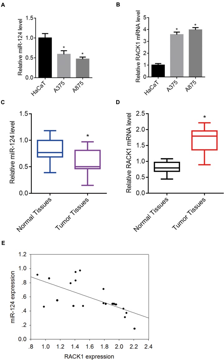
Real-time qPCR was used to evaluate the expression profiles of miR-124 and RACK1 in melanoma cell lines and tissue samples. The results showed that miR-124 levels were lower and RACK1 mRNA levels were higher in A375 and A875 cells (melanoma cell lines) compared with those in HaCaT cells (normal skin cell line) (P<0.05) (Figure 1A and B). Similar results were obtained when comparing melanoma tumor tissues with adjacent non-tumor skin tissues. Compared with non-tumor skin tissues, miR-124 levels were lower and RACK1 levels were higher in melanoma tissue samples (P<0.05) (Figure 1C and D). Spearman’s rank correlation analysis indicated that the expression of miR-124 was negatively correlated with that of RACK1 in melanoma tissue samples (r = −0.646, P<0.05) (Figure 1E). This negative correlation suggested that RACK1 might be a target gene of miR-124.
Figure 1.
Expression of miR-124 and RACK1 in melanoma cell lines and tissue samples. (A–B) The mRNA levels of miR-124 and RACK1 in A375, A875, and HaCaT cells were determined using real-time-qPCR. *P<0.05, compared with HaCaT cells. (C–D) The mRNA levels of miR-124 and RACK1 in melanoma tumor tissues and peripheral non-tumor skin tissues were determined using real-time-qPCR. *P<0.05, compared with normal tissues. (E) Correlation between the expression of miR-124 and RACK1 in patients with melanoma.
RACK1 Was A Direct Target Of miR-124
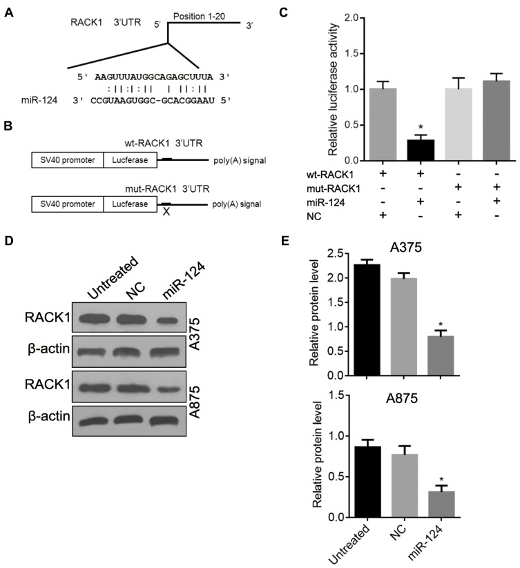
The binding site of miR-124 on the 3′UTR of RACK1 was predicted using software (Figure 2A). To evaluate the ability of miR-124 to bind to RACK1, wt-RACK1 and mut-RACK1 luciferase reporter vectors were constructed (Figure 2B). Dual-Luciferase reporter assay indicated that transfection of HEK293 cells with miR-124 mimic decreased the luciferase activity of wt-RACK1 compared with the NC-transfected group (lane 2 vs. lane 1, P<0.05, Figure 2C). In addition, no differences were observed between the mut-RACK1 and miR-124 mimic co-transfection groups, the mut-RACK-1 and NC co-transfection groups, or the wt-RACK1 and NC co-transfection groups.
Figure 2.
Inhibitory effects of miR-124 on the expression of RACK1 in melanoma cells. (A) The binding site of miR-124 on the 3′UTR of RACK1 was predicted using a software. (B) Design and construction of double luciferase reporter plasmid. (C) The effects of miR-124 on RACK1 expression were determined using the Dual-Luciferase reporter assay. *P<0.05, compared with the other three groups. (D–E) RACK1 protein level was inhibited by miR-124 in A375 and A875 cells. *P<0.05, compared with NC-treated or untreated cells. miR-124 mimics are abbreviated as miR-124 in the following figures.
Overexpression Of miR-124 Downregulated RACK1 Expression In Melanoma Cells
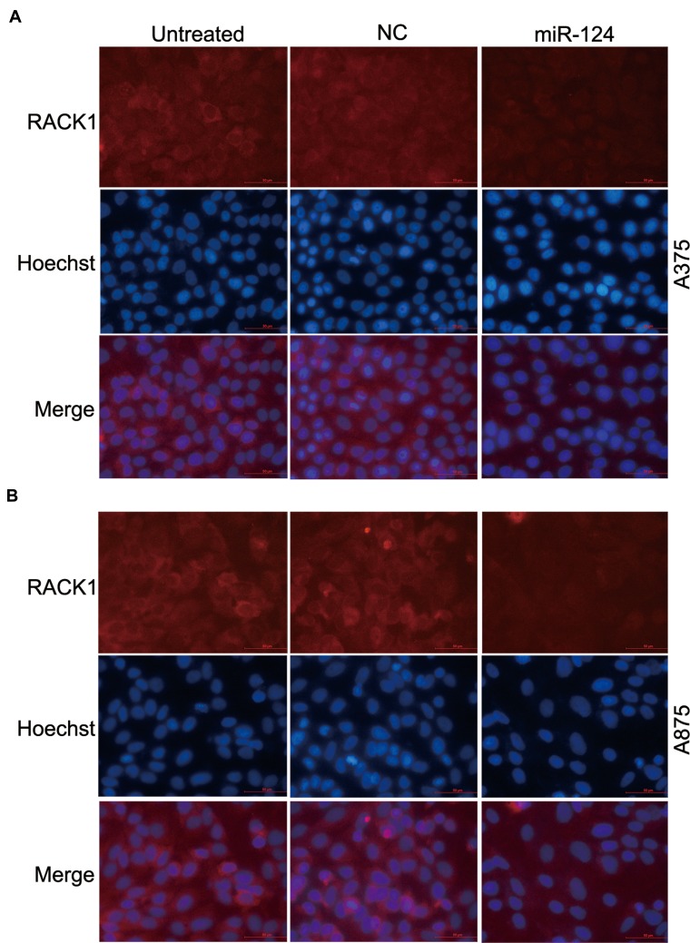
To investigate the inhibitory effects of miR-124 on the expression of RACK1 in melanoma cells, A375 and A875 cells were transfected with a miR-124 mimic to upregulate endogenous miR-124 levels. Western blot (P<0.05, Figure 2D and E) and fluorescence microscopy (P<0.05, Figure 3) analysis showed that miR-124 significantly inhibited RACK1 expression in A375 and A875 cells compared with NC-treated or untreated cells.
Figure 3.
Inhibition of RACK1 protein expression by miR-124 in melanoma cells. Cells were transfected with miR-124 mimic and NC, and the inhibitory effect of miR-124 was evaluated using fluorescence microscopy. (A) RACK1 staining in A375 cells. (B) RACK1 staining in A875 cells. Red staining indicated RACK1 expression, and the cell nucleus was stained with Hoechst 33258 (blue). Scale bar=50 μm.
Overexpression Of miR-124 Inhibited Proliferation Of Melanoma Cells
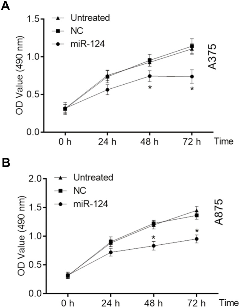
We used the MTT assay to verify the inhibitory effects of miR-124 on melanoma cell growth. The results showed that miR-124 inhibited the growth of A375 and A875 cells at 48 hrs and 72 hrs compared with NC-treated or untreated cells (P<0.05, Figure 4).
Figure 4.
Inhibitory effects of miR-124 on proliferation of melanoma cells. MTT analysis was performed on untreated, NC-treated, and miR-124 mimic-transfected melanoma cells. Data at each time point were recorded and represented using line graphs. (A) A375 cells; (B) A875 cells. *P<0.05, compared with NC-treated or untreated cells.
Overexpression Of miR-124 Inhibited Migration And Invasion Of Melanoma Cells
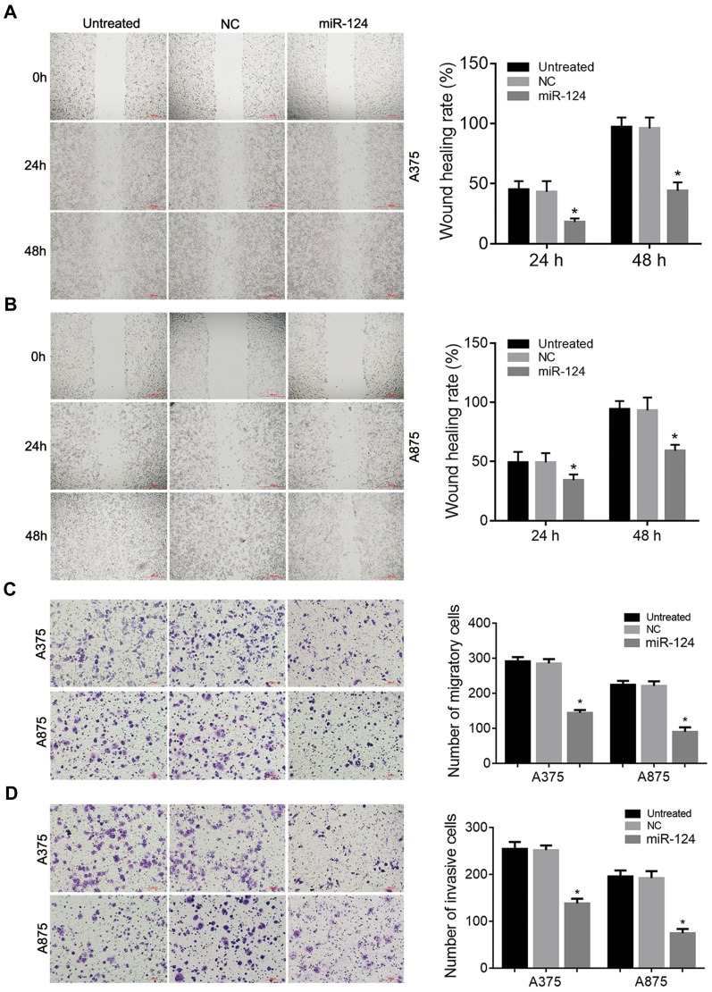
The inhibitory effect of miR-124 on migration of melanoma cells was evaluated using the wound-healing and transwell assays, and invasion of melanoma cells was evaluated using the matrigel-based transwell assay. The results showed that overexpression of miR-124 significantly decreased migration (P<0.05, Figure 5A–C) and invasion of A375 and A875 cells compared with NC-treated or untreated cells (P<0.05, Figure 5D).
Figure 5.
Inhibitory effects of miR-124 on migration and invasion of melanoma cells. Untreated, NC-reated, and miR-124 mimic-transfected melanoma cells (A375 and A875) were used in these experiments. (A–B) Cell migration was evaluated using the wound-healing assay. The scale bar indicates 500 μm. (C) Cell migration was evaluated using the transwell assay. The scale bar indicates 100 μm. (D) Invasion of cells was evaluated using the matrigel transwell assay. The scale bar indicates 100 μm. *P<0.05, compared with NC-treated or untreated cells.
Overexpression Of miR-124 Induced Melanoma Cell Apoptosis
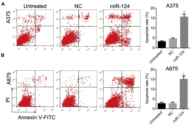
Annexin-V-FITC/PI followed by FCM was used to evaluate the effect of miR-124 on melanoma cell apoptosis. Overexpression of miR-124 induced A375 and A875 cell apoptosis (P<0.05, Figure 6).
Figure 6.
miR-124-induced apoptosis of melanoma cells. The apoptosis rates of untreated, NC-treated, and miR-124 mimic-transfected melanoma cells were determined using flow cytometry with Annexin V-FITC/PI staining. (A) miR-124 promoted A375 apoptosis. (B) miR-124 promoted A875 apoptosis. *P<0.05, compared with untreated or NC-treated cells.
Discussion
Melanoma, the most serious type of skin cancer, is one of the most rapidly growing malignant tumors.3,4 Traditional treatments for melanoma include surgery, chemotherapy, and radiotherapy. Recently, immunotherapy and targeted therapy have become frontline treatments for patients with advanced melanoma. miRNAs are small noncoding RNAs that cannot serve as templates for protein translation. However, by guiding the splitting of target genes or inhibiting translation, miRNAs act as posttranscriptional regulators and play key roles in development and disease progression.27 Studies have shown that aberrant expression of miRNAs may be associated with the development and progression of cancers.7,8 miRNAs have great potential as targets for the treatment of cancer because they interact with multiple targets. Future studies should focus on the identification of miRNAs as therapeutic targets in melanoma.
miR-124 is the most abundant miRNA in the adult brain and central nervous system of mammals.8,28,29 Previous studies showed that the expression level of miR-124 was significantly lower in a number of human cancers, and miR-124 has been identified as an important modulator of carcinogenesis.9–19 miR-124 can target IGF2BP1, resulting in decreased proliferation, migration, and invasion of cervical cancer.9 In addition, miR-124 can target Ras-related C3 botulinum toxin substrate 1 and specificity protein 1,10 resulting in reduced gastric cancer cell viability and tumor growth. Furthermore, miR-124 has been shown to suppress glioblastoma,11 hepatocellular carcinoma, pancreatic cancer,13 and lung cancer.14 In the present study, we found that miR-124 levels were significantly lower in A375 and A875 melanoma cell lines than those in the normal skin cell line HaCaT. In addition, miR-124 levels were significantly lower in melanoma tumor tissue than those in adjacent normal skin, which suggested that miR-124 might be involved in onset or progression of melanoma.
High metastasis and invasion rates are the main determinants of poor melanoma prognosis. To better understand the role of miR-124 in melanoma, we overexpressed miR-124 in A375 and A875 cells. Overexpression of miR-124 resulted in impaired cell proliferation, migration, invasion, and increased apoptosis. miR-124 targets a large number of genes, resulting in the modulation of transcriptional regulation, cell transport, and signal transduction. In this study, RACK1 was predicted as a target of miR-124 using online software. The interaction between miR-124 and RACK1 was confirmed using the Dual-Luciferase reporter assay. Further experiments showed that miR-124 expression was negatively correlated with RACK1 expression in melanoma tumor tissue, and A375 and A875 cells. In addition, the upregulation of miR-124 inhibited RACK1 expression in A375 and A875 cells, which further suggested that miRNA may modulate melanoma through targeting RACK1.
RACK1 plays an important role in cellular protein transport, anchoring proteins in specific subcellular locations, and regulating protein activity.23 As a classic scaffold protein for kinases and receptors, RACK1 regulates signal transduction between receptors and cytoskeleton proteins, which contributes to cell migration and invasion. Dysregulation of RACK1 has been reported in various tumors and has been identified as a carcinogenic molecule in pulmonary adenocarcinoma, colorectal carcinoma, glioma, breast cancer, and hepatocellular carcinoma.22 Furthermore, RACK1 regulates migration and invasion of tumor cells.23 In addition, RACK1 has been shown to control the biogenesis of a subset of miRNAs, including let-7, and has been shown to affect the development of Caenorhabditis elegans.30 In mammals, RACK1 contributes to the recruitment of miRISC to the site of translation.31,32 Chen reported that RACK1 bound to miR-302 in gastric cancer cell lines and tumor tissues.33 miR-124 and RACK1 are involved in many biological processes and regulation of cancers. As such, our results showed that miR-124 and RACK1 are potential targets for the diagnosis and treatment of melanoma.
In summary, this study showed that miR-124 may suppress melanoma and that low expression of miR-124 contributed to proliferation, migration, and invasion of tumor cells, and inhibited apoptosis. Furthermore, low miR-124 levels were associated with poor prognosis of patients with melanoma. We showed that RACK1 was a direct target of miR-124 and that miR-124/RACK1 may be involved in the onset and progression of melanoma.
Acknowledgment
This study was supported by the grant of National Natural Science Foundation of China (No. 81101178) and the Science Foundation of Nantong City, Jiangsu Province, China (No. yyz15023).
Informed Consent
Written informed consent was obtained from all individual participants included in the study.
Author Contributions
CS and XC contributed to conception and design of the study; SC collected the clinical samples and patient information; CS, HH, LG, HC, and XY performed the experiments; SC, HH, LG, CS, HC, and XY acquired, analyzed, and interpreted the data; CS and XC drafted the manuscript; HH, LG, SC, HC, and XY revised the manuscript. All authors contributed to data analysis, drafting and revising the article, gave final approval of the version to be published, and agree to be accountable for all aspects of the work.
Disclosure
The authors report no conflicts of interest in this work.
References
- 1.Atkinson V. Recent advances in malignant melanoma. Intern Med J. 2017;47(10):1114–1121. doi: 10.1111/imj.2017.47.issue-10 [DOI] [PubMed] [Google Scholar]
- 2.Fischer GM, Vashisht Gopal YN, McQuade JL, Peng W, DeBerardinis RJ, Davies MA. Metabolic strategies of melanoma cells: mechanisms, interactions with the tumor microenvironment, and therapeutic implications. Pigment Cell Melanoma Res. 2018;31(1):11–30. doi: 10.1111/pcmr.12661 [DOI] [PMC free article] [PubMed] [Google Scholar]
- 3.Moran B, Silva R, Perry AS, Gallagher WM. Epigenetics of malignant melanoma. Semin Cancer Biol. 2018;51:80–88. doi: 10.1016/j.semcancer.2017.10.006 [DOI] [PubMed] [Google Scholar]
- 4.Gladfelter P, Darwish NHE, Mousa SA. Current status and future direction in the management of malignant melanoma. Melanoma Res. 2017;27(5):403–410. doi: 10.1097/CMR.0000000000000379 [DOI] [PubMed] [Google Scholar]
- 5.Visvanathan J, Lee S, Lee B, Lee JW, Lee SK. The microRNA miR-124 antagonizes the anti-neural REST/SCP1 pathway during embryonic CNS development. Genes Dev. 2007;21(7):744–749. doi: 10.1101/gad.1519107 [DOI] [PMC free article] [PubMed] [Google Scholar]
- 6.Gregory RI, Yan KP, Amuthan G, et al. The microprocessor complex mediates the genesis of microRNAs. Nature. 2004;432(7014):235–240. doi: 10.1038/nature03120 [DOI] [PubMed] [Google Scholar]
- 7.Osada H, Takahashi T. MicroRNAs in biological processes and carcinogenesis. Carcinogenesis. 2007;28(1):2–12. doi: 10.1093/carcin/bgl185 [DOI] [PubMed] [Google Scholar]
- 8.Svoronos AA, Engelman DM, Slack FJ. OncomiR or tumor suppressor? The duplicity of MicroRNAs in cancer. Cancer Res. 2016;76(13):3666–3670. doi: 10.1158/0008-5472.CAN-16-0359 [DOI] [PMC free article] [PubMed] [Google Scholar]
- 9.Wang P, Zhang L, Zhang J, Xu G. microRNA-124-3p inhibits cell growth and metastasis in cervical cancer by targeting IGF2BP1. Exp Ther Med. 2018;15(2):1385–1393. doi: 10.3892/etm.2017.5528 [DOI] [PMC free article] [PubMed] [Google Scholar] [Retracted]
- 10.Liu F, Hu H, Zhao J, et al. miR-124-3p acts as a potential marker and suppresses tumor growth in gastric cancer. Biomed Rep. 2018;9(2):147–155. doi: 10.3892/br.2018.1113 [DOI] [PMC free article] [PubMed] [Google Scholar]
- 11.Zhang G, Chen L, Khan AA, et al. miRNA-124-3p/neuropilin-1(NRP-1) axis plays an important role in mediating glioblastoma growth and angiogenesis. Int J Cancer. 2018;143(3):635–644. doi: 10.1002/ijc.v143.3 [DOI] [PubMed] [Google Scholar]
- 12.Lu Y, Yue X, Cui Y, Zhang J, Wang K. MicroRNA-124 suppresses growth of human hepatocellular carcinoma by targeting STAT3. Biochem Biophys Res Commun. 2013;441(4):873–879. doi: 10.1016/j.bbrc.2013.10.157 [DOI] [PubMed] [Google Scholar]
- 13.Wang P, Chen L, Zhang J, et al. Methylation-mediated silencing of the miR-124 genes facilitates pancreatic cancer progression and metastasis by targeting Rac1. Oncogene. 2014;33(4):514–524. doi: 10.1038/onc.2012.598 [DOI] [PubMed] [Google Scholar]
- 14.Li X, Yu Z, Li Y, et al. The tumor suppressor miR-124 inhibits cell proliferation by targeting STAT3 and functions as a prognostic marker for postoperative NSCLC patients. Int J Oncol. 2015;46(2):798–808. doi: 10.3892/ijo.2014.2786 [DOI] [PubMed] [Google Scholar]
- 15.Zo RB, Long Z. MiR-124-3p suppresses bladder cancer by targeting DNA methyltransferase 3B. J Cell Physiol. 2018;234(1):464–474. doi: 10.1002/jcp.26591 [DOI] [PubMed] [Google Scholar]
- 16.Ma J, Weng L, Wang Z, et al. MiR-124 induces autophagy-related cell death in cholangiocarcinoma cells through direct targeting of the EZH2-STAT3 signaling axis. Exp Cell Res. 2018;366(2):103–113. doi: 10.1016/j.yexcr.2018.02.037 [DOI] [PubMed] [Google Scholar]
- 17.Tian F, Chen J, Zheng S, et al. miR-124 targets GATA6 to suppress cholangiocarcinoma cell invasion and metastasis. BMC Cancer. 2017;17(1):175. doi: 10.1186/s12885-017-3166-z [DOI] [PMC free article] [PubMed] [Google Scholar]
- 18.Cai WL, Huang WD, Li B, et al. microRNA-124 inhibits bone metastasis of breast cancer by repressing Interleukin-11. Mol Cancer. 2018;17(1):9. doi: 10.1186/s12943-017-0746-0 [DOI] [PMC free article] [PubMed] [Google Scholar]
- 19.Yu B, Jiang K, Zhang J. MicroRNA-124 suppresses growth and aggressiveness of osteosarcoma and inhibits TGF-β-mediated AKT/GSK-3β/SNAIL-1 signaling. Mol Med Rep. 2018;17(5):6736–6744. doi: 10.3892/mmr.2018.8637 [DOI] [PubMed] [Google Scholar]
- 20.Ron D, Chen CH, Caldwell J, Jamieson L, Orr E, Mochly-Rosen D. Cloning of an intracellular receptor for protein kinase C: a homolog of the beta subunit of G proteins. Proc Natl Acad Sci U S A. 1994;91(3):839–843. doi: 10.1073/pnas.91.3.839 [DOI] [PMC free article] [PubMed] [Google Scholar]
- 21.Adams DR, Ron D, Kiely PA. RACK1, a multifaceted scaffolding protein: structure and function. Cell Commun Signal. 2011;9:22. doi: 10.1186/1478-811X-9-22 [DOI] [PMC free article] [PubMed] [Google Scholar]
- 22.Li JJ, Xie D. RACK1, a versatile hub in cancer. Oncogene. 2015;34(15):1890–1898. doi: 10.1038/onc.2014.127 [DOI] [PubMed] [Google Scholar]
- 23.Duff D, Long A. Roles for RACK1 in cancer cell migration and invasion. Cell Signal. 2017;35:250–255. doi: 10.1016/j.cellsig.2017.03.005 [DOI] [PubMed] [Google Scholar]
- 24.Chou CH, Shrestha S, Yang CD, et al. miRTarBase update 2018: a resource for experimentally validated microRNA-target interactions. Nucleic Acids Res. 2018;46(D1):D296–D302. doi: 10.1093/nar/gkx1067 [DOI] [PMC free article] [PubMed] [Google Scholar]
- 25.Chen C, Ridzon DA, Broomer AJ, et al. Real-time quantification of microRNAs by stem-loop RT-PCR. Nucleic Acids Res. 2005;33(20):e179. doi: 10.1093/nar/gni178 [DOI] [PMC free article] [PubMed] [Google Scholar]
- 26.Livak KJ, Schmittgen TD. Analysis of relative gene expression data using real-time quantitative PCR and the 2(-delta delta C(T)) method. Methods. 2001;25(4):402–408. doi: 10.1006/meth.2001.1262 [DOI] [PubMed] [Google Scholar]
- 27.Valentina I, Gerber AP. Combinatorial control of mRNA fates by RNA-binding proteins and non-coding RNAs. Biomolecules. 2015;5(4):2207–2222. doi: 10.3390/biom5042207 [DOI] [PMC free article] [PubMed] [Google Scholar]
- 28.Lagos-Quintana M, Rauhut R, Yalcin A, Meyer J, Lendeckel W, Tuschl T. Identification of tissue-specific microRNAs from mouse. Curr Biol. 2002;12(9):735–739. doi: 10.1016/S0960-9822(02)00809-6 [DOI] [PubMed] [Google Scholar]
- 29.Krichevsky AM, Sonntag KC, Isacson O, Kosik KS. Specific microRNAs modulate embryonic stem cell-derived neurogenesis. Stem Cells. 2006;24(4):857–864. doi: 10.1634/stemcells.2005-0441 [DOI] [PMC free article] [PubMed] [Google Scholar]
- 30.Chu YD, Wang WC, Chen SA, et al. RACK-1 regulates let-7 microRNA expression and terminal cell differentiation in Caenorhabditis elegans. Cell Cycle. 2014;13(12):1995–2009. doi: 10.4161/cc.29017 [DOI] [PMC free article] [PubMed] [Google Scholar]
- 31.Speth C, Laubinger S. RACK1 and the microRNA pathway: is it deja-vu all over again? Plant Signal Behav. 2014;9(2):e27909. doi: 10.4161/psb.27909 [DOI] [PMC free article] [PubMed] [Google Scholar]
- 32.Jannot G, Bajan S, Giguère NJ, et al. The ribosomal protein RACK1 is required for microRNA function in both C. elegans and humans. EMBO Rep. 2011;12(6):581–586. doi: 10.1038/embor.2011.66 [DOI] [PMC free article] [PubMed] [Google Scholar]
- 33.Chen L, Min L, Wang X, et al. Loss of RACK1 promotes metastasis of gastric cancer by inducing a miR-302c/IL8 signaling loop. Cancer Res. 2015;75(18):3832–3841. doi: 10.1158/0008-5472.CAN-14-3690 [DOI] [PubMed] [Google Scholar]


