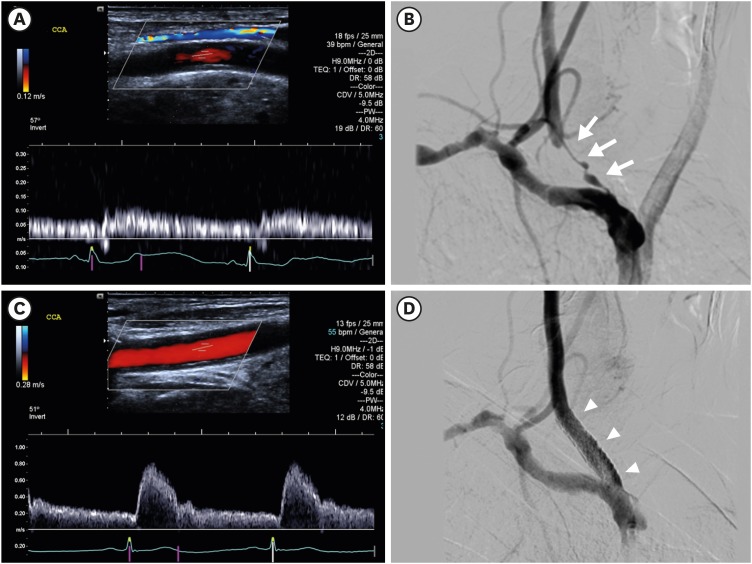
An 81-year-old man, with a history of radiation therapy (RTx) 40 years ago, presented a sudden blindness and was diagnosed as acute ocular ischemic syndrome. On carotid Doppler ultrasonography, right common carotid artery (CCA) showed increased intima-media thickness (IMT) without a significant stenosis. Doppler examination revealed a delayed slow-rising upstroke and decreased flow velocity (pulsus parvus et tardus, Figure 1A). Magnetic resonance angiography showed critical narrowing of proximal portion of right CCA (arrows, Figure 2A) with diffuse irregular narrowing of right subclavian artery (filled arrowheads) and whole left CCA (open arrowheads, Figure 2A). He underwent a stent implantation. Pre-procedural angiogram demonstrated critical narrowing of right CCA (arrows, Figure 1B), and post-procedural angiogram revealed no stenotic component (arrowheads, Figure 1D). Pre-procedural intravascular ultrasound examination revealed increased carotid IMT and mixed echogenic plaque without an acoustic shadowing (Figure 2B). Follow-up ultrasound showed normalized flow velocities (Figure 1C).
Figure 1. Carotid Doppler ultrasonography shows increased intima-media thickness of the right CCA without a significant stenosis. Doppler examination reveals a delayed slow-rising upstroke and decreased flow velocity (pulsus parvus et tardus, A). Pre-procedural angiogram demonstrates critical narrowing of right CCA (arrows, B). Post-procedural carotid Doppler ultrasonography shows normalized flow velocities of CCA (C) and post-procedural angiogram reveals no stenotic component (arrowheads, D).
CCA = common carotid artery.
Figure 2. (A) Magnetic resonance angiography shows critical narrowing of proximal portion of right CCA (arrows) with diffuse irregular narrowing of right subclavian artery (filled arrowheads) and whole left CCA (open arrowheads). (B) Pre-procedural intravascular ultrasound examination reveals increased carotid IMT and mixed echogenic plaque without an acoustic shadowing.
CCA = common carotid artery; IMT = intima-media thickness.
RTx can cause vascular injury and subsequent stenosis by enhancing atherosclerosis and elastic degradation1) and consequently increase the risk of ischemic stroke.2) Stenotic lesions associated with RTx tend to be longer, usually bilateral involvement, and the maximal stenotic lesion is inclined to distribute at proximal portion of CCA. Imaging studies can demonstrate characteristic findings including diffuse narrowing of the involved artery with increased IMT and proximal narrowing with decreased flow velocities and a decreased pulse. The treatment can be done with carotid endarterectomy or carotid artery stenting (CAS).3),4) In our patient, the stenotic lesion was successfully treated with CAS because of surgical difficulty.
Footnotes
Conflict of Interest: The authors have no financial conflicts of interest.
- Conceptualization: Park JH, Lee JH.
- Data curation: Shin JW.
- Writing - original draft: Park JH.
- Writing - review & editing: Park JH, Lee JH.
References
- 1.Venkatesulu BP, Mahadevan LS, Aliru ML, et al. Radiation-induced endothelial vascular injury: a review of possible mechanisms. JACC Basic Transl Sci. 2018;3:563–572. doi: 10.1016/j.jacbts.2018.01.014. [DOI] [PMC free article] [PubMed] [Google Scholar]
- 2.Murros KE, Toole JF. The effect of radiation on carotid arteries. A review article. Arch Neurol. 1989;46:449–455. doi: 10.1001/archneur.1989.00520400109029. [DOI] [PubMed] [Google Scholar]
- 3.Houdart E, Mounayer C, Chapot R, Saint-Maurice JP, Merland JJ. Carotid stenting for radiation-induced stenoses: a report of 7 cases. Stroke. 2001;32:118–121. doi: 10.1161/01.str.32.1.118. [DOI] [PubMed] [Google Scholar]
- 4.Park JH, Lee JH. Carotid artery stenting. Korean Circ J. 2018;48:97–113. doi: 10.4070/kcj.2017.0208. [DOI] [PMC free article] [PubMed] [Google Scholar]


