Abstract
Scytalidium auriculariicola is the causative pathogen of slippery scar disease in the cultivated cloud ear fungus, Auricularia polytricha. In the present study, the mitogenome of S. auriculariicola was sequenced and assembled by next-generation sequencing technology. The circular mitogenome is 96,857 bp long and contains 56 protein-coding genes, 2 ribosomal RNA genes, and 30 transfer RNA genes (tRNAs). The high frequency of A and T used in codons contributed to the high AT content (73.70%) of the S. auriculariicola mitogenome. Comparative analysis indicated that the base composition and the number of introns and protein-coding genes in the S. auriculariicola mitogenome varied from that of other Leotiomycetes mitogenomes, including a uniquely positive AT skew. Five distinct groups were found in the gene arrangements of Leotiomycetes. Phylogenetic analyses based on combined gene datasets (15 protein-coding genes) yielded well-supported (BPP = 1) topologies. A single-gene phylogenetic tree indicated that the nad4 gene may be useful as a molecular marker to analyze the phylogenetic relationships of Leotiomycetes species. This study is the first report on the mitochondrial genome of the genus Scytalidium, and it will contribute to our understanding of the population genetics and evolution of S. auriculariicola and related species.
Subject terms: Evolution, Microbiology
Introduction
Scytalidium auriculariicola is the causative pathogen of slippery scar on the cloud ear fungus Auricularia polytricha (Mont.) Sacc1. The pathogen only infects the mycelia of A. polytricha and not the fruiting body2. Slippery scar first appeared in cultivated A. polytricha in Sichuan Province in the 1990s, with the infection rate being over 40%2. The identity of the pathogen of slippery scar has been controversial and has undergone a process of re-identification and re-establishment. Sun et al.2 initially identified the pathogen as the Ascomycete S. lignicola according to Koch’s postulates, morphological observations, rDNA-internal transcribed spacer (ITS), and 18S sequence analysis. Peng et al.1 further investigated the causative pathogen of this disease by morphological observations, in vivo pathogenicity tests, and molecular evidence from rRNA ITS and RNA polymerase II subunit (RPB2) sequences. Their results showed that the pathogen is a new species in the Scytalidium genus differing from S. lignicola, which they named S. auriculariicola1. Peng et al.1 also pointed out that S. auriculariicola strains from diseased A. polytricha mycelium in China consist of two clonal groups. Previously, ITS rDNA sequences, 18S rDNA sequences, RPB2 genes, and ribosomal large subunit (LSU) genes have been used as molecular markers to determine species relationships between Scytalidium and other species within the Leotiomycetes1,3–5. However, limited genetic information has prevented a comprehensive understanding of phylogenetic relationships between Scytalidium and its related species6. Thus, more available molecular markers are needed to assess the relationships between Scytalidium and other taxa within the Leotiomycetes, especially between the important S. auriculariicola and its related species.
Mitochondria are organelles necessary for the life of eukaryotic cells, and their dysfunction is associated with disease, the aging process, development, and various biological traits7,8. Mitochondria are thought to originate from symbiotic bacteria and supply most of the energy for eukaryotes9. Mitochondrial genomes are independent from nuclear genomes and have maternal inheritance, a high copy number, larger sequence length than the rRNA gene, which has made them widely used as a tool for studying evolution, phylogenetics, population genetics, and comparative or evolutionary genomics10,11. Mitochondrial gene rearrangements and the secondary structure of tRNAs are also widely used for deep-level phylogenetic studies in eukaryotes12–14. However, the mitogenomes of fungi are not as well studied as those of animals and plants15. The limited available studies have shown that most fungal mitochondrial genomes are circular, with the exception of very few species with linear mitogenomes16,17. The size of fungal mitogenomes varies greatly, ranging from 18.84 kb (Hanseniaspora uvarum) to 235.85 kb18. Fungal mitochondrial DNA usually contains 14 conserved protein-coding genes (atp6, atp8, atp9, cob, cox1, cox2, cox3, nad1, nad2, nad3, nad4, nad4L, nad5, and nad6), 1 ribosomal protein S3 gene (rps3), 2 ribosomal RNA genes (rnl and rns), and a relatively constant set of tRNA genes19. Fungal mitogenomes sometimes include homing endonuclease genes, plasmid-derived genes, genes transferred from the nuclear genome, and genes of unknown function. The number and variation of mitochondrial genes, genome rearrangement, and the presence or absence of large intronic and intergenic sequences all contribute to the considerable variation in gene content, structure, and size of fungal mitogenomes. To date, no mitochondrial genomes are available in the Scytalidium, and only 16 species from the Leotiomycetes class, including Cairneyella variabilis, Marssonina brunnea, Phialocephala subalpina, 2 Glarea lozoyensis subspecies, 4 Pseudogymnoascus species, 4 Rhynchosporium species, 2 Sclerotinia species, and Botrytis cinera (teleomorph Botryotinia fuckeliana), have been sequenced20 (https://www.ncbi.nlm.nih.gov/genome/browse#!/organelles/Leotiomycetes). More mitogenomes are needed, especially from the Scytalidium genus, which contains multiple pathogens, to reveal species and genus-level phylogenetic relationships in Leotiomycetes.
In the present study, the pathogen S. auriculariicola was isolated, identified, and sequenced. We assembled and annotated the complete mitochondrial genome of S. auriculariicola and assessed its gene content, tRNA structure, and genome organization. The mitochondrial genome size, base composition, number of protein-coding genes, tRNA genes, and gene arrangement of S. auriculariicola and previously sequenced species within Leotiomycetes were compared. We then analyzed the phylogenetic relationships among various Leotiomycetes species based on mitochondrial genes. The mitochondrial genome of S. auriculariicola will allow further investigations into the taxonomy, phylogenetics, conservation genetics, and evolutionary biology of this important genus as well as other closely related species.
Results
Protein coding genes, rRNA genes, tRNA genes, intergenic regions, and codon analyses
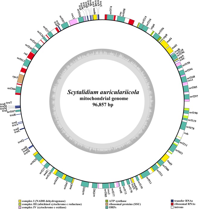
The mitogenome of S. auriculariicola is composed of a circular DNA molecule of 96,857 bp (Fig. 1). The GC content of the S. auriculariicola mitogenome is 26.30%. The AT skew and GC skew were both positive in the S. auriculariicola mitogenome (Table 1). A total of 56 protein-coding genes were identified in the mitogenome of S. auriculariicola, including 15 core mitochondrial genes (atp6, atp8, atp9, cob, cox1, cox2, cox3, nad1, nad2, nad3, nad4, nad4L, nad5, nad6, and rps3) and 41 ORFs (Table S1). Among these ORFs, we found 32 ORFs located in introns and the remaining 9 ORFs were regarded as free-standing. Twenty-one out of the 32 intronic ORFs were predicted to encode proteins that exhibit similarities to the homing endonucleases from the LAGLIDADG family. Eight ORFs are similar to homing endonucleases in the family GIY-YIG, and 3 ORFs were predicted to encode hypothetical proteins. We also found seven free-standing ORFs that were predicted to encode proteins that exhibit similarities to homing endonucleases in the LAGLIDADG (four ORFs) and GIY-YIG (three ORFs) families. In addition, two free-standing ORFs were predicted to encode hypothetical proteins (Table S1). All of the 56 detected mitochondrial genes were located on the sense strand.
Figure 1.
Circular map of the Scytalidium auriculariicola mitochondrial genome. Various genes are represented with different color blocks. The mitochondrial circular map was drawn using the OGDRAW software58.
Table 1.
Comparison of Leotiomycetes mitogenomes.
| Item | Accession number | Genome size (bp) | GC content (%) | AT skew | GC skew | conserved PCGs | free-standing ORFs | No. of introns | Intronic ORFs | No. of tRNAs |
|---|---|---|---|---|---|---|---|---|---|---|
| Scytalidium auriculariicola | MK111108 | 96,857 | 26.30 | 0.030 | 0.127 | 14 | 9 | 27 | 32 | 30 |
| Cairneyella variabilis | NC_029759 | 27,186 | 26.27 | −0.043 | 0.133 | 14 | 1 | 1 | 1 | 29 |
| Glarea lozoyensis | NC_031375 | 45,501 | 29.76 | −0.041 | 0.081 | 14 | 4 | 5 | 2 | 30 |
| Marssonina brunnea | NC_015991 | 70,379 | 29.34 | −0.009 | 0.068 | 14 | 8 | 18 | 13 | 28 |
| Phialocephala subalpina | NC_015789 | 43,742 | 27.95 | −0.029 | 0.091 | 14 | 9 | 0 | 0 | 27 |
| Pseudogymnoascus destructans | NC_033907 | 32,181 | 28.53 | −0.048 | 0.116 | 13 | 3 | 2 | 2 | 28 |
| Pseudogymnoascus pannorum | NC_027422 | 26,918 | 28.10 | −0.063 | 0.113 | 13 | 1 | 1 | 1 | 27 |
| Pseudogymnoascus_sp._04NY16 | CM004376 | 32,146 | 28.66 | −0.050 | 0.118 | 13 | 4 | 2 | 2 | 28 |
| Pseudogymnoascus_sp._BL308 | CM004375 | 32148 | 28.55 | −0.045 | 0.112 | 13 | 4 | 2 | 2 | 29 |
| Rhynchosporium agropyri | NC_023125 | 68,904 | 29.36 | −0.006 | 0.066 | 14 | 9 | 19 | 25 | 29 |
| Rhynchosporium commune | NC_023126 | 69,581 | 29.40 | −0.006 | 0.068 | 14 | 9 | 18 | 25 | 29 |
| Rhynchosporium orthosporum | NC_023127 | 49,539 | 28.80 | −0.028 | 0.086 | 14 | 9 | 3 | 2 | 32 |
| Rhynchosporium secalis | NC_023128 | 68,729 | 29.33 | −0.007 | 0.069 | 14 | 8 | 18 | 25 | 29 |
| Sclerotinia borealis | NC_025200 | 203,051 | 32.01 | −0.001 | 0.084 | 15 | 18 | 61 | 62 | 31 |
| Sclerotinia sclerotiorum | NC_035155 | 128,852 | 30.90 | −0.007 | 0.103 | 14 | 21 | 36 | 32 | 35 |
| Botrytis cinerea | Broad Institute | 82,212 | 29.90 | −0.008 | 0.100 | 14 | 7 | 22 | 20 | 33 |
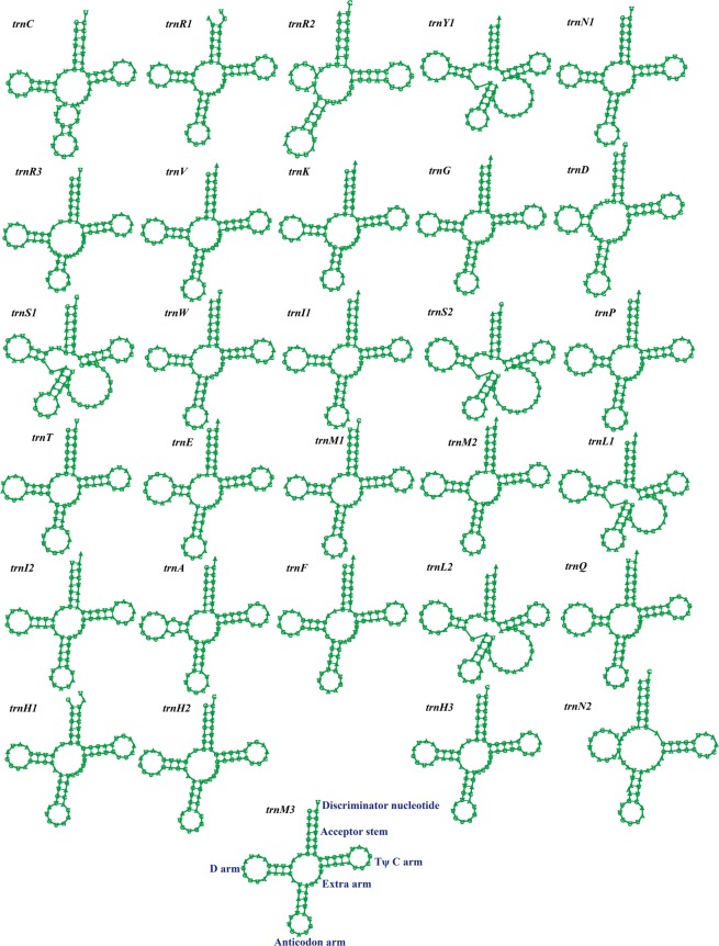
The mitogenome of S. auriculariicola contains two rRNA genes, a large subunit ribosomal RNA gene (rnl), and a small subunit ribosomal RNA gene (rns). A total of 30 tRNA genes were detected. The length of individual tRNAs ranges from 71 to 86 bp, mainly due to the different sizes of the extra arms (Fig. 2). The main cluster of tRNA genes (16 tRNAs) is located around the rnl gene. The other two clusters, including ten and two tRNA genes, are located around the nad6 and atp9 genes, respectively. In addition, there are two other tRNA genes located between the nad4L and cox2 genes (Fig. 1). There are mispairings of bases in 27 out of the 30 tRNAs, with a total of 45 base mispairings, all of which are G-U mispairings (Table S2). The S. auriculariicola mitogenome contains two tRNAs that code for asparagine with the same anticodons, three tRNAs coding for methionine with the same anticodons, and three tRNAs that code for histidine with the same anticodons. In addition, there are four amino acids that have more than one anticodon coding for them (Table S3).
Figure 2.
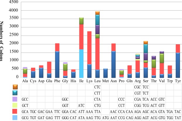
Codon usage in the Scytalidium auriculariicola mitochondrial genome. Codon families are plotted on the X axis and represented by different color patches. Frequency of codon usage is plotted on the Y axis.
The mitogenome of S. auriculariicola contains three pairs of overlapping genes: orf196/orf118 overlap by 23 nucleotides, nad4L/nad5 and nad2/orf447 overlap by 1 bp. In addition, the S. auriculariicola mitogenome contains 55 intergenic regions with lengths ranging from 5 to 1986 bp, with a total length of 20,534 bp (Table S1). Intergenic nucleotides account for 21.20% of the mitogenome.
All of the 15 conserved protein-coding genes start with the canonical translation initiation codon ATG except for cox1 and nad4, which start with TTG (Table S4). Five genes (atp9, cox3, nad3, cox2, and rps3) use TAG as the stop codon while the others use TAA. Codon usage analysis indicated that the most frequently used codons in the S. auriculariicola mitogenome are AAA (6.53%, for lysine; Lys), TTA (6.23%, leucine; Leu), ATA (5.68%, for isoleucine; Ile), AAT (5.34%, for asparagine; Asn), TTT (5.31%, for phenylalanine; Phe), and TAT (5.02%, for tyrosine; Tyr) (Fig. 3; Table S5). The high frequency of A and T use in codons contributes to the high AT content (73.70%) of the S. auriculariicola mitochondrial genome.
Figure 3.
Putative secondary structures of the 30 tRNA genes from Scytalidium auriculariicola mitochondrial genome. The tRNAs are labeled with the abbreviations of their corresponding amino acids. The tRNA arms are illustrated as for trnM3. The map of tRNA structures was drawn using the mitos software69.
Repetitive elements analysis
There are 33 repeat regions that were identified by a BLASTn search of the S. auriculariicola mitogenome against itself. The size of the repeats ranges from 39 to 610 bp; 19 repeat regions are over 100 bp, and 10 are over 200 bp. The similarities of these repeated sequences are between 74.81% and 100%. The longest repeat regions are located in the intergenic region between nad2 and orf173. The repeat sequences account for 5.65% of the entire S. auriculariicola mitogenome (Table S6).
We detected 26 tandem repeats in the mitogenome of S. auriculariicola. The length of the tandem repeats ranges from 6 to 77 bp. Copy numbers of each tandem repeat were between 2.0 and 16.5. The longest tandem sequence occurs with two copies. The tandem sequences account for 1.58% of the entire mitogenome (Table S7). We identified 47 forward, 1 palindromic, and 2 reverse repeats in the mitogenome of S. auriculariicola, accounting for 3.70% of the entire mitogenome (Table S8).
Comparative genome analysis
The size of S. auriculariicola mitogenome is the third largest genome among the 16 mitogenomes in the Leotiomycetes (Table 1), being only smaller than those of Sclerotinia borealis and Sclerotinia sclerotiorum, which belong to the family Sclerotiniaceae, order Helotiales. The GC content of the S. auriculariicola mitogenome is very low (26.30%) and is only higher than that of C. variabilis (26.27%). Both the AT skew and GC skew of the S. auriculariicola mitogenome are positive, while the AT skew is negative and GC skew positive in the other mitogenomes from the Leotiomycetes. The number of protein-coding genes and introns is closely related to the size of mitogenomes. The S. borealis with the largest mitogenome has 95 protein-coding genes and 61 introns, while the P. pannorum with the smallest mitogenome has only 15 protein-coding genes and one intron. The number of tRNA genes in the mitogenomes of Leotiomycetes varies from 27 to 35 (Table 1).
The composition of Leotiomycetes mitogenomes is shown in Table 2. The size of RNA genes and conserved protein-coding genes is relatively conservative between different species. The size of introns and intergenic regions is found closely related to the size variations of mitogenomes. Free-standing ORFs also contribute to the size variation of Leotiomycetes mitogenomes, which varies greatly between different species. The mitogenome of S. sclerotiorum contained the largest size of free-standing ORFs, followed by S. borealis, R. orthosporum and P. subalpina. The mitogenome of S. borealis contains the largest repeated sequences (exceeding 20 kb), while C. variabilis mitogenome contains the smallest (only 30 bp).
Table 2.
The size of different mitochondrial regions and their proportion to the whole mitogenome.
| ID | Mitogenome | RNA genes | Introns | Conserved PCGs | Free-standing ORFs | Intergenic regions | Repeated sequences | ||||||
|---|---|---|---|---|---|---|---|---|---|---|---|---|---|
| size (bp) | size (bp) | Proportion | size (bp) | proportion | size (bp) | proportion | size (bp) | proportion | size (bp) | proportion | size (bp) | proportion | |
| Sbo | 203051 | 7000 | 3.45 | 125696 | 61.90 | 13935 | 6.86 | 10461 | 5.15 | 45959 | 22.63 | 23122 | 11.39 |
| Ssc | 128852 | 7016 | 5.45 | 53723 | 41.69 | 12757 | 9.90 | 12690 | 9.85 | 42666 | 33.11 | 12321 | 9.56 |
| Sau | 96857 | 6865 | 7.09 | 48702 | 50.28 | 13668 | 14.11 | 7113 | 7.34 | 20509 | 21.17 | 5470 | 5.65 |
| Bci | 82212 | 7343 | 8.93 | 31793 | 38.67 | 13242 | 16.11 | 6351 | 7.73 | 23483 | 28.56 | 4723 | 5.74 |
| Mbr | 70379 | 6916 | 9.83 | 19289 | 27.41 | 15098 | 21.45 | 6246 | 8.87 | 22830 | 32.44 | 6402 | 9.10 |
| Rco | 69581 | 7073 | 10.17 | 27385 | 39.36 | 14118 | 20.29 | 6633 | 9.53 | 14372 | 20.66 | 3551 | 5.10 |
| Rag | 68904 | 7074 | 10.27 | 28435 | 41.27 | 13529 | 19.63 | 6024 | 8.74 | 13842 | 20.09 | 2694 | 3.91 |
| Rse | 68729 | 7073 | 10.29 | 27385 | 39.84 | 15099 | 21.97 | 6255 | 9.10 | 12917 | 18.79 | 3466 | 5.04 |
| Ror | 49539 | 7310 | 14.76 | 4016 | 8.11 | 13419 | 27.09 | 9177 | 18.52 | 15617 | 31.52 | 7363 | 14.86 |
| Glo | 45501 | 7432 | 16.33 | 5525 | 12.14 | 15456 | 33.97 | 2886 | 6.34 | 14202 | 31.21 | 5726 | 12.58 |
| Psu | 43742 | 7411 | 16.94 | 0 | 0.00 | 13938 | 31.86 | 8217 | 18.79 | 14176 | 32.41 | 2838 | 6.49 |
| Pde | 32181 | 6495 | 20.18 | 3334 | 10.36 | 12681 | 39.41 | 3429 | 10.66 | 6242 | 19.40 | 1144 | 3.55 |
| Psp.1 | 32148 | 6564 | 20.42 | 3332 | 10.36 | 12488 | 38.85 | 2967 | 9.23 | 6797 | 21.14 | 1112 | 3.46 |
| Psp.2 | 32146 | 6493 | 20.20 | 3332 | 10.37 | 12359 | 38.45 | 2925 | 9.10 | 7037 | 21.89 | 1218 | 3.79 |
| Cva | 27186 | 7147 | 26.29 | 1905 | 7.01 | 13176 | 48.47 | 753 | 2.77 | 4205 | 15.47 | 30 | 0.11 |
| Ppa | 26918 | 6407 | 23.80 | 1999 | 7.43 | 12621 | 46.89 | 363 | 1.35 | 5528 | 20.54 | 742 | 2.76 |
Species information in this table can be found in Supplementary Table S9.
Gene rearrangements
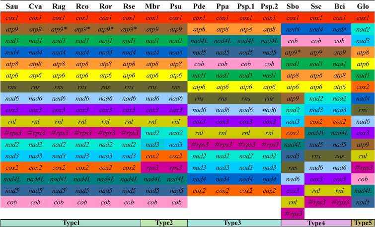
Five different groups of gene arrangement were detected in the 16 Leotiomycetes mitogenomes (Fig. 4). The results indicated that Leotiomycetes have undergone large-scale gene rearrangements in their mitogenomes over the course of evolution. The gene order of the S. auriculariicola mitogenome is identical to that of C. variabilis, which belongs to the Helotiaceae, order Helotiales. However, large-scale gene rearrangements were observed in the G. lozoyensis mitogenome, another species of the Helotiaceae. The mitochondrial gene order of four Rhynchosporium species, R. agropyri, R. commune, R. orthosporum, and R. secalis, is the same as that of S. auriculariicola. However, the atp9 genes of Rhynchosporium sp. are likely dysfunctional due to a premature stop codon in a conserved domain21. The mitochondrial gene order of S. auriculariicola is also similar to those of M. brunnea and P. subalpine, with the position of the rps3 gene in their mitogenomes differing. In the Pseudogymnoascus genus, we observed an identical mitochondrial gene order between the four Pseudogymnoascus species, P. destructans, P. pannorum, P. sp.04NY16, and P. sp.BL308. In the Sclerotiniaceae family, the order of mitochondrial genes of three Sclerotiniaceae species, S. sclerotiorum, S. borealia, and B. cinerea, is identical, except for minor differences (the location of atp9) in the S. borealia mitogenome due to replication events of atp9 genes. In addition, large-scale gene rearrangements were observed between the mitogenomes of S. auriculariicola, the Sclerotiniaceae, and Pseudogymnoascus.
Figure 4.
Comparison of gene order across 16 Leotiomycetes mitogenomes. Cva, Cairneyella variabilis (NC_029759); Glo, Glarea lozoyensis (NC_031375); Mbr, Marssonina brunnea f. sp. Multigermtubi (NC_015991); Psu, Phialocephala subalpine (NC_015789); Pde, Pseudogymnoascus destructans (NC_033907); Ppa, Pseudogymnoascus pannorum (NC_027422); Psp1, Pseudogymnoascus_sp._04NY16 (CM004376); Psp2, Pseudogymnoascus_sp._BL308 (CM004375); Rag, Rhynchosporium agropyri (NC_023125); Rco, Rhynchosporium commune (NC_023126); Ror, Rhynchosporium orthosporum (NC_023127); Rse, Rhynchosporium secalis (NC_023128); Sbo, Sclerotinia borealis (NC_025200); Ssc, Sclerotinia sclerotiorum 1980 UF-70 (NC_035155); Bci, Botrytis cinerea (Broad Institute); Sau, Scytalidium auriculariicola (MK111108). The # symbol indicates the rps3 gene is located in the intron of the rnl gene.
Compared with the arrangement of rps3 genes in type 1, the rps3 genes in the gene arrangement type 2 become free-standing ORFs, and are transferred to the downstream of the cox2 gene. The gene arrangement type 3 is characterized by the loss of atp9 gene, and the relocation of nad4, cox2, atp8, nad4L, nad5, cob, and nad1 genes. For the gene arrangement type 4, the rns, nad6, cox3, rnl and rps3 genes are transferred to the downstream of nad5 gene; nad4 and cob genes are transferred to the upstream of the atp9 gene. The type5 involves multiple gene repositions, including atp9, cox2, cox3, nad1, nad2, nad3, and nad4 genes.
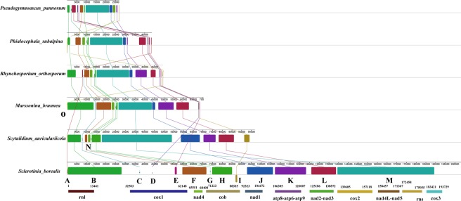
Synteny analysis indicated that the six Leotiomycetes mitogenomes can be divided into fifteen homologous regions, where the sizes and relative positions of these regions are highly variable (Fig. 5). Nine of the fifteen homologous regions were detected in all six mitogenomes. The R. orthosporum mitogenome has all 15 homologous regions while the other mitogenomes lack one to three homologous regions. M. brunnea lacks the homologous region ‘A’, S. borealia lacks the homologous regions ‘N’ and ‘O’, and S. auriculariicola lacks the homologous regions ‘D’, ‘E’, and ‘O’.
Figure 5.
Mitogenome synteny among six Leotiomycetes species. Synteny analyses were generated in Mauve 2.4.0. Fifteen homologous regions were identified among the six mitogenomes. The sizes and relative positions of the homologous fragments varied across the mitogenomes.
Phylogenetic analysis
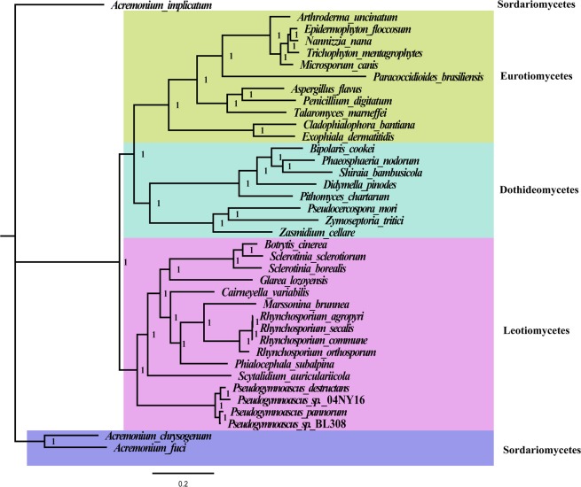
We used the Bayesian inference method to establish the phylogenetic relationships of 16 species within Leotiomycetes, 8 species from Dothideomycetes, and 11 species from Eurotiomycetes based on the combined mitochondrial gene set (15 typical protein-coding genes) using three Acremonium species, A. chrysogenum, A. fuci, and A. implicatum, as outgroups. The best-fit evolutionary model for the phylogenetic analysis is “GTR + I + G”. We obtained a stable evolutionary tree topology (Fig. 6) with all of the recovered clades well supported (Bayesian posterior probability (BPP) = 1). Based on the phylogenetic analysis, the 38 Pezizomycotina species could be divided into four major clades corresponding to the classes Sordariomycetes, Eurotiomycetes, Dothideomycetes, and Leotiomycetes20. The 16 Leotiomycetes species were divided into three clades in the phylogenetic tree. The genus Pseudogymnoascus containing four species, P. pannorum, P. destructans, P. sp.04NY16, and P. sp.BL308, is the first clade. S. auriculariicola is the second single clade. The third clade contains 11 fungal species within the order Helotiales. In the order Helotiales, the relationships of the three families were consistently recovered as (Sclerotiniaceae + (Helotiaceae + (Dermateaceae)).
Figure 6.
The phylogenetic tree was calculated from the multiple sequence alignment of the combined mitochondrial gene set (15 PCGs) of 38 fungal species. Topology was inferred using Bayesian method. Species analysed are shown in Table S9. The phylogenetic tree was drawn using the Figtree v1.4.3 software (https://mac.softpedia.com/get/Graphics/FigTree.shtml).
Single-gene tree topologies varied (Fig. S1), which indicates incongruent phylogenetic signals among different genes. However, the BI phylogenies based on the nad4 gene were consistent with the all-protein-coding genes phylogeny. Therefore, the nad4 gene may be a useful barcode sequence for species identification and phylogenetic analysis within the Leotiomycetes.
The phylogenetic analysis of nuclear multi-locus polygenes yielded evolutionary trees different from that of mitochondrial genes (Fig. S2). The BPP value of the nuclear multi-locus phylogenetic tree is between 0.43 and 1. Pseudocercospora mori, belonging to the Dothideomycetes class, is incorrectly clustered into the Eurotiomycetes class in the nuclear multi-locus phylogenetic tree. Exophiala dermatitidis and Cladophialophora bantiana,both belonging to the Eurotiomycetes class, are also incorrectly clustered into the Dothideomycetes class. In addition, M. brunnea, belonging to the order of Helotiales, forms an independent clade separated from other Helotiales species. The results further prove that mitochondrial genes are suitable as reliable tools for analyzing phylogenetic relationships of Leotiomycetes species.
Discussion
Mitochondrial markers have been successfully applied in phylogenetic taxonomy and evolutionary biology due to the advantage of their faster evolution22,23. The advent of next-generation sequencing technology has promoted the sequencing of fungal mitochondrial genomes. However, the mitogenomes of fungi are still less well-understood than those of animals and plants24. As of July 2019, only 612 mitogenomes of fungi have been reported in the NCBI database, 488 of which belong to the Ascomycetes. However, most species from the Ascomycetes belong to the Saccharomycetes and Sordariomycetes, which account for more than 78%, while the Leotiomycetes account for less than 4%. More mitogenomes belonging to Leotiomycetes are needed to facilitate our understanding of the mitochondrial characteristics and evolution of this group. In the present study, we first sequenced and analyzed the S. auriculariicola mitogenome, which is the first reported sequencing of a mitogenome of the Scytalidium genus. The mitogenome size of S. auriculariicola is larger than that of other Leotiomycetes, with the exception of S. borealis and S. sclerotiorum20. Variability in the length of mitogenomes has been frequently observed in the Leotiomycetes class, which is consistent with the variable mitogenomes in many eukaryotes25,26. The largest mitogenome in Leotiomycetes fungi is 203.05 kb20, while P. pannorum contains the smallest mitogenome at 26.92 kb (Table 1). The S. borealis mitogenome has the largest number of introns (61) in the Leotiomycetes, while only one intron was detected in the P. pannorum mitogenome. Another species, C. variabilis, which contained a mitogenome of less than 30 kb, also had only one intron. Therefore, the number of introns was considered one of the main factors contributing to the mitogenome size variation27,28. Interestingly, the intron-free species, P. subalpina, has a mitogenome size of 43.74 kb, higher than that of P. destructans, P. sp.04NY16, and P. sp.BL308, which have two introns each. Similarly, the number of introns in the R. orthosporum mitogenome is less than that of G. lozoyensis, which contains a much smaller mitogenome. Previous studies have shown that the size of fungal mitogenomes is related to the number of introns, intergenic regions, repetitive elements, plasmid-derived regions, and gene transfer events29,30. In the present study, we found that the number of free-standing ORFs of the P. subalpina mitogenome is higher than that of P. destructans, Pseudogymnoascus sp.04NY16, and Pseudogymnoascus sp.BL308. Furthermore, the number of free-standing ORFs of the R. orthosporum mitogenome is more than that of G. lozoyensis. The size of free-standing ORFs in P. subalpina and R. orthosporum mitogenomes accounts for 18.79% and 18.52% of their total mitogenomes, respectively (Table 2). The results reveal that free-standing ORFs are also an important factor affecting the mitogenome sizes of the Leotiomycetes.
Mitogenomes have an independent evolutionary origin relative to nuclear genomes31 and are widely believed to originate from endosymbiotic bacteria9. In the course of evolution, most mitochondrial genes of eukaryotes have been transferred to the nuclear genome32,33, a process that has been frequent, sporadic, and episodic32. The transfer of nucleic acids from the mitochondrion to the nucleus is an ongoing process in most eukaryotes, resulting in the transfer of functional genes32,34. The presence of genes in mitochondria has its advantages, such as the production of hydrophobic proteins in mitochondria to avoid long-distance transport from the nucleus, and the maintenance of mitochondrial structure35,36. The mitogenome of S. auriculariicola retains all 15 typical protein-coding genes (atp6, atp8, atp9, cob, cox1, cox2, cox3, nad1, nad2, nad3, nad4, nad4L, nad5, nad6, and rps3) for energy metabolism and transcriptional regulation. However, four species of Pseudogymnoascus have lost the mitochondrial atp9 gene. In addition, the S. auriculariicola mitogenome has 31 intronic ORFs and 10 independent ORFs. Most of these ORFs are homing endonuclease genes (25 LAGLIDADG, 11 GIY-YIG), but there are five ORFs with unknown functions. ORFs have been widely found in fungal mitogenomes, though their origin and function have not been elucidated37. More studies on the mitogenomes of S. auriculariicola and related species are needed to recover the exact functions of these ORF genes.
tRNA genes are important nexus molecules between mRNAs and protein, and are essential for translation38. The S. auriculariicola mitogenome contains 30 tRNAs, most of which are clustered around rnl, nad6, and atp9. This is similar to S. borealis, P. subalpina, and G. lozoyensis, which also belong to the Leotiomycetes. The number of mitochondrial tRNAs of Leotiomycetes ranges from 27 to 35, which is higher than that of most fungi with 22–26 tRNAs19,39,40. These tRNA genes can adequately satisfy the need to decode and predict all of the codons in mitochondrial ORFs, thus reducing the need for tRNAs to enter mitochondria from the cytoplasm41. All of the tRNAs in the mitogenome of S. auriculariicola have distinctive primary and secondary structures, of which 27 have base mutations and mismatches, with all of them being G-U mismatches. In addition to G-U mismatches, C-U and A-C mismatches also exist in other eukaryotic mitogenomes42. Mitochondrial tRNA mutations have been demonstrated to be associated with metabolism and various diseases43,44. However, little research has been done on tRNA mutations in fungal mitogenomes. Further studies are needed to investigate the effects of mitochondrial tRNA mutations on the growth and pathogenicity of pathogenic fungi.
The GC content of mitogenomes varies among organisms and is thought to be influenced by mutation bias, selection, and reconstruction-related DNA repair bias45. The GC content of the S. auriculariicola mitogenome is lower than that of all of the Leotiomycetes except C. variabilis, suggesting that the S. auriculariicola mitogenome has undergone a large number of variations during its evolution. In addition, the S. auriculariicola mitogenome has a uniquely positive AT skew, while the AT skews are negative in the mitogenomes of other Leotiomycetes. According to the second parity rule, as long as there is no mutation or selection bias, each base in the complementary DNA strand exists at approximately equal frequencies46. The presence of AT or GC skews on the same DNA strand from different species indicated that mitogenomes of different species underwent different mutations or environmental selection. Compared with other Leotiomycetes, the S. auriculariicola mitogenome has a unique A-over-T situation, indicating that it had undergone a unique genetic mutations or evolutionary selection.
Because all of the mitochondria originate from common ancestors, mitochondrial gene rearrangements have been widely used to study the origin and evolution of eukaryotes. Plant and animal mitogenomes have been extensively studied with regard to gene rearrangement, and several models have been established to explain its causes47,48. In recent years, there have been many studies on mitochondrial gene rearrangement in fungi39,49, which exists even within the same genus6. Andrey et al.20 compared the mitochondrial gene recombination of seven fungi in the Helotiales order and found that the mitochondrial genes can be divided into two main groups. In the present study, we analyzed the mitochondrial gene rearrangement in 16 Leotiomycetes fungi, including S. auriculariicola, and divided them into 5 distinct groups. Further, large-scale rearrangements of mitochondrial genes in the Leotiomycetes were confirmed by synteny analysis, indicating that the mitochondrial genes of Leotiomycetes have undergone different evolutionary patterns.
Mitochondrial genomes are widely used in fungal phylogenetic analysis due to their useful and informative markers50. Because many fungi are very similar in morphology and difficult to distinguish, fungal classification is easily confused. Phylogenetic and taxonomic analyses of fungi therefore need to be combined with molecular markers, and those of the mitochondrial genome could to be a good complement. In the present study, we constructed phylogenetic trees with 15 protein-coding genes genes. The high support rate indicated that these mitochondrial gene data can be used as reliable molecular markers. However, most single-gene phylogenetic trees exhibited different branches of evolution, and some even failed to distinguish fungi of different classes. This is due to insufficient evolutionary signals provided by individual genes51. However, the single-gene evolutionary tree of the nad4 gene is consistent with that of combined mitochondrial gene set, suggesting that the nad4 gene can be used as a potential molecular marker for evaluating the taxonomy of Leotiomycetes fungi. The nad4 gene had relatively conserved gene length (ranging from 1464 bp to 1485 bp) and intron number (zero or one) between different Leotiomycete species. In addition, the nad4 gene may be subjected to low oxidative stress in mitochondria compared with other mitochondrial genes, which leads to different selective pressures on nad4 gene. All of these promote nad4 to become a potential molecular marker to analyze phylogenetic relationships of Leotiomycete species.
In conclusion, this study enriches the mitochondrial database of Leotiomycetes fungi and fills in a gap for the mitogenomes of the Scytalidium genus. The gene content, structure, gene rearrangement, and phylogenetic analysis of the S. auriculariicola mitogenome will provide a basis for population genetics, taxonomy, and evolutionary biology of the Leotiomycetes and related groups.
Materials and Methods
Sampling and DNA extraction
The symptomatic mycelium of the pathogen of slippery scar from A. polytricha was collected from Jintang, Sichuan Province, China. The isolation of the causative pathogen was conducted according to Peng et al.1. Suspected fungi were first cultured on PDA medium for 3 days, and then inoculated into cultivation bags with healthy A. polytricha mycelia. The inoculated cultivation bags were cultured at 25 °C for 20 days. Then the pathogenic fungi were re-isolated from the cultivation bags with infected A. polytricha, which showed the symptoms of slippery scar. The strain was identified as S. auriculariicola based on the Koch’s postulates, morphology, and ITS sequences. The mycelium of S. auriculariicola was cultured in liquid potato dextrose medium for 4 days and then collected for DNA extraction. Total DNA was extracted from the mycelia using the fungal DNA Kit D3390-00 (Omega Bio-Tek, Norcross, GA, USA) according to the manufacturer’s instructions. The quality of extracted DNA was checked by electrophoresis, and DNA was stored at −20 °C until sequencing. The S. auriculariicola strain was stored in Sichuan Academy of Agricultural Sciences (No. SAAS_Sau), and is available from Cheng Chen and Daihua Lu of the Sichuan Academy of Agricultural Sciences, China.
Sequencing, assembly, and annotation of the mitochondrial genome
Purified DNA was used to construct sequencing libraries following the instructions of the NEBNext Ultra II DNA Library Prep Kit (NEB, Beijing, China). Whole genome shotgun sequencing was performed using an Illumina HiSeq 2500 Platform (Illumina, San Diego, CA, USA). We performed quality control and de novo assembly of the mitogenome according to Bi52. SPAdes 3.9.0 software53 was used for de novo assembly of the mitogenome, and the MITObim V1.9 program54 was used to fill in the gaps between contigs.
MFannot (http://megasun.bch.umontreal.ca/cgi-bin/mfannot/mfannotInterface.pl) and MITOS55 tools were used for mitogenome annotation of S. auriculariicola, both of which are based on Genetic Code 4. Uncertain results were adjusted manually by sequence alignments with orthologous genes without intron from the closely related species. The initially annotated protein-coding genes, rRNA, or tRNA genes of S. auriculariicola were also modified by alignment with previously published Leotiomycetes mitogenomes. ORFs were functionally annotated by InterProScan software56. The tRNAscan-SE 2.0 program was used to predict tRNA genes57. Finally, we used the OrganellarGenomeDraw (OGDRAW) tool58 to draw a map of the S. auriculariicola complete mitogenome.
Analysis of the mitogenomic organization
We used the Lasergene v7.1 (DNASTAR; http://www.dnastar.com/) tool with default settings to analyze the base composition of the mitogenome of S. auriculariicola. Strand asymmetry of the mitogenome was assessed using the following formulas: AT skew = [A − T]/[A + T], and GC skew = [G − C]/[G + C]59. We calculated the codon usage using Sequence Manipulation Suite software60 based on genetic code 4. We compared the arrangement of genes in S. auriculariicola with those of other published Leotiomycetes species. Genomic synteny analysis of mitogenomes from six representative species within the Leotiomycetes was conducted with Mauve v2.4.061.
Repetitive elements analysis
We searched the entire mitogenome of S. auriculariicola by BLASTn searches against itself using Circoletto62 (http://tools.bat.infspire.org/circoletto/) with an E-value of <10−10, aiming to identify large intragenomic replications of sequences and interspersed repeats. The Tandem Repeats Finder63 (http://tandem.bu.edu/trf/trf.advanced.sub-mit.html) with default settings was used to analyze the tandem repeats. We searched for repeated sequences including forward, reverse, complementary, and reverse complementary sequences in S. auriculariicola using the REPuter64 tool with E-values <10−5.
Phylogenetic analysis
For the phylogenetic analysis, we constructed a phylogenetic tree based on 15 common mitochondrial genes from S. auriculariicola and other 15 species in Leotiomycetes, 8 species in Dothideomycetes, 11 species in Eurotiomycetes, and 3 species in Sordariomycetes (outgroup). The MAFFT algorithm within the TranslatorX online platform65 was used to align the 15 conserved protein-coding genes. The Sequence Matrix 1.7.8 program66 was used to combine the individual genes into a combined matrix. We used the Modelgenerator v85167 tool to determine the best-fit evolutionary model for the phylogenetic analysis.
The Bayesian inference (BI) method was used for phylogenetic analysis based on the combined gene dataset with the MrBayes 3.2.668 program. Two independent runs were performed for 2 × 10⁶ generations sampling per 100 generations. Each run was sampled every 100 generations. Stationarity was assumed to have been reached when the estimated sample size (ESS) was >100, and the potential scale reduction factor (PSRF) approached 1.0. After the analysis was stable, the first 25% of the yielded trees were discarded as burn-in, and a 50% majority-rule consensus tree with posterior probability (PP) values was generated from the remaining trees. In order to compare mitochodrial phylogeny with nuclear multi-locus phylogeny, we downloaded internal transcribed spacer (ITS), RNA polymerase II second largest subunit (RPB2), translation elongation factor-1 alpha (EF1-α) and beta-tubulin (β-TUB) genes of 38 species from the NCBI database. Phylogenetic trees were constructed using the same method as mitochondrial genes. We also used the BI method to analyze the phylogenetic relationships of S. auriculariicola and related species using individual mitochondrial genes (15 core protein-coding genes); the purpose of which is to test whether these genes were useful as molecular markers for the phylogenetic analysis of Leotiomycetes species.
Supplementary information
Acknowledgements
We thank LetPub (www.letpub.com) for its linguistic assistance during the preparation of this manuscript. This research was funded by the Foundation Program of the Financial & Innovational Capacity Building Project of Sichuan (2019LWJJ-007& 2016GYSH-014) and Traction project of Chengdu industrial upgrading (2015-NY02-00063-NC).
Author contributions
Conceived and designed experiments: C.C., Q.L. and D.H.L. Performed the experiments: C.C., Q.L., J.W. and R.T.F. Analyzed the data: C.C., Q.L., C.X., and D.H.L. Contributed reagents/materials/analysis tools: C.C., Q.L., C.X., Z.H.F., H.Z., R.P.H. and D.H.L. Wrote the paper: C.C.
Data availability
The newly sequenced S. auriculariicola mitogenome was deposited to the GenBank database as accession number MK111108.
Competing interests
The authors declare no competing interests.
Footnotes
Publisher’s note Springer Nature remains neutral with regard to jurisdictional claims in published maps and institutional affiliations.
These authors contributed equally: Cheng Chen and Qiang Li.
Supplementary information
is available for this paper at 10.1038/s41598-019-53941-5.
References
- 1.Peng W, et al. A new species of Scytalidium causing slippery scar on cultivated Auricularia polytricha in China. Fems Microbiology Letters. 2015;359:72–80. doi: 10.1111/1574-6968.12564. [DOI] [PubMed] [Google Scholar]
- 2.Jie S, Yinbing B. Slippery Scar: A New Mushroom Disease in Auricularia polytricha. Mycobiology. 2012;40:129–133. doi: 10.5941/MYCO.2012.40.2.129. [DOI] [PMC free article] [PubMed] [Google Scholar]
- 3.Machouart M, et al. Polymorphisms and intronic structures in the 18S subunit ribosomal RNA gene of the fungi Scytalidium dimidiatum and Scytalidium hyalinum. Evidence of an IC1 intron with an His-Cys endonuclease gene. Fems Microbiology Letters. 2004;238:455–467. doi: 10.1016/j.femsle.2004.08.011. [DOI] [PubMed] [Google Scholar]
- 4.Crous PW, et al. Phylogenetic lineages in the Botryosphaeriaceae. Studies in Mycology. 2006;55:235–253. doi: 10.3114/sim.55.1.235. [DOI] [PMC free article] [PubMed] [Google Scholar]
- 5.Pavlov, I. Y. et al. Scytalidium candidum 3C is a new name for the Geotrichum candidum Link 3C strain. Journal of Basic Microbiology, 1–9 (2018). [DOI] [PubMed]
- 6.Li Q, et al. Characterization and comparative mitogenomic analysis of six newly sequenced mitochondrial genomes from ectomycorrhizal fungi (Russula) and phylogenetic analysis of the Agaricomycetes. International Journal of Biological Macromolecules. 2018;119:792–802. doi: 10.1016/j.ijbiomac.2018.07.197. [DOI] [PubMed] [Google Scholar]
- 7.Mcfarland R, Taylor RW, Turnbull DM. Mitochondrial Disease-Its Impact, Etiology, and Pathology. Current Topics in Developmental Biology. 2007;77:113–155. doi: 10.1016/S0070-2153(06)77005-3. [DOI] [PubMed] [Google Scholar]
- 8.Yamato KT. Heteroplasmy and homoplasmy for maize mitochondrial mutants: a rare homoplasmic nad4 deletion mutant plant. Journal of Heredity. 1999;90:369–373. doi: 10.1093/jhered/90.3.369. [DOI] [Google Scholar]
- 9.Muñozgómez SA, Wideman JG, Roger AJ, Slamovits CH. The origin of mitochondrial cristae from alphaproteobacteria. Molecular Biology Evolution. 2017;34:943–956. doi: 10.1093/molbev/msw298. [DOI] [PubMed] [Google Scholar]
- 10.Zhang, Y. J., Zhang, H. Y., Liu, X. Z. & Zhang, S. Mitochondrial genome of the nematode endoparasitic fungus Hirsutella vermicola reveals a high level of synteny in the family Ophiocordycipitaceae. 101, 3295–3304 (2017). [DOI] [PubMed]
- 11.Li Q, et al. Characterization and comparative analysis of six complete mitochondrial genomes from ectomycorrhizal fungi of the Lactarius genus and phylogenetic analysis of the Agaricomycetes. International Journal of Biological Macromolecules. 2019;121:249–260. doi: 10.1016/j.ijbiomac.2018.10.029. [DOI] [PubMed] [Google Scholar]
- 12.Matsumoto N, et al. Inheritance and recombination of mitochondrial genomes in plants, fungi and animals. New Phytologist. 2010;168:39–50. doi: 10.1111/j.1469-8137.2005.01492.x. [DOI] [PubMed] [Google Scholar]
- 13.Brankovics B, et al. Mitochondrial genomes reveal recombination in the presumed asexual Fusarium oxysporum species complex. Bmc Genomics. 2017;18:735. doi: 10.1186/s12864-017-4116-5. [DOI] [PMC free article] [PubMed] [Google Scholar]
- 14.Liu H, et al. Novel insights into mitochondrial gene rearrangement in thrips (Insecta: Thysanoptera) from the grass thrips, Anaphothrips obscurus. Scientific Reports. 2017;7:4284. doi: 10.1038/s41598-017-04617-5. [DOI] [PMC free article] [PubMed] [Google Scholar]
- 15.Chatre L, Ricchetti M. Are mitochondria the Achilles’ heel of the Kingdom Fungi? Current Opinion in Microbiology. 2014;20:49–54. doi: 10.1016/j.mib.2014.05.001. [DOI] [PubMed] [Google Scholar]
- 16.Forget L, Ustinova J, Zhang W, Huss VAR, Lang BF. Hyaloraphidium curvatum: A Linear Mitochondrial Genome, tRNA Editing, and an Evolutionary Link to Lower Fungi. Molecular Biology. Evolution. 2002;19:310–319. doi: 10.1093/oxfordjournals.molbev.a004084. [DOI] [PubMed] [Google Scholar]
- 17.Peter K, Matus V, Lubomir T, Wolfe KH, Jozef N. Complete DNA sequences of the mitochondrial genomes of the pathogenic yeasts Candida orthopsilosis and Candida metapsilosis: insight into the evolution of linear DNA genomes from mitochondrial telomere mutants. Nucleic acids research. 2006;34:2472–2481. doi: 10.1093/nar/gkl327. [DOI] [PMC free article] [PubMed] [Google Scholar]
- 18.Liliana L, et al. Mobile elements and mitochondrial genome expansion in the soil fungus and potato pathogen Rhizoctonia solani AG-3. Fems Microbiology Letters. 2014;352:165–173. doi: 10.1111/1574-6968.12387. [DOI] [PubMed] [Google Scholar]
- 19.Kang X, Hu L, Shen P, Li R, Liu D. SMRT Sequencing Revealed Mitogenome Characteristics and Mitogenome-Wide DNA Modification Pattern inOphiocordyceps sinensis. Frontiers in Microbiology. 2017;8:1422. doi: 10.3389/fmicb.2017.01422. [DOI] [PMC free article] [PubMed] [Google Scholar]
- 20.Mardanov AV, Beletsky AV, Kadnikov VV, Ignatov AN, Ravin NV. The 203 kbp Mitochondrial Genome of the Phytopathogenic Fungus Sclerotinia borealis Reveals Multiple Invasions of Introns and Genomic Duplications. Plos One. 2014;9:e107536. doi: 10.1371/journal.pone.0107536. [DOI] [PMC free article] [PubMed] [Google Scholar]
- 21.Torriani SFF, et al. Comparative analysis of mitochondrial genomes from closely related Rhynchosporium species reveals extensive intron invasion. Fungal Genetics and Biology. 2014;62:34–42. doi: 10.1016/j.fgb.2013.11.001. [DOI] [PubMed] [Google Scholar]
- 22.Liu J, Yu J, Zhou M, Yang J. Complete mitochondrial genome of Japalura flaviceps: Deep insights into the phylogeny and gene rearrangements of Agamidae species. International Journal of Biological Macromolecules. 2019;125:423–431. doi: 10.1016/j.ijbiomac.2018.12.068. [DOI] [PubMed] [Google Scholar]
- 23.Vanhove MPM, Briscoe AG, Jorissen MWP, Littlewood DTJ, Huyse T. The first next-generation sequencing approach to the mitochondrial phylogeny of African monogenean parasites (Platyhelminthes: Gyrodactylidae and Dactylogyridae) Bmc Genomics. 2018;19:520. doi: 10.1186/s12864-018-4893-5. [DOI] [PMC free article] [PubMed] [Google Scholar]
- 24.Qiang L, et al. The first complete mitochondrial genome from the family Hygrophoraceae (Hygrophorus russula) by next-generation sequencing and phylogenetic implications. International Journal of Biological Macromolecules. 2019;122:1313–1320. doi: 10.1016/j.ijbiomac.2018.09.091. [DOI] [PubMed] [Google Scholar]
- 25.Sandor S, Zhang Y, Xu J. Fungal mitochondrial genomes and genetic polymorphisms. Applied Microbiology Biotechnology. 2018;102:9433–9448. doi: 10.1007/s00253-018-9350-5. [DOI] [PubMed] [Google Scholar]
- 26.Zhang S, Hao A, Zhao Y, Zhang X, Zhang Y. Comparative mitochondrial genomics toward exploring molecular markers in the medicinal fungus Cordyceps militaris. Scientific Reports. 2017;7:40219. doi: 10.1038/srep40219. [DOI] [PMC free article] [PubMed] [Google Scholar]
- 27.Zhang Y, et al. The complete mitochondrial genome of the Chan-hua fungus Isaria cicadae: a tale of intron evolution in Cordycipitaceae. Fungal Genet. Biol. 2015;77:95–107. doi: 10.1016/j.fgb.2015.04.009. [DOI] [PubMed] [Google Scholar]
- 28.Lang BF, Laforest MJ, Burger G. Mitochondrial introns: a critical view. Trends in Genetics. 2007;23:119–125. doi: 10.1016/j.tig.2007.01.006. [DOI] [PubMed] [Google Scholar]
- 29.Kajsa H, Ake O, Mikael BMD, Magnus K, Jan S. Intronic and plasmid-derived regions contribute to the large mitochondrial genome sizes of Agaricomycetes. Current Genetics. 2014;60:303–13. doi: 10.1007/s00294-014-0436-z. [DOI] [PMC free article] [PubMed] [Google Scholar]
- 30.Li Q, et al. Characterization and phylogenetic analysis of the complete mitochondrial genome of the medicinal fungus Laetiporus sulphureus. Scientific Reports. 2018;8:9104. doi: 10.1038/s41598-018-27489-9. [DOI] [PMC free article] [PubMed] [Google Scholar]
- 31.Thielsch A, Knell A, Mohammadyari A, Petrusek A, Schwenk K. Divergent clades or cryptic species? Mito-nuclear discordance in a Daphnia species complex. Bmc Evolutionary Biology. 2017;17:227. doi: 10.1186/s12862-017-1070-4. [DOI] [PMC free article] [PubMed] [Google Scholar]
- 32.Adams KL, Palmer JD. Evolution of mitochondrial gene content: gene loss and transfer to the nucleus. Molecular Phylogenetics. Evolution. 2003;29:380–395. doi: 10.1016/s1055-7903(03)00194-5. [DOI] [PubMed] [Google Scholar]
- 33.Petersen G, et al. Mitochondrial genome evolution in Alismatales: Size reduction and extensive loss of ribosomal protein genes. Plos One. 2017;12:e0177606. doi: 10.1371/journal.pone.0177606. [DOI] [PMC free article] [PubMed] [Google Scholar]
- 34.Kubo N, Takano M, Nishiguchi M, Kadowaki K. Mitochondrial sequence migrated downstream to a nuclear V-ATPase B gene is transcribed but non-functional. Gene. 2001;271:193–201. doi: 10.1016/S0378-1119(01)00537-6. [DOI] [PubMed] [Google Scholar]
- 35.Allen JF. Why chloroplasts and mitochondria retain their own genomes and genetic systems: Colocation for redox regulation of gene expression. Proc Natl Acad Sci USA. 2015;112:10231–10238. doi: 10.1073/pnas.1500012112. [DOI] [PMC free article] [PubMed] [Google Scholar]
- 36.Björkholm P, Harish A, Hagström E, Ernst AM, Andersson SGE. Mitochondrial genomes are retained by selective constraints on protein targeting. Proceedings of the National Academy of Sciences. 2015;112:10154–10161. doi: 10.1073/pnas.1421372112. [DOI] [PMC free article] [PubMed] [Google Scholar]
- 37.Duò A, Bruggmann R, Zoller S, Bernt M, Grünig CR. Mitochondrial genome evolution in species belonging to the Phialocephala fortinii s.l. - Acephala applanata species complex. Bmc Genomics. 2012;13:166. doi: 10.1186/1471-2164-13-166. [DOI] [PMC free article] [PubMed] [Google Scholar]
- 38.Lant JT, et al. Visualizing tRNA-dependent mistranslation in human cells. Rna Biology. 2017;15:567–575. doi: 10.1080/15476286.2017.1379645. [DOI] [PMC free article] [PubMed] [Google Scholar]
- 39.Qiang L, et al. Characterization of the mitochondrial genomes of three species in the ectomycorrhizal genus Cantharellus and phylogeny of Agaricomycetes. International Journal of Biological Macromolecules. 2018;118:756–769. doi: 10.1016/j.ijbiomac.2018.06.129. [DOI] [PubMed] [Google Scholar]
- 40.Li Q, et al. Characterization and comparison of the mitochondrial genomes from two Lyophyllum fungal species and insights into phylogeny of Agaricomycetes. International Journal of Biological Macromolecules. 2019;121:364–372. doi: 10.1016/j.ijbiomac.2018.10.037. [DOI] [PubMed] [Google Scholar]
- 41.Kolesnikova OA, et al. Suppression of mutations in mitochondrial DNA by tRNAs imported from the cytoplasm. Science. 2000;289:1931–1933. doi: 10.1126/science.289.5486.1931. [DOI] [PubMed] [Google Scholar]
- 42.Yang M, et al. Complete mitochondrial genome of the soybean leaffolder, Omiodes indicata (Lepidoptera: Pyraloidea: Crambidae), and phylogenetic analysis for Pyraloidea. International Journal of Biological Macromolecules. 2018;115:53–60. doi: 10.1016/j.ijbiomac.2018.03.041. [DOI] [PubMed] [Google Scholar]
- 43.Blakely EL, et al. Pathogenic mitochondrial tRNA point mutations: nine novel mutations affirm their importance as a cause of mitochondrial disease. Human Mutation. 2013;34:1260–1268. doi: 10.1002/humu.22358. [DOI] [PMC free article] [PubMed] [Google Scholar]
- 44.Fakruddin M, et al. Defective Mitochondrial tRNA Taurine Modification Activates Global Proteostress and Leads to Mitochondrial Disease. Cell Reports. 2018;22:482–496. doi: 10.1016/j.celrep.2017.12.051. [DOI] [PubMed] [Google Scholar]
- 45.Qian L, Wang X, Chen X, Han B. Complete mitochondrial genome of the tea looper caterpillar, Ectropis obliqua (Lepidoptera: Geometridae) with a phylogenetic analysis of Geometridae. International Journal of Biological Macromolecules. 2018;114:491–496. doi: 10.1016/j.ijbiomac.2018.02.038. [DOI] [PubMed] [Google Scholar]
- 46.Chen H, Sun S, Norenburg JL, Sundberg P. Mutation and Selection Cause Codon Usage and Bias in Mitochondrial Genomes of Ribbon Worms (Nemertea) Plos One. 2014;9:e85631. doi: 10.1371/journal.pone.0085631. [DOI] [PMC free article] [PubMed] [Google Scholar]
- 47.Boore JL. Animal mitochondrial genomes. Nucleic Acids Research. 1999;27:1767–1780. doi: 10.1093/nar/27.8.1767. [DOI] [PMC free article] [PubMed] [Google Scholar]
- 48.Perseke M, et al. Evolution of mitochondrial gene orders in echinoderms. Molecular Phylogenetics. 2008;47:855–864. doi: 10.1016/j.ympev.2007.11.034. [DOI] [PubMed] [Google Scholar]
- 49.Pogoda CS, Keepers KG, Lendemer JC, Kane NC, Tripp EA. Reductions in Complexity of Mitochondrial Genomes in Lichen-Forming Fungi Shed Light on Genome Architecture of Obligate Symbioses. Molecular Ecology. 2018;27:1155–1169. doi: 10.1111/mec.14519. [DOI] [PubMed] [Google Scholar]
- 50.Liang X, et al. Comparative analysis of the mitochondrial genomes of Colletotrichum gloeosporioides sensu lato: insights into the evolution of a fungal species complex interacting with diverse plants. Bmc Genomics. 2017;18:171. doi: 10.1186/s12864-016-3480-x. [DOI] [PMC free article] [PubMed] [Google Scholar]
- 51.Wang J, et al. Comparative mitogenomic analysis of mirid bugs (Hemiptera: Miridae) and evaluation of potential DNA barcoding markers. Peerj. 2017;5:e3661. doi: 10.7717/peerj.3661. [DOI] [PMC free article] [PubMed] [Google Scholar]
- 52.Bi, G. The complete mitochondrial genome of northern grasshopper mouse (Onychomys leucogaster). 2, 393–394 (2017). [DOI] [PMC free article] [PubMed]
- 53.Bankevich A, et al. SPAdes: a new genome assembly algorithm and its applications to single-cell sequencing. Journal of Computational Biology. 2012;19:455–477. doi: 10.1089/cmb.2012.0021. [DOI] [PMC free article] [PubMed] [Google Scholar]
- 54.Christoph H, Lutz B, Bastien C. Reconstructing mitochondrial genomes directly from genomic next-generation sequencing reads—a baiting and iterative mapping approach. Nucleic Acids Res. 2013;41:e129–e129. doi: 10.1093/nar/gkt371. [DOI] [PMC free article] [PubMed] [Google Scholar]
- 55.Bernt M, et al. MITOS: improved de novo metazoan mitochondrial genome annotation. Molecular Phylogenetics & Evolution. 2013;69:313–319. doi: 10.1016/j.ympev.2012.08.023. [DOI] [PubMed] [Google Scholar]
- 56.Philip J, et al. InterProScan 5: genome-scale protein function classification. Bioinformatics. 2014;30:1236–40. doi: 10.1093/bioinformatics/btu031. [DOI] [PMC free article] [PubMed] [Google Scholar]
- 57.Lowe TM, Chan PP. tRNAscan-SE On-line: integrating search and context for analysis of transfer RNA genes. Nucleic Acids Res. 2016;44:W54–W57. doi: 10.1093/nar/gkw413. [DOI] [PMC free article] [PubMed] [Google Scholar]
- 58.Marc L, Oliver D, Sabine K, Ralph B. OrganellarGenomeDRAW–a suite of tools for generating physical maps of plastid and mitochondrial genomes and visualizing expression data sets. Nucleic Acids Res. 2013;41:W575–W581. doi: 10.1093/nar/gks979. [DOI] [PMC free article] [PubMed] [Google Scholar]
- 59.Liu QN, et al. The complete mitochondrial genome of fall armyworm Spodoptera frugiperda (Lepidoptera:Noctuidae) Genes & Genomics. 2016;38:205–216. doi: 10.1007/s13258-015-0346-6. [DOI] [Google Scholar]
- 60.Stothard P. The sequence manipulation suite: JavaScript programs for analyzing and formatting protein and DNA sequences. Biotechniques. 2000;28(1102):1104. doi: 10.2144/00286ir01. [DOI] [PubMed] [Google Scholar]
- 61.Darling ACE, Mau B, Blattner FR, Perna NT. Mauve: Multiple Alignment of Conserved Genomic Sequence With Rearrangements. Genome Research. 2004;14:1394–1403. doi: 10.1101/gr.2289704. [DOI] [PMC free article] [PubMed] [Google Scholar]
- 62.Darzentas N. Circoletto: visualizing sequence similarity with Circos. Bioinformatics. 2010;26:2620–2621. doi: 10.1093/bioinformatics/btq484. [DOI] [PubMed] [Google Scholar]
- 63.Benson G. Tandem repeats finder: a program to analyze DNA sequences. Nucleic Acids Research. 1999;27:573–580. doi: 10.1093/nar/27.2.573. [DOI] [PMC free article] [PubMed] [Google Scholar]
- 64.Kurtz S, et al. REPuter: the manifold applications of repeat analysis on a genomic scale. Nucleic Acids Res. 2001;29:4633–42. doi: 10.1093/nar/29.22.4633. [DOI] [PMC free article] [PubMed] [Google Scholar]
- 65.Abascal F, Zardoya R, Telford MJ. TranslatorX: multiple alignment of nucleotide sequences guided by amino acid translations. Nucleic Acids Res. 2010;38:W7–13. doi: 10.1093/nar/gkq291. [DOI] [PMC free article] [PubMed] [Google Scholar]
- 66.Lanfear R, Frandsen PB, Wright AM, Senfeld T, Calcott B. PartitionFinder 2: New Methods for Selecting Partitioned Models of Evolution for Molecular and Morphological Phylogenetic Analyses. Molecular Biology & Evolution. 2017;34:772–773. doi: 10.1093/molbev/msw260. [DOI] [PubMed] [Google Scholar]
- 67.Keane TM, Creevey CJ, Pentony MM, Naughton TJ, Mclnerney JO. Assessment of methods for amino acid matrix selection and their use on empirical data shows that ad hoc assumptions for choice of matrix are not justified. BMC Evol Biol. 2006;6:29. doi: 10.1186/1471-2148-6-29. [DOI] [PMC free article] [PubMed] [Google Scholar]
- 68.Ronquist F, et al. MrBayes 3.2: Efficient Bayesian Phylogenetic Inference and Model Choice Across a Large Model Space. Systematic Biology. 2012;61:539–542. doi: 10.1093/sysbio/sys029. [DOI] [PMC free article] [PubMed] [Google Scholar]
- 69.Bernt M, et al. MITOS: Improved de novo metazoan mitochondrial genome annotation. Molecular Phylogenetics. Evolution. 2013;69:313–319. doi: 10.1016/j.ympev.2012.08.023. [DOI] [PubMed] [Google Scholar]
Associated Data
This section collects any data citations, data availability statements, or supplementary materials included in this article.
Supplementary Materials
Data Availability Statement
The newly sequenced S. auriculariicola mitogenome was deposited to the GenBank database as accession number MK111108.


