Abstract
Background
Phosphatidylserine (PS) is normally confined in an energy-dependent manner to the inner leaflet of the lipid cell membrane. During cellular stress PS is exteriorized to the outer layer, initiating a cascade of events. Because cellular stress is often accompanied by decreased energy levels and because maintaining PS asymmetry is an energy-dependent process, it would make sense that cellular stress associated with decreased energy levels is also associated with PS exteriorization that ultimately leads to endothelial cell dysfunction. Our hypothesis was that anoxia-reoxygenation (A-R) is associated with decreased ATP levels, increased PS exteriorization on endothelial cell membranes, and increased endothelial cell membrane permeability.
Methods
The effect on ATP levels during anoxia-reoxygenation was measured via colorimetric assay in cultured cells. To measure the effect of A-R on PS levels, cultured cells underwent A-R and exteriorized PS levels and also total cell PS were measured via biofluorescence assay. Finally, we measured endothelial cell monolayer permeability to albumin after A-R.
Results
ATP levels in cell culture decreased 27% from baseline after A-R (p<0.02). There was over a 2-fold increase in exteriorized PS as compared to controls (p<0.01). Interestingly, we found that during A-R, the total amount of cellular PS increased (p<0.01). The finding that total PS changed 2-fold over normal cells suggested that not only is there a change in the distribution of PS across the cell membrane, but there may also be an increase in the amount of PS inside the cell. Finally, A-R increased endothelial cell monolayer permeability (p<0.01).
Conclusions
We found that endothelial cell dysfunction during A-R is associated with decreased ATP levels, increased PS exteriorization, and increased in monolayer permeability. This supports the idea that phosphatidylserine exteriorization may a key event during clinical scenarios involving oxygen lack and may one day lead to novel therapies in these situations.
Level of Evidence
Basic Science Paper
Keywords: phosphatidyl serine, microvascular permeability, ischemia reperfusion, anoxia-reoxygenation
Background
The cell membrane consists of a lipid bilayer that demonstrates polarity in lipid moiety distribution(1). This polarity is preserved across eukaryotic cell lines and is now known to play a role in cellular functions, including cell activation and cell death(2). Specifically, phosphatidyl serine (PS) in the lipid bilayer of cell membranes is not randomly distributed and the maintenance of this PS asymmetry is an energy-dependent process(3). Normally, PS is exclusively confined to the inner layer of the cell membrane, while phosphatidyl choline and sphingomyelin moieties are found in greater amounts on the outer layer of the cell membrane(3). However, in times of stress or cellular activation this asymmetric distribution is lost and PS is exteriorized to the external cell surface(4).
Since cell membranes consist of a lipid bilayer with a hydrophobic core, transport of lipid moieties must confront a thermodynamic barrier(5). Enzymes have been identified which facilitate the transport of lipids in cells under different conditions(6). The ATP-dependent enzymes, flippase and floppase, are responsible for maintaining the polarity of phospholipids across the cell membrane(1). Flippase requires energy to concentrate phosphatidyl serine on the inner layer of the cell membrane(7). Floppase plays a similar ATP-dependent role in transporting phosphatidyl choline and sphingomyelin to the outer cell membrane(3). Scramblase is an energy-independent enzyme which randomly transports phospholipids across cell membranes in both directions(7). Thus, the net effect is a concentration gradient across the cell membrane with PS moieties on the cytosolic or inner side of the cell membrane and phosphatidyl choline and sphingomyelin on the apical or outer layer. As apoptosis proceeds, evaginations of the cell membrane and blebs in the cells develop containing cytoplasmic proteins(8). These apoptotic bodies have PS expressed on their membranes which are recognized by macrophages and then result in consumption by macrophages which have PS receptors(8, 9).
As clinicians, we are acutely aware of the endothelial cell dysfunction that occurs as a result of hypoxia, ischemia, and ischemia-reperfusion(10). Ischemia and hypoxia lead to cellular dysfunction via many mechanisms. A common pathway for these mechanisms results in mitochondrial dysfunction and a decrease in adenosine triphosphate (ATP) production(8). The decrease in ATP production causes failure of multiple intracellular processes including maintenance of Na-K pump activity. Anaerobic metabolism results not only in decreased production of ATP, but also results in intracellular acidosis and dysfunction of enzyme dependent processes, compounding the derangements of energy dependent cellular processes. The now inefficient transmembrane ion pumps lead to the accumulation of sodium, hydrogen, and calcium ions within the cellular cytoplasm resulting in an intracellular hyperosmolar state. Cellular swelling soon develops causing further derangements in the cells abilities to maintain homeostasis(8). As cellular dysfunction increases, this is reflected clinically as worsening end-organ function which presents a significant clinical burden in critically ill patients.
Because cellular stress is often accompanied by decreased energy levels and because maintaining PS asymmetry is an energy-dependent process, it would make sense that cellular stress associated with decreased energy levels is also associated with PS exteriorization that ultimately leads to endothelial cell dysfunction as evidenced by an increase in endothelial cell permeability. Using an anoxia-reoxygenation cell model we sought to further investigate the interplay of oxygen lack, PS asymmetry and endothelial cell dysfunction. Our hypothesis was that anoxia-reoxygenation is associated with decreased ATP levels, increased PS exteriorization on endothelial cell membranes, and increased endothelial cell membrane permeability.
Methods
Anoxia-Reoxygenation (A-R) Protocol
The protocol for A-R has been described previously(11). Briefly, confluent bovine pulmonary artery endothelial cells (BPAECs) [American Type Culture Collection (ATCC), Manassas, VA] or human umbilical vein endothelial cells (HUVECs) [American Type Culture Collection (ATCC), Manassas, VA] monolayers were exposed to anoxia by incubation in a vacuum chamber that was continuously purged with an anoxic gas mixture of (93% N2, 5% CO2, 2% H2), with continuous oxygen monitoring. After 45 minutes of anoxia, endothelial cells were re-exposed to room air conditions (21% O2, 5% CO2 and 74% N2) and incubated for 4 hours.
Effect of Anoxia-Reoxygenation on ATP levels
The effect on ATP levels of was measured with cultured HUVECs. HUVECs were cultured and grown in 25cm2 tissue culture flasks using EBM-2 media. After exposure to anoxia or anoxia-reoxygenation, cells were lysed in ATP assay buffer for 2 minutes. ATP levels were measured at 570 nm using a standard ATP Colorimetric Assay from BioVision, according to the manufacturer’s specifications. The following groups were tested: 1) Control cells: cells serving as time controls, 2) Anoxia cells: Cells that underwent 45 minutes of anoxia (0% O2) via nitrogen gas displacement using an oxygen sensor for verification of anoxia, and 3) Anoxia-Reoxygenation cells: Cells that underwent 45 minutes of anoxia followed by 240 minutes of normoxic (reoxygenation) conditions. ATP concentrations in the samples were quantified against a standard ATP curve obtained using standards provided in the kit from the manufacturer.
Effect of Anoxia-Reoxygenation on Phosphatidyl exteriorization
Next, to measure the effect of A-R on PS exteriorization, BPAECs and HUVECs were grown separately to confluence and then underwent the A-R treatment as above. To measure changes in exposed phosphatidyl serine due to anoxia-reoxygenation BPAECs and HUVECs were incubated separately with 680-IRDye Annexin-V in culture medium with 2 mM CaCl2 for 1 hour; annexin-V binds to external or exposed PS, but not internal or unexposed PS. Subsequently, cells were fixed with 4% paraformaldehyde (PFA), washed with PBS and annexin-V fluorescence was read at 680nm on the Odyssey SA (Licor Biosciences). Comparisons were made with two controls: a negative control, which was an empty well, and a positive control which is cultured cells that serve as time controls that do not undergo anoxia-reoxygenation but are incubated with 10 μM oligomycin A to promote phosphatidyl serine exposure.
Effect of Anoxia-Reoxygenation on total cell Phosphatidyl Serine
Next, we were interested in the effect of A-R on total cell PS quantity, not just exteriorized PS. To measure total PS, we felt that disrupting cell membrane integrity with a detergent which and opening the cell membrane with a detergent would and allowed Annexin-V to permeate and bind intracellularly exposed PS (PSinner.). A-R was induced as described above in both BPAEC and HUVEC monolayers, and after the 4 hours of reoxygenation, monolayers were treated with 1% Triton X-100 to open the cell membrane, thereby allowing Annexin-V intracellular access and providing the ability to measure total PS (PSinner+PSouter). Anoxia-reoxygenation alone and treatment with 1% Triton X without anoxia served as controls. All cell groups underwent experimental manipulation for the same amount of time. Finally, we used diannexin to selectively block PS. Diannexin blocks exposed PS from interacting with proteins that are attracted to the negatively charged moieties of PS, and as such, it is a selective phosphatidylserine blocker. Cells were incubated with diannexin (100 μM) both prior and after the A-R protocol.
Effect of Anoxia-Reoxygenation on Endothelial Cell monolayer permeability
Finally, to measure endothelial cell permeability, cultured BPAECs and HUVECs were seeded separately onto collagen-coated transwells to form monolayers with Eagle’s minimal essential media supplemented with 2mM L-glutamine and Earle’s balanced salt solution. To induce anoxia, monolayers were sealed and nitrogen gas was infused until 0% oxygen conditions were achieved. Cells were incubated under these oxygen-free conditions for 45 minutes. Cells were then reoxygenated by incubating them in normal culture conditions of 5% CO2 for 4 hours. Monolayer formation was confirmed by measuring electrical resistance to confirm tight junction integrity. Resistance of over 1KΩ indicates tight junction integrity and monolayer formation. The apical side of the cells was treated with either PBS (controls) or PBS with biotinylated-bovine serum albumen (BSA), while the basolateral surface contained normal medium. To measure the amount of leaked BSA, solution from the basolateral compartment was incubated with Streptavidin-Horseradish Peroxidase. Color reaction was performed with tetramethylbenzidine, the reaction was stopped with 2.5M H2SO4 and the absorbance measured at 450 nm as previously described. BPAECs and HUVECs that were not exposed to 0% oxygen conditions served as time controls.
Statistical Analysis
Comparisons between control and sample means for in vitro endothelial monolayer permeability measurements were made with single-factor analysis-of-variance. Differences between measures were evaluated with a paired Student’s t-test or repeated measures ANOVA, as appropriate. Statistical significance was assigned at a P value of < 0.05.
Results
Anoxia-Reoxygenation decreased ATP levels
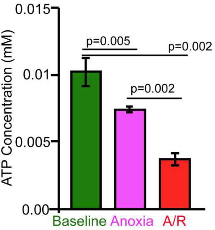
Adenosine triphosphate (ATP) levels were measured in human umbilical vein endothelial cells (HUVECs) at baseline and following exposure to anoxia reoxygenation. ATP concentrations decreased 27% from baseline after 45 minutes of anoxia and decreased further by 63% from baseline after 45 minutes of anoxia and 4 hours of reoxygenation (Fig 1, p<0.02).
Fig. 1. Energy dependence of PS exposure.
ATP levels decrease after 45 minutes of anoxia and decrease further after 45 minutes of anoxia followed by 4 hours of reoxygenation in human umbilical vein endothelial cells.
Anoxia-Reoxygenation increased Phosphatidyl exteriorization
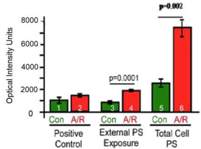
Bovine pulmonary artery endothelial cells (BPAEC) and HUVECs were exposed to anoxia reoxygenation as described previously. Anoxia-reoxygenation resulted in an over 2-fold increase in the amount of exteriorized phosphatidyl serine as measured by emittance. This was observed in both HUVECs (Fig 2, p < 0.01) and BPAECs (Fig. 3, p < 0.05).
Fig. 2. PS exteriorization occurs in HUVECs.
Anoxia-reoxygenation increased PS exposure on the exterior cell membrane and total PS in HUVECs
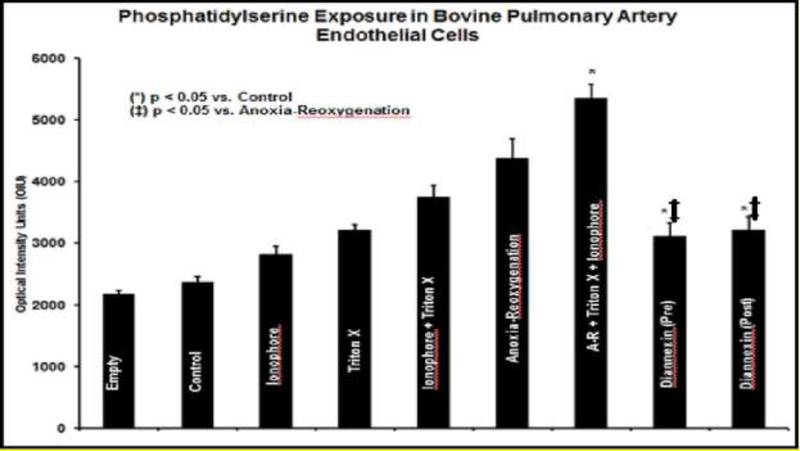
Fig. 3. Phosphatidylserine Exposure in Bovine Pulmonary Endothelial Cells.
While the PS signal intensity increased for ionophore and Triton X alone (suggesting that both activate PS exteriorization on the endothelial membrane), the highest signal intensity and PS cell content was seen with the addition of A/R (p < 0.001). Overall, A/R increased intracellular PS exposure by 19% compared to controls (p < 0.001). When BPAECs underwent A/R in the presence of Diannexin (pre-treatment) and Diannexin (post-treatment), Diannexin reduced PS signal by 34% pre-A/R (p < 0.001) and 30% post-A/R (p < 0.001). Units = Optical Intensity Units (OIU) = Ratio signal Annexin-V-PS/beta-actin x 103. Values are represented as mean ± SD.
Anoxia-Reoxygenation increased total cell Phosphatidyl Serine
Treatment of BPAECs and HUVECs with 1% Triton X-100 results in cell membranes lysis and allows the measurement of total cellular phosphatidyl serine. Following anoxia-reoxygenation the total amount of cellular PS increased 2-fold in BPAECs (p < 0.01) and 3-fold in HUVECs (p < 0.01). This suggests that A-R does not just result in exteriorization of PS but may also increase de novo production within the affected cells (Fig 3). While the PS signal intensity increased for ionophore and Triton X alone, suggesting that both activate PS exteriorization on the endothelial membrane, the highest signal intensity and PS cell content was seen with the addition of A/R (p < 0.001). Overall, A/R increased intracellular PS exposure by 19% compared to controls (p < 0.001). When BPAECs underwent A/R in the presence of Diannexin (pre-treatment) and Diannexin (post-treatment), Diannexin reduced PS signal by 34% pre-A/R (p < 0.001) and 30% post-A/R (p < 0.001). This is displayed in Figure 3.
Anoxia-Reoxygenation increased Endothelial Cell monolayer permeability
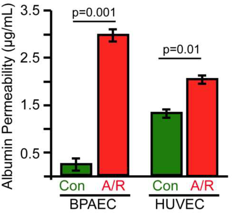
Cultured BPAECs underwent anoxia-reoxygenation as described and had their monolayer’s permeability determined. Anoxia-reoxygenation resulted in a 12-fold increase in albumin permeability in BPAEC monolayers (Fig. 4, p < 0.01). Under the same anoxia-reoxygenation conditions HUVECs demonstrated an increase in albumin permeability of 37% compared to controls (Fig. 4, p < 0.01). This data corroborates the increase in permeability seen in BPAECs suggesting that this phenomenon occurs in different cell lines and in at least two mammalian species.
Fig. 4. A/R increases monolayer permeability.
Anoxia-reoxygenation increased albumin efflux reflecting increased monolayer permeability of both cultured bovine pulmonary artery endothelial cells (BPAECs) and human umbilical vein endothelial cells (HUVECs).
Discussion
Clinicians are often confronted with shock as a consequence of hemorrhage and infection that may result in circulatory failure. In this clinical situation, the focus is to correct the shock as quickly as possible in order to prevent end organ ischemia and damage(12). However, correction of shock and ischemia does not necessarily result in improvement of end organ function(13). The correction of shock may lead to reperfusion injury which can lead to worsening of clinical outcomes despite adequate oxygen delivery(14). The reintroduction of oxygen to cells after a period of oxygen lack presents an additional obstacle to end-organ and patient recovery and as such has been a major target for investigation(15). The effects of anoxia-reoxygenation have become a topic of investigation with the hope of finding therapeutic targets to help mitigate deleterious effects on patient outcomes(14). We sought to further investigate the interplay of oxygen lack and cellular dysfunction, specifically looking at the PS asymmetry that may occur and endothelial cell dysfunction that follows. Our hypothesis was that anoxia-reoxygenation is associated with decreased ATP levels in human umbilical vein endothelial cells, increased PS exteriorization on endothelial cell membranes, and increased endothelial cell membrane permeability. We found that anoxia-reoxygenation is associated with decreased ATP levels in human umbilical vein endothelial cells, increased phosphatidyl serine exposure, and increased endothelial cell membrane permeability in two different cell lines.
We used two different mammalian cell cultures lines in order to investigate the mechanisms and effects of an anoxia-reoxygenation scenario which is a clinical correlate to ischemia-reperfusion. Bovine pulmonary artery endothelial cells are a good first choice when looking at endothelial cell function. Adding the human umbilical vein line was important so that we not only have a human cell line to make the study more applicable to patient care, but also because veins are where the majority of trans-endothelial fluid movement occurs(16). Findings between human umbilical vein endothelial cells and bovine pulmonary artery endothelial cells were consistent, as anoxia-reoxygenation resulted in an increase in monolayer permeability in both cell lines. These data at the least suggest a correlation between decreased ATP levels and endothelial dysfunction. The reason for this correlation is unclear. Prior work on apoptosis has shown that autophagy via cell membrane evagination and endosome production are part of the mechanism of apoptosis(8). The production of endosomes and cell membrane changes to expose PS could be an energy-dependent process requiring ATP(17). Therefore, anoxia resulting in autophagy consumes energy and may lead to diminished ATP levels.
Our work does not determine the time sequence of diminished ATP levels and increased PS exposure. It is possible that decrease in ATP levels leads to cellular dysfunction by starving the energy-dependent mechanism for producing PS asymmetry versus ATP being required for an energy dependent process to degrade cell membranes. Hypoxia is known to lead to decreased ATP production in human patients(18), but we needed to prove that this also occurs in mammalian cell culture. This depletion in ATP levels can lead to a decrease function in flippase and floppase. The net result of scramblase activity, which is an energy independent protein, would be to degrade the polarity of lipid asymmetry as it would act unopposed by the energy dependent flippase and floppase. As scramblase randomly distributes PS and PC moieties across the cell membrane, and the specificity of flippase and floppase activity would be lost, the membrane polarity would decrease.
Anoxia-reoxygenation resulted in an increase in the exteriorization of phosphatidyl serine. This has been previously reported by other investigators as well and is suggestive of cellular membrane dysfunction(19). Phosphatidyl serine exposure has been found to be a signal to induce apoptosis of the cell by macrophages(20). Anoxia-reoxygenation activates many cell signaling pathways which lead to autophagy, apoptosis, and worsening inflammation. Exteriorization of PS targets the cell for apoptosis, potentially leading to increased end-organ dysfunction(22). Our data supports this notion as we found an increase in permeability in endothelial cell monolayers under condition where PS was exteriorized to the outer leaflet.
Interestingly, by lysing cells in culture, we also found that the total amounts of cellular phosphatidyl serine (whether bound to the intra- or extra-cellular portion of the cell membrane) was increased. This was an unexpected finding. The current thinking is that most of the stress induced exposed phosphatidylserine on the outer cell membrane is due to “flipping” of PS from the inner cell membrane layer to the outer cell membrane layer. If this hypothesis is valid, the total amount of phosphatidyl serine should be conserved (PStotal=PSinner+PSouter). The reason for total increase in PS levels is not clear. The total amount of cellular phosphatidylserine is dependent on the biosynthesis of phosphatidylserine, its transport across the cellular membrane and its degradation, though the contribution of each of these components is unknown(23, 24). Phosphatidylserine moieties are known to be part of subcellular organelles, albeit at different amounts and not necessarily with the same gradients across organelle membranes that is seen across cell membranes(25, 26). Anoxia/Reoxygenation stress and subsequent cell damage may lead to the release of phospholipid moieties from within organelles such as the mitochondria thus affecting the total amounts of the various phospholipid moieties. A-R may lead to an increase in PS production in the Golgi body or its exposure along the surface of the endoplasmic reticulum(8). Additionally, work in animal models found that animals under stress that are provided with omega-3 polyunsaturated fatty acids show an increase in phosphatidylserine synthesis which may activate prosurvival genes(27). This increase in survival gene activity may be a defensive mechanism for the cell against apoptosis under stress conditions.(27) Work by other investigators have demonstrated that cellular stress does result in an increase in biosynthesis of PS(24). Phosphatidylserine production occurs via the action of two known enzymes—phosphatidylserine synthase I (PSSI) and II (PSSII) (23), however, the exact regulation of the activity of these enzymes is not clear(23). Nevertheless, de novo synthesis of PS occurs in stressed cells (24). It has been suggested that due to the strict regulation of PS levels within cells there may exist a negative feedback loop regulating the activity of PSSI and PSSII(28, 29). Once phosphatidylserine moieties become transported across the cell membrane, the inhibition of PS production may be diminished leading to new biosynthesis of PS. The net result of these actions is an increase in total cellular PS and its exteriorization on the external leaflet of the cell membrane.
A question that remains is whether the energy depletion and increased PS exposure and total PS amounts we observed are a marker of anoxia-reoxygenation itself or are a marker of cellular damage as a result of hypoxic stress. Shorter conditions of anoxia that do not result in cellular damage may result in the same effects on ATP levels, and phosphatidyl serine exteriorization(30). Shorter durations of anoxia which do not result in increased monolayer cell permeability may allow us to discern whether phosphatidyl serine amounts are a marker for cellular stress or for apoptosis itself. Further investigation would be required to determine if the depletion in ATP which is coincident with increased permeability is a result of as of yet unrecognized transmembrane proteins. One potential are transmembrane proteins which are energy dependent that become activated under cellular stress or hypoxia and lead to active transport of lipid moieties and reverse the polarity seen between the inner and outer cell membrane layers under physiologic conditions in cell culture.
Although both bovine pulmonary artery endothelial cells and human umbilical vein cells in culture showed a similar increase in phosphatidylserine exposure and total amounts there was a difference in microvascular permeability between these cell lines. BPAEC anoxia-reoxygenation resulted in a 12-fold increase in permeability while an increase of 37% was observed in HUVECs following anoxia-reoxygenation. Although anoxia-reoxygenation did result in increased microvascular permeability in both cell lines, there is a difference in the degree of increase. The reasons for this are not clear from our experiments. The reasons may include different biologic response among different species or different endothelial cell lines.
In human forearm muscle, ischemia-reperfusion results in increased exteriorization of PS on muscle cell membranes (31). Additionally, in vivo rat models demonstrated a decrease in microvascular permeability when rats were treated with diannexin, a phosphatidyl serine blocker, as compared to untreated animals following ischemia-reperfusion simulating shock conditions(32). These data suggest that anoxia-reoxygenation effects on phosphatidyl serine are not limited to cell culture but may be seen in vivo and potentially offer therapeutic targets to mitigate the effects of ischemia reperfusion injury(32).
In summary, anoxia-reoxygenation in cultured endothelial cells results in ATP depletion, increased phosphatidyl serine on the outer leaflet of the cell membrane, increased total cellular PS levels, and increased monolayer cell permeability. Whether this is a consequence of de novo production of PS, a dynamic energy dependent process of cell membrane derangement required for apoptosis, a loss of function of energy dependent lipid transporters required to maintain a lipid moiety gradient across a cell membrane, or a combination of any or all of these mechanisms is unclear and warrants further investigation. There is a potential to identify further targets for cellular protection from dysfunction and/or apoptosis in cells exposed to hypoxia and reoxygenation injury.
Acknowledgments
Research for this paper was funded by NIH Grant#: KO8 GMO81361
Footnotes
There are no conflicts of interest to report.
References
- 1.Tanaka K, Fujimura-Kamada K, Yamamoto T. Functions of phospholipid flippases. J Biochem.. 2011;149(2):131–43. [DOI] [PubMed] [Google Scholar]
- 2.Fadok VA, Bratton DL, Frasch SC, Warner ML, Henson PM. The role of phosphatidylserine in recognition of apoptotic cells by phagocytes. Cell Death Differ. 1998;5(7):551–62. [DOI] [PubMed] [Google Scholar]
- 3.Pomorski T, Hrafnsdottir S, Devaux PF, van Meer G. Lipid distribution and transport across cellular membranes. Semin Cell Dev Biol. 2001;12(2):139–48. [DOI] [PubMed] [Google Scholar]
- 4.Lhermusier T, Chap H, Payrastre B. Platelet membrane phospholipid asymmetry: from the characterization of a scramblase activity to the identification of an essential protein mutated in Scott syndrome. J Thromb Haemost. 2011;9(10):1883–91. [DOI] [PubMed] [Google Scholar]
- 5.Daleke DL. Regulation of transbilayer plasma membrane phospholipid asymmetry. J Lipid Res. 2003;44(2):233–42. [DOI] [PubMed] [Google Scholar]
- 6.Herrmann A, Zachowski A, Devaux PF. Protein-mediated phospholipid translocation in the endoplasmic reticulum with a low lipid specificity. Biochemistry. 1990;29(8):2023–7. [DOI] [PubMed] [Google Scholar]
- 7.Pomorski TG, Menon AK. Lipid somersaults: Uncovering the mechanisms of protein-mediated lipid flipping. Prog Lipid Res. 2016;64:69–84. [DOI] [PMC free article] [PubMed] [Google Scholar]
- 8.Wu MY, Yiang GT, Liao WT, Tsai AP, Cheng YL, Cheng PW, Li CY, Li CJ.. Current Mechanistic Concepts in Ischemia and Reperfusion Injury. Cell Phystiol Biochem. 2018;46(4):1650–67. [DOI] [PubMed] [Google Scholar]
- 9.Li MO, Sarkisian MR, Mehal WZ, Rakic P, Flavell RA. Phosphatidylserine receptor is required for clearance of apoptotic cells. Science. 2003;302(5650):1560–3. [DOI] [PubMed] [Google Scholar]
- 10.Cai H, Harrison DG. Endothelial dysfunction in cardiovascular diseases: the role of oxidant stress. Circ Res. 2000;87(10):840–4. [DOI] [PubMed] [Google Scholar]
- 11.Yoshida N, Granger DN, Anderson DC, Rothlein R, Lane C, Kvietys PR. Anoxia/reoxygenation-induced neutrophil adherence to cultured endothelial cells. Am J Physiol Heart Circ Physiol. 1992;262(6 Pt 2):H1891–8. [DOI] [PubMed] [Google Scholar]
- 12.Dellinger RP, Levy MM, Rhodes A, Annane D, Gerlach H, Opal SM, Sevransky JE, Sprung CL, Douglas IS, Jaeschke R, et al. Surviving sepsis campaign: international guidelines for management of severe sepsis and septic shock: 2012. Crit Care Med. 2013;41(2):580–637. [DOI] [PubMed] [Google Scholar]
- 13.Singer M Critical illness and flat batteries. CritCare. 2017;21(Suppl 3):309. [DOI] [PMC free article] [PubMed] [Google Scholar]
- 14.Proteasome Oliva J. and Organs Ischemia-Reperfusion Injury. Int J Mol Sci. 2017;19(1). [DOI] [PMC free article] [PubMed] [Google Scholar]
- 15.Valko M, Leibfritz D, Moncol J, Cronin MT, Mazur M, Telser J. Free radicals and antioxidants in normal physiological functions and human disease. Int J Biochem Cell Biol. 2007;39(1):44–84. [DOI] [PubMed] [Google Scholar]
- 16.Sumagin R, Sarelius IH. Emerging understanding of roles for arterioles in inflammation. Microcirculation. 2013;20(8):679–92. [DOI] [PMC free article] [PubMed] [Google Scholar]
- 17.Harrison MA, Muench SP. The Vacuolar ATPase - A Nano-scale Motor That Drives Cell Biology. Subcell Biochem. 2018;87:409–59. [DOI] [PubMed] [Google Scholar]
- 18.Consolini AE, Ragone MI, Bonazzola P, Colareda GA. Mitochondrial Bioenergetics During Ischemia and Reperfusion. Adv Exp Med Biol. 2017;982:141–67. [DOI] [PubMed] [Google Scholar]
- 19.Kodigepalli KM, Bowers K, Sharp A, Nanjundan M. Roles and regulation of phospholipid scramblases. FEBS Lett. 2015;589(1):3–14. [DOI] [PubMed] [Google Scholar]
- 20.Nonaka S, Shiratsuchi A, Nagaosa K, Nakanishi Y. Mechanisms and Significance of Phagocytic Elimination of Cells Undergoing Apoptotic Death. Biol Pharm Bull. 2017;40(11):1819–27. [DOI] [PubMed] [Google Scholar]
- 21.Ryan S, McNicholas WT. Intermittent hypoxia and activation of inflammatory molecular pathways in OSAS. Arch Physiol Biochem. 2008;114(4):261–6. [DOI] [PubMed] [Google Scholar]
- 22.Chaurio RA, Janko C, Munoz LE, Frey B, Herrmann M, Gaipl US. Phospholipids: key players in apoptosis and immune regulation. Molecules. 2009;14(12):4892–914. [DOI] [PMC free article] [PubMed] [Google Scholar]
- 23.Vance JE, Tasseva G. Formation and function of phosphatidylserine and phosphatidylethanolamine in mammalian cells. Biochim Biophys Acta. 2013;1831(3):543–54. [DOI] [PubMed] [Google Scholar]
- 24.Aussel C, Pelassy C, Breittmayer JP. CD95 (Fas/APO-1) induces an increased phosphatidylserine synthesis that precedes its externalization during programmed cell death. FEBS Lett. 1998;431(2):195–9. [DOI] [PubMed] [Google Scholar]
- 25.Hankins HM, Baldridge RD, Xu P, Graham TR. Role of flippases, scramblases and transfer proteins in phosphatidylserine subcellular distribution. Traffic. 2015;16(1):35–47. [DOI] [PMC free article] [PubMed] [Google Scholar]
- 26.Arumugam S, Kaur A. The Lipids of the Early Endosomes: Making Multimodality Work. Chembiochem. 2017;18(12):1053–60. [DOI] [PubMed] [Google Scholar]
- 27.Zhang W, Liu J, Hu X, Li P, Leak RK, Gao Y, Chen J.. n-3 Polyunsaturated Fatty Acids Reduce Neonatal Hypoxic/Ischemic Brain Injury by Promoting Phosphatidylserine Formation and Akt Signaling. Stroke. 2015;46(10):2943–50. [DOI] [PubMed] [Google Scholar]
- 28.Kuge O, Hasegawa K, Saito K, Nishijima M. Control of phosphatidylserine biosynthesis through phosphatidylserine-mediated inhibition of phosphatidylserine synthase I in Chinese hamster ovary cells. Proc Natl Acad Sci U S A. 1998;95(8):4199–203. [DOI] [PMC free article] [PubMed] [Google Scholar]
- 29.Nishijima M, Kuge O, Akamatsu Y. Phosphatidylserine biosynthesis in cultured Chinese hamster ovary cells. I. Inhibition of de novo phosphatidylserine biosynthesis by exogenous phosphatidylserine and its efficient incorporation. J Biol Chem. 1986;261(13):5784–9. [PubMed] [Google Scholar]
- 30.Kulkarni AC, Kuppusamy P, Parinandi N. Oxygen, the lead actor in the pathophysiologic drama: enactment of the trinity of normoxia, hypoxia, and hyperoxia in disease and therapy. Antioxid Redox Signal. 2007;9(10):1717–30. [DOI] [PubMed] [Google Scholar]
- 31.Wouters CW, Meijer P, Janssen CI, Frederix GW, Oyen WJ, Boerman OC, Smits P, Rongen GA. Atorvastatin does not affect ischaemia-induced phosphatidylserine exposition in humans in-vivo. J Atheroscler Thromb. 2012;19(3):285–91. [DOI] [PubMed] [Google Scholar]
- 32.Strumwasser A, Bhargava A, Victorino GP. Attenuation of endothelial phosphatidylserine exposure decreases ischemia-reperfusion induced changes in microvascular permeability. J Trauma Acute Care Surg. 2018;84(6):838–46. [DOI] [PMC free article] [PubMed] [Google Scholar]


