Abstract
Purpose:
The aim of the present study was to evaluate the efficacy of arthrocentesis with and without sodium hyaluronate (SH) injection in the treatment of patients with temporomandibular joint (TMJ) internal derangement.
Materials and Methods:
The study consisted of 20 patients with chief complaints of limited mouth opening, TMJ pain, and jaw deviation. Patients with disc displacement with reduction and closed lock were randomly divided into two groups. In Group 1, only arthrocentesis was performed, and in Group 2, arthrocentesis plus intra-articular injection of SH was performed. Arthrocentesis was performed under aseptic conditions using normal saline. Clinical evaluation was done for maximum mouth opening (MMO), TMJ pain, and jaw deviation before the procedure and 1 week, 2 weeks, 1 month, and 3 months following arthrocentesis.
Results:
The mean visual analog scale (VAS) score change was statistically significant in Group 1 and Group 2 for within the group analysis. There was statistically significant difference in VAS score between Group 1 and Group 2 at all time intervals postoperatively. The increase in MMO from preoperative to 3 months postoperatively was statistically significant for within the group analysis. There was a reduction in mandibular deviation in both Group 1 and Group 2, but the difference was not statistically significant. There was no statistically significant difference in deviation between the two groups.
Conclusion:
Arthrocentesis with SH is superior to arthrocentesis alone in treating patients suffering with TMJ internal derangement, who are refractory to conservative treatment.
Keywords: Arthrocentesis, internal derangement, normal saline, sodium hyaluronate
INTRODUCTION
Temporomandibular joint (TMJ) internal derangement is a disruption within the internal aspects of the TMJ in which there is a displacement of the disc from its normal functional relationship with the mandibular condyle and articular portion of the temporal bone. Conventionally, TMJ internal derangement has been described as a progressive disorder. The fibrocartilage disc is typically displaced anteromedially.
Internal derangement is usually treated with nonsurgical methods initially such as diet modification, occlusal splint therapy, physiotherapy, pharmacotherapy, transcutaneous electrical nerve stimulation, and stress reduction techniques followed by surgical methods such as arthroscopy, reconstruction arthroplasty (disc repositioning), meniscectomy (discectomy), eminectomy, and repair of perforation of disc.[1] These surgical procedures are aggressive and invasive and may even lead to more serious symptoms. Arthrocentesis of the TMJ seems to meet the requirement as a minimally invasive procedure. It has an intermediate place between the medical and surgical forms of treatment.
TMJ arthrocentesis refers to lavage of the upper joint space, hydraulic pressure, and manipulation to release adhesions or the “anchored disc phenomenon” and improve motion. It is a simple and efficient procedure that can be performed under local anesthesia and carries no reported complications. It has proved to be highly efficacious in releasing even long-standing severe closed lock of TMJ with no relapse after extended follow-up. Lavage of the upper joint space reduces pain by removing mediators from the joint, increasing mandibular mobility by removing intra-articular adhesions, eliminating the negative pressure within the joint, recovering disc and fossa space, and improving disc mobility, which reduces the mechanical obstruction caused by the anterior position of the disc.[2]
Intra-articular corticosteroid injection alone or after arthrocentesis provides long-term palliative effects on subjective symptoms and clinical signs of TMJ pain. Unfortunately, intra-articular corticosteroid injection has an unpredictable prognosis and can cause local side effects on joint tissues. Recently, sodium hyaluronate (SH) has been proposed as an alternative therapeutic agent.[3] SH has a lubricating, protective, and repairing effect on the joint surfaces. It also has an analgesic and anti-inflammatory action.[4]
The aim of the present study was to compare arthrocentesis with normal saline versus arthrocentesis with SH in the treatment of TMJ internal derangement patients.
MATERIALS AND METHODS
The present study was done on twenty patients with age ranging from 16 to 67 years who were diagnosed with TMJ internal derangement. The inclusion criteria were TMJ pain, joint sound, or limited mouth opening. All patients were refractory to conservative treatment (muscle relaxant, diet, physical therapy, and compresses). The exclusion criteria included any previous invasive procedure of the TMJ, bony or fibrous ankylosis, extracapsular causes of pain and dysfunction, osteoarthritis, rheumatoid arthritis, and gout.
All patients were informed about the procedure, its possible complications, and the materials used. Informed consent was also obtained from the Institutional Ethical Clearance Committee. All the patients were randomly divided into two groups – Group 1 included patients with arthrocentesis using normal saline and Group 2 included patients with arthrocentesis using normal saline followed by an intra-articular injection of 1 ml of SH.
The preoperative and postoperative clinical assessments were done by a single clinician for signs and symptoms of TMJ disorders which included pain, MMO, and jaw deviation. Pain was assessed using VAS 0–10. Zero reading of VAS was taken as the absence of pain and 10 as the maximum pain. The initial MMO was measured as the distance in millimeters between the incisal edges of the upper and lower central incisors. The effect of treatment on deviation was decided on the basis of proportion of improvement at the end of the treatment by noting their presence or absence. Arthrocentesis was performed using an aseptic procedure.
Procedure
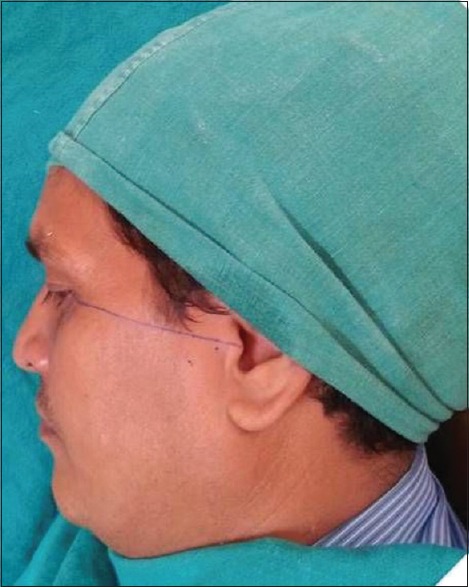
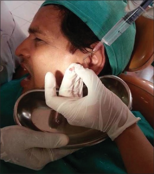
A patient was seated comfortably at 45° angle on the dental chair with the head turned toward the unaffected side. The target site was prepared, scrubbed, and isolated with sterile drapes. The points of needle insertion were marked on the skin according to the method suggested by McCain.[1] A line was drawn from the middle of the tragus to the outer canthus of the eye, and entry points were marked along this canthotragal line. The first point (posterior entrance point) which corresponds to the glenoid fossa was marked 10 mm from the midtragus and 2 mm below the line. The distance is about 25 mm from the skin to the center of the joint space.[5] The second point (anterior entrance point) which corresponds to the articular eminence was marked 10 mm from the first point and 10 mm below the line [Figure 1]. Two percent lignocaine was injected at the planned entrance points. A patient was asked to open the mouth wide. An 18-gauge needle was introduced at the first point, and 2–3-ml normal saline was injected through this needle to distend the joint space. Another 18-gauge needle was then inserted at the second point to establish a free flow of the solution through the joint space. A 10-ml syringe filled with normal saline was injected into the superior joint space through the first needle, and the second needle provided an outflow for normal saline. A total of 80–90-ml solution was used to lavage the superior joint space [Figure 2]. On termination of the procedure, 1 ml of commercially available SH (Hyalgan) was injected in the upper joint space for patients in the second group only once, after the first arthrocentesis. Once the needles were removed, a patient's lower jaw was gently manipulated in the vertical, protrusive, and lateral directions to facilitate the lysis of adhesions and to further free up the disc.
Figure 1.

Photograph demonstrating markings for landmarks for anterior and posterior needle insertion
Figure 2.

Photograph demonstrating procedure of lavage with normal saline solution
Patients were kept on soft diet, and analgesics (ibuprofen + paracetamol combination) were advised as necessary for a week. Arthrocentesis procedure was done twice at an interval of 1 week for all the patients. The patients were assessed for all parameters preoperatively and postoperatively at 1 week, 2 weeks, 1 month, and 3 months following the first arthrocentesis.
RESULTS
The age of patients in Group 1 ranged from 16 to 67 years with a mean age of 37 years. It included seven males and three females. The age of patients in Group 2 ranged from 15 to 65 years with a mean age of 34.2 years. It included seven males and three females [Table 1]. All patients were followed up for 3 months.
Table 1.
Mean age of the study sample groups
| n | Mean age±SD | P | |
|---|---|---|---|
| Group 1 | Group 2 | ||
| 10 | 37±16.11 | 34.2±12.54 | 0.67 |
SD: Standard deviation
The preoperative mean VAS score was 6.75 in Group 1 and 6.9 in Group 2. The postoperative mean VAS score at 3 months was 2.4 in Group 1 and 0.95 in Group 2. The mean VAS change was 4.35 ± 0.91 in Group 1 and 5.95 ± 1.52 in Group 2, which was statistically significant for within the group analysis [Table 2]. There was statistically significant difference (P < 0.05) in the VAS score between Group 1 and Group 2 at all time intervals postoperatively [Table 3].
Table 2.
Comparison of visual analog scale change between Group 1 and Group 2
| Variable | n | Mean±SD | P | |
|---|---|---|---|---|
| Group 1 | Group 2 | |||
| VAS change | 10 | 4.35±0.91 | 5.95±1.52 | 0.01 |
SD: Standard deviation, VAS: Visual analog scale
Table 3.
Comparison of pain between the two groups at different time intervals
| Variable | n | Mean±SD | P | |
|---|---|---|---|---|
| Group 1 | Group 2 | |||
| VAS preoperative | 10 | 6.75±0.89 | 6.9±1.05 | 0.734 |
| VAS 1 week | 10 | 4.85±1.29 | 3.35±1.16 | 0.014 |
| VAS 2 weeks | 10 | 4.25±1.32 | 2.6±1.15 | 0.008 |
| VAS 1 month | 10 | 3±1.29 | 1.35±0.75 | 0.003 |
| VAS 3 months | 10 | 2.4±0.88 | 0.95±0.72 | 0.001 |
SD: Standard deviation, VAS: Visual analog scale
The preoperative mean MMO was 35.2 ± 5.55 mm in Group 1 and 28.8 ± 7.97 mm in Group 2. The postoperative mean MMO at 3 months was 44.8 ± 2.30 mm in Group 1 and 41.4 ± 6.64 mm in Group 2. The mean MMO increased by 9.6 ± 4.67 mm for Group 1 and 12.6 ± 9.01 mm for Group 2, which was statistically significant for within the group analysis [Table 4]. There was no statistically significant difference in mean MMO between Group 1 and Group at any given time during the study [Table 5].
Table 4.
Change in mouth opening in Group 1 and Group 2
| Variable | n | Mean±SD | P | |
|---|---|---|---|---|
| Group 1 | Group 2 | |||
| MO change | 10 | 9.6±4.67 | 12.6±9.01 | 0.362 |
SD: Standard deviation, MO: Mouth opening
Table 5.
Comparison of mouth opening between the two groups at different time intervals
| Variable | n | Mean±SD | P | |
|---|---|---|---|---|
| Group 1 | Group 2 | |||
| MO preoperative | 10 | 35.2±5.55 | 28.8±7.97 | 0.052 |
| MO 1 week | 10 | 38.8±4.26 | 37.3±7.67 | 0.596 |
| MO 2 weeks | 10 | 40.6±3.78 | 38.7±6.90 | 0.455 |
| MO 1 month | 10 | 43.5±2.59 | 40.8±6.73 | 0.252 |
| MO 3 months | 10 | 44.8±2.30 | 41.4±6.64 | 0.143 |
SD: Standard deviation, MO: Mouth opening
The effect of treatment on deviation was evaluated by noting their presence or absence at the end of the treatment. Preoperatively, deviation was present in six patients in Group 1 and six patients in Group 2. Postoperatively, at 3-month follow-up, deviation was present in three patients in Group 1 and two patients in Group 2. There was a reduction in mandibular deviation in both Group 1 and Group 2, but the difference was not statistically significant. There was no statistically significant difference in deviation between the two groups [Table 6].
Table 6.
Comparison of mandibular deviation in Group 1 and Group 2 at different time intervals
| Group | Total | χ2 | P | ||
|---|---|---|---|---|---|
| Group 1 | Group 2 | ||||
| Deviation preoperative | |||||
| Present | 6 | 6 | 12 | 0 | 1 |
| Absent | 4 | 4 | 8 | ||
| Total | 10 | 10 | 20 | ||
| Deviation 1 week | |||||
| Present | 6 | 5 | 11 | 0.202 | 0.653 |
| Absent | 4 | 5 | 9 | ||
| Total | 10 | 10 | 20 | ||
| Deviation 2 weeks | |||||
| Present | 5 | 3 | 8 | 0.833 | 0.361 |
| Absent | 5 | 7 | 12 | ||
| Total | 10 | 10 | 20 | ||
| Deviation 1 month | |||||
| Present | 3 | 2 | 5 | 0.267 | 0.61 |
| Absent | 7 | 8 | 15 | ||
| Total | 10 | 10 | 20 | ||
| Deviation 3 months | |||||
| Present | 3 | 2 | 5 | 0.267 | 0.61 |
| Absent | 7 | 8 | 15 | ||
| Total | 10 | 10 | 20 | ||
DISCUSSION
Temporomandibular disorders represent a wide range of functional changes and pathological conditions affecting both the jaw joint and the chewing muscles and ultimately all the other components of the oromaxillofacial system.[6] Internal derangement, a type of disc interference disorder, is cited as one of the most common. Dolwick defined internal derangement as “an abnormal relationship of the articular disc to the mandibular condyle, fossa and articular eminence.” This disorder has clinical features such as pain, joint sounds, restriction of joint function during movements, and irregular or deviating jaw function.[7] Different approaches have been proposed to control such disorders. They include conservative treatments (drugs, physiotherapy, and stabilizing and repositioning occlusal devices), minimally invasive treatments (SH or corticosteroid infiltrations and arthrocentesis), and invasive treatments (arthroscopy, arthroplasty, and arthrotomy).[8]
The pathogenesis of internal derangement of the TMJ has shifted focus from disc displacement theory to an increased emphasis on the biochemical causes. It has been suggested that TMJ internal derangement often progresses from a stage of clicking with normal MMO to a stage where clicking gradually ceases with varying degrees for restriction in mouth opening. Ultimately, it leads to a stage of closed lock. The closed lock is customarily attributed to a clinical state of nonreducible anteriorly displaced disc acting as an obstacle to the gliding condyle. In the past, the treatment of TMJ dysfunction that did not respond to conservative treatment was surgical disc repair and repositioning to re-establish normal MMO.[9]
A turning point occurred in 1997, when Nitzan described another category that resulted in limitation of mouth opening, namely the anchored disc phenomenon. This disorder causes the disc to stick tightly to the fossa, thus preventing the gliding movement of the condyle.[10]
Lysis and lavage of the TMJ were first done using arthroscopy by Ohinishi, but because it was found that visualization of the joint is not necessary to accomplish these objectives, arthrocentesis was developed as a modification of TMJ arthroscopy.[11] TMJ arthrocentesis is understood to include lavage of the upper joint space, hydraulic pressure and manipulation to release adhesions, or the “anchored disc phenomenon” or the suction cup effect and improve motion. Besides being the least invasive of all surgical procedures, arthrocentesis is claimed to have minimum morbidity and is relatively easy to accomplish on an outpatient basis under local anesthesia alone or in combination with conscious sedation.
The increase in MMO from preoperative to 3 months postoperatively was 9.6 ± 4.67 mm for Group 1 and 12.6 ± 9.01 mm for Group 2, which was statistically significant for within the group analysis. This was in accordance with the study done by Cavalcanti do Egito Vasconcelos et al. 2006.[12] They reported an increase in mouth opening in both the groups (arthrocentesis only and arthrocentesis with SH), but the increase was more in arthrocentesis with SH group. The increase in MMO can be attributed to a reduction in mediators of inflammation from the joint, removal of adhesions, recovering the disc fossa space, and improving disc mobility, which reduces the mechanical obstruction caused by the anterior position of the disc.
The mean VAS change was 4.35 ± 0.91 in Group 1 and 5.95 ± 1.52 in Group 2. There was a statistically significant difference between the mean VAS change in between the groups. The reduction in pain was in accordance with the studies done by Hosaka et al. 1996,[13] Sato et al. 1997,[14] Alpaslan and Alpaslan 2001,[3] and Cavalcanti do Egito Vasconcelos et al. 2006[12] who documented that lavage of the upper joint space reduces pain by removing inflammation mediators from the joint space, and instillation of a therapeutic substance such as SH further enhances this relief.
There was a reduction in mandibular deviation in both Group 1 and Group 2, but the difference was not statistically significant from preoperative period to 3-month follow-up. There was no statistically significant difference observed between the groups for deviation.
Complications such as transient facial paresis due to local anesthetic or swelling of the neighboring tissue caused by perfusion of solution may occur during arthrocentesis. There were no severe complications observed in our study. One patient complained of altered motor function on the side of arthrocentesis. This could be due to paresis of the facial nerve, a transient phenomenon. Swelling or puffiness in the preauricular region was observed after arthrocentesis which was due to perfusion of normal saline into surrounding tissues. Both the complications were transient and resolved in a few hours. Patients in both the groups experienced some tenderness over the treated TMJ in the immediate postoperative phase, due to trauma from the needle and its manipulation, which resolved in 2–3 days.
Quinn and Bazan identified prostaglandin E2 and leukotriene B4 in the synovial fluid from patients with painful dysfunctional TMJs. They observed a strong correlation between the levels of these chemical mediators of pain and inflammation and an index of clinical pain pathology. In our patients, rinsing of the superior joint space with normal saline might have excluded these chemical mediators which led to reduction in pain.
The volume of solution used for TMJ lavage varies widely and ranges from 50 to 500 ml. Kaneyama et al. reported that 200 ml of perfusate was required to significantly decrease the concentration of protein in the joint cavity and only 50 ml was required for bradykinin and interleukin-6, whereas Zardenata et al. reported that approximately 100 ml of total perfusate is sufficient for therapeutic lavage.[15] In our study, 70–100 ml of normal saline was used to irrigate the joint space which is sufficient to remove the pain mediators.
On the other hand, hyaluronic acid is a major natural component of synovial fluid that plays an important role in lubrication of synovial tissues. SH has been reported to improve joint pain and prevent intra-articular adhesions. Injected SH might have shown its analgesic effect by covering pain mediators in synovial tissue and endogenous pain substances in its molecules.[16] In Group 2 patients of our study, following arthrocentesis, 1 ml of SH was injected into the superior joint space which significantly reduced the joint pain.
In summary, TMJ arthrocentesis, the least invasive and simplest of all surgical techniques, has proven to be highly successful in re-establishing a normal range of mouth opening in patients with TMJ internal derangements. However, arthrocentesis with SH seems to be superior to arthrocentesis alone.
CONCLUSION
In the present study, the performance of arthrocentesis and hydraulic distension was associated with a significant reduction in TMJ pain and a significant increase in MMO and reduction of mandibular deviation. Thus, it may be the preferred treatment for patients suffering with TMJ internal derangement, who are refractory to conservative management. Based on our results, arthrocentesis with SH injection seems to be superior to arthrocentesis alone. However, a study using a larger sample size and a longer follow-up period is desirable.
Declaration of patient consent
The authors certify that they have obtained all appropriate patient consent forms. In the form, the patients have given their consent for their images and other clinical information to be reported in the journal. The patients understand that names and initials will not be published and due efforts will be made to conceal identity, but anonymity cannot be guaranteed.
Financial support and sponsorship
Nil.
Conflicts of interest
There are no conflicts of interest.
REFERENCES
- 1.Giraddi GB, Siddaraju A, Kumar B, Singh C. Internal derangement of temporomandibular joint: An evaluation of effect of corticosteroid injection compared with injection of sodium hyaluronate after arthrocentesis. J Maxillofac Oral Surg. 2012;11:258–63. doi: 10.1007/s12663-011-0324-8. [DOI] [PMC free article] [PubMed] [Google Scholar]
- 2.Nitzan Dorrit W. Arthrocentesis for management of severe closed lock of the temporomandibular joint. Oral Maxillofac Surg Clin North Am. 1994;6:245–55. [Google Scholar]
- 3.Alpaslan GH, Alpaslan C. Efficacy of temporomandibular joint arthrocentesis with and without injection of sodium hyaluronate in treatment of internal derangements. J Oral Maxillofac Surg. 2001;59:613–8. doi: 10.1053/joms.2001.23368. [DOI] [PubMed] [Google Scholar]
- 4.Morey-Mas MA, Caubet-Biayna J, Varela-Sende L, Iriarte-Ortabe JI. Sodium hyaluronate improves outcomes after arthroscopic lysis and lavage in patients with wilkes stage III and IV disease. J Oral Maxillofac Surg. 2010;68:1069–74. doi: 10.1016/j.joms.2009.09.039. [DOI] [PubMed] [Google Scholar]
- 5.Tozoglu S, Al-Belasy FA, Dolwick MF. A review of techniques of lysis and lavage of the TMJ? Br J Oral Maxillofac Surg. 2011;49:302–9. doi: 10.1016/j.bjoms.2010.03.008. doi: 10.1016/j.bjoms.2010.03.008. [Epub 2010 May 14] [DOI] [PubMed] [Google Scholar]
- 6.Tvrdy P, Heinz P, Pink R. Arthrocentesis of the temporomandibular joint: A review. Biomed Pap Med Fac Univ Palacky Olomouc Czech Repub. 2015;159:31–4. doi: 10.5507/bp.2013.026. [DOI] [PubMed] [Google Scholar]
- 7.Kuruvilla VE, Prasad K. Arthrocentesis in TMJ internal derangement: A Prospective study. J Maxillofac Oral Surg. 2012;11:53–6. doi: 10.1007/s12663-011-0288-8. [DOI] [PMC free article] [PubMed] [Google Scholar]
- 8.Grossman E, Januzzi E, Filho LI. The use of sodium hyaluronate in the treatment of temporomandibular joint disorders. Rev Dor Sao Paulo. 2013;14:301–6. [Google Scholar]
- 9.Yeung RW, Chow RL, Samman N, Chiu K. Short-term therapeutic outcome of intra-articular high molecular weight hyaluronic acid injection for nonreducing disc displacement of the temporomandibular joint. Oral Surg Oral Med Oral Pathol Oral Radiol Endod. 2006;102:453–61. doi: 10.1016/j.tripleo.2005.09.018. [DOI] [PubMed] [Google Scholar]
- 10.Dhaif G, Ali T. TMJ Arthrocentesis for acute closed lock: Retrospective analysis of 40 consecutive cases. Saudi Dent J. 2001;13:123–7. [Google Scholar]
- 11.Tozoglu S, Al-Belasy FA, Dolwick MF. A review of techniques of lysis and lavage of the TMJ. Br J Oral Maxillofac Surg. 2011;49:302–9. doi: 10.1016/j.bjoms.2010.03.008. [DOI] [PubMed] [Google Scholar]
- 12.Cavalcanti do Egito Vasconcelos B, Bessa-Nogueira RV, Rocha NS. Temporomandibular joint arthrocententesis: Evaluation of results and review of the literature. Braz J Otorhinolaryngol. 2006;72:634–8. doi: 10.1016/S1808-8694(15)31019-3. [DOI] [PMC free article] [PubMed] [Google Scholar]
- 13.Hosaka H, Murakami K, Goto K, Iizuka T. Outcome of arthrocentesis for temporomandibular joint with closed lock at 3 years follow-up. Oral Surg Oral Med Oral Pathol Oral Radiol Endod. 1996;82:501–4. doi: 10.1016/s1079-2104(96)80193-4. [DOI] [PubMed] [Google Scholar]
- 14.Sato S, Kawamura H, Nagasaka H, Motegi K. The natural course of anterior disc displacement without reduction in the temporomandibular joint: Follow-up at 6, 12, and 18 months. J Oral Maxillofac Surg. 1997;55:234–8. doi: 10.1016/s0278-2391(97)90531-0. [DOI] [PubMed] [Google Scholar]
- 15.Alkan A, Baş B. The use of double-needle canula method for temporomandibular joint arthrocentesis: Clinical report. Eur J Dent. 2007;1:179–82. [PMC free article] [PubMed] [Google Scholar]
- 16.Sato S, Kawamura H. Evaluation of mouth opening exercise after pumping of the temporomandibular joint in patients with nonreducing disc displacement. J Oral Maxillofac Surg. 2008;66:436–40. doi: 10.1016/j.joms.2007.09.004. [DOI] [PubMed] [Google Scholar]


