Abstract
Objectives
Many patients with papillary thyroid cancer (PTC) have a high recurrence risk and poor prognosis, and the main obstacle to the clinical diagnosis and treatment of PTC is lack of effective predictive molecular markers. The purpose of this study was to investigate the clinicopathological and prognostic implications of WW domain binding protein 5 (WBP5) expression in PTC.
Materials and Methods
Immunohistochemistry of WBP5 was performed using tissue microarrays of 131 patients with PTC who underwent surgery during January 2006 and January 2010 in the Zhejiang Cancer Hospital. Statistical analyses were conducted to evaluate the association between WBP5 expression and the clinicopathological features and to analyze the disease-free survival (DFS) and prognostic factors.
Results and Conclusion
The positive expression rate of WBP5 in PTC and the adjacent normal tissues was 42.75% (56/131) and 45.45% (10/22), respectively. WBP5 expression was significantly correlated with bilaterality, capsule invasion, and N-stage, and it was a favorable factor of DFS. Moreover, patients with a high WBP5 expression exhibited reduced risk of disease recurrence compared with that in patients with low WBP5 expression in the univariate analysis, whereas the multivariate analysis suggested that WBP5 was not an independent prognostic factor. Our results indicate that WBP5 might be a favorable prognosis indicator of PTC.
1. Introduction
Thyroid cancer (TC) is one of the most common endocrine malignancies, and its global incidence has tripled during the last three decades [1–3]. In the 2018 Global Cancer Statistics, TC was ranked the fifth most common malignancy in women, only behind breast, lung, rectal and cervical cancers [4]. Papillary thyroid carcinoma (PTC) is the most common subtype of TC, constituting approximately 80–85% of all thyroid cancer cases. Patients with PTC are typically treated by surgical resection and radioactive iodine therapy, with a five-year survival rate of over 95% [1]. In spite of the slow progression of PTC with effective treatments, approximately 15% of patients with PTC relapse within 10 years after the initial treatment, leading to aggressive disease and poor survival outcomes [5, 6]. Several clinical and molecular studies have been performed to assess the risk of PTC recurrence. The BRAFV600E mutation has received great attention owing to its potential utility in identifying aggressive clinicopathological features and a high risk of recurrence in patients with PTC [7–9]. However, significant differences in the frequency of genetic alterations exist among the histologic variants of PTC [10], which might limit its clinical value in certain histologic variants. Therefore, it is important to explore novel biomarkers associated with PTC metastasis and progression.
WW domain binding protein 5 (WBP5) belongs to the WW domain binding protein family. It contains the proline-rich region and mediates the interaction of proteins [11]. WBP5 was the first of the eight ligands to be identified (WBP3 through WBP10), and it had been shown to bind to the FBP11 WW domain in a mouse limb bud expression library [12]. Studies have shown that WBP5 might induce small cell lung cancer (SCLC) multidrug resistance through the WBP5-Abl-MST2-YAP1 pathway [13–15]. In addition, WBP5 is also one of the 15 candidate oncogenes in human colorectal cancer with microsatellite instability [16]. Recently, however, WBP5 has been reported to be a possible tumor suppressor gene in gastric carcinogenesis [17]. Thus, the role of WBP5 in tumors remains controversial. In this study, we aimed to investigate the clinicopathological and prognostic implications of WW domain binding protein 5 (WBP5) expression in PTC.
2. Materials and Methods
2.1. Patients and Tissue Samples
Retrospective analysis data of patients who received primary surgical treatment for PTC between January 2006 and January 2010 were obtained. A total of 153 tissue samples were collected for this study, comprising tumor samples from 131 patients diagnosed with PTC and the adjacent normal tissue samples from 22 patients. All pathologic sections were reconfirmed by three expert pathologists. A final diagnosis was made based on postoperative histopathological examination, and some were reconfirmed by immunohistochemistry (IHC). This study had excluded patients with other types of malignancies or undergone preoperative anticancer therapy. The clinicopathological characteristics, treatment methods, and clinical outcomes were summarized according to the medical records (Table 1). The tumor-node-metastasis (TNM) stage of the patients with PTC was determined according to the eighth American Joint Committee on Cancer recommendations [18]. The surgeon decided whether or not to perform total thyroidectomy according to preoperative ultrasonography and ultrasound-guided fine needle aspiration and intraoperative exploration. All patients were treated with levothyroxine sodium tablets for thyroid hormone replacement and thyroid stimulating hormone suppression after surgery. And all the patients provied informed consent before surgical treatment, and the study was approved by the Ethics Committee of Zhejiang Cancer Hospital. The prognosis of the 131 patients with primary PTC was evaluated by regular follow-up after completion of treatment at three-month intervals in the first two years, and six-month intervals thereafter. Follow-up evaluations included clinical examination, ultrasonography, and blood tests (T3, T4, TgAb, Tg, etc.,). A chest radiograph or computed tomography (CT) was performed once yearly. Through regular review and patients with clinical suspicions of relapse were admitted to the hospital for further treatment. All cases of relapse were confirmed by pathology or imaging. We calculated the survival time interval between the diagnosis day and the latest follow-up (or relapse or death). The median duration of follow-up was 108 months (range 1–155 months). Disease-free survival (DFS) data were obtained for all the 131 (100%) patients, and 20 patients (15.3%) relapsed after surgery.
Table 1.
Association of WBP5 expression with clinicopathological factors in 131 PTC patients.
| Variables | WBP5 | P | |
|---|---|---|---|
| Low (n = 75) | High (n = 56) | ||
| Age (years) | |||
| <55 | 65 | 52 | 0.257 |
| ≥55 | 10 | 4 | |
|
| |||
| Gender | |||
| Men | 18 | 11 | 0.301 |
| Women | 47 | 45 | |
|
| |||
| Histological variants | |||
| PTC | |||
| Classical variant | 63 | 52 | 0.054 |
| Follicular variant (infiltrative) | 12 | 3 | |
| Solid variant | 0 | 1 | |
|
| |||
| Bilaterality | |||
| Unilateral | 53 | 48 | 0.043∗ |
| Bilateral | 22 | 8 | |
|
| |||
| Tumor number | |||
| Solitary | 47 | 43 | 0.085 |
| Multiple | 28 | 13 | |
|
| |||
| Maximal tumor diameter | |||
| <1 (cm) | 17 | 21 | 0.111 |
| 1~4 (cm) | 51 | 33 | |
| >4 (cm) | 7 | 2 | |
|
| |||
| Caspsule invasion | |||
| Absent | 33 | 37 | 0.042∗ |
| Present | 10 | 4 | |
| Extracapsular | 32 | 15 | |
|
| |||
| Intrathyroidal dissemination | |||
| Absent | 61 | 50 | 0.211 |
| Present | 14 | 6 | |
|
| |||
| T staging | |||
| pT1 | 35 | 35 | 0.195 |
| pT2 | 6 | 4 | |
| pT3 | 18 | 12 | |
| pT4 | 16 | 5 | |
|
| |||
| N staging | |||
| pN0/Nx | 24 | 29 | 0.026∗ |
| pN1a | 29 | 20 | |
| pN1b | 22 | 7 | |
|
| |||
| M staging | |||
| M0 | 74 | 54 | 0.797 |
| M1 | 1 | 2 | |
|
| |||
| TNM staging | |||
| Ⅰ | 67 | 54 | 0.562 |
| Ⅱ | 6 | 2 | |
| Ⅲ | 1 | 0 | |
| Ⅳ | 1 | 0 | |
|
| |||
| Total thyroidectomy | |||
| Not done | 49 | 46 | 0.033∗ |
| Done | 26 | 10 | |
|
| |||
| Lymph node dissection | |||
| Not done | 9 | 4 | 0.261 |
| CCND only | 35 | 34 | |
| CCND with MRND | 31 | 18 | |
|
| |||
| Iodine radiotherapy | |||
| Not done | 47 | 52 | 0∗ |
| Done | 28 | 4 | |
|
| |||
| Recurrence of disease | |||
| Absent | 58 | 53 | 0.006∗ |
| Present | 17 | 3 | |
aPTC, papillary thyroid carcinoma, bCCND, central compartment node dissection, cMRND, modified radical neck dissection, ∗Significantly different by the χ2 test.
2.2. Tissue Microarray Construction and Immunohistochemistry
The hematoxylin and eosin-stained tissue sections were performed in the construction of tissue microarray, and 30 adjacent normal tissues and 131 PTC tissues were evaluated by three senior pathologists independently. Representative regions were selected and extracted using the TM-1 Tissue Microarray Kit (Changzhou Ruipin Precision Instrument Co. Ltd., Jiangsu, China) from each paraffin-embedded tissue block. They were subsequently embedded into blank paraffin blocks to construct the tissue microarray. The paraffin-embedded tissue microarray was incubated at 60°C for approximately 30 minutes and cooled at 25°C for better tissue fixation. A series of 3-μm-thick sections were sectioned from the tissue microarray blocks and used for IHC. All paraffin-embedded sections were deparaffinized with xylene and dehydrated with alcohol. Following dehydration, microwave pre-treatment for 15 min in citrate buffer (pH 6.0) was used to retrieve the antigen. After neutralizing endogenous peroxidase with 5% hydrogen peroxide for 5 min, the sections were preincubated with WBP5 (HPA011790, ATLAS) (1 : 50 dilution) antibody at 4°C overnight. After washing with phosphate buffered saline (PBS) three times, the sections were incubated with horseradish peroxidase-labeled goat anti-rabbit secondary antibody (Dako, Glostrup, Denmark) for 50 min at 25°C, followed by three more washes in PBS. Subsequently, all the sections were visualized with 3,3′-diaminobenzidine (DAB) for 5 min, and then counterstained with hematoxylin. Immunohistochemical assessment of WBP5 expression was further evaluated by two experienced pathologists. Semiquantitative expression level of WBP5 was evaluated based on both staining intensity and percentage of positively stained cells. The staining intensity was scored as: 0 for no staining, 1for light-brown, 2 for medium-brown, and 3 for dark-brown. The percentage of staining was graded as: 0 if <5%, 1if 5% and <25%, 2 if ≥25% and <50%, 3 if ≥50% and <75%, and 4 if ≥75%. The results were classified semiquantitatively based on the multiplication of the intensity and distribution scores. Statistical analysis of the P values with scores of 2 (P = 0.0253), 3 (P = 0.0089), 4 (P = 0.0183), 5 (P = 0.0047) and 6 (P = 0.2262), indicated 5 is the best cut-off value for low expression and high expression. And a final score <5 was considered low expression and ≥5 was considered high expression.
2.3. RNA Extraction, Purification, Reverse Transcription, and Quantitative Real-Time Polymerase Chain Reaction (qPCR)
Human tissue sample (PTC and adjacent normal tissues) was separated and homogenized using TRIzol reagent (Invitrogen, Carlsbad, CA, USA) for the extraction of total RNA. The quality and quantity of the total RNA extracted from each sample were determined using agarose gel electrophoresis and spectrophotometry using a Nanodrop ND-1000 Spectrophotometer (Thermo Fisher Scientific Inc., USA), respectively. The mRNA was converted to cDNA using the PrimeScript RT Master Mix (TaKaRa Biotechnology, Dalian, China) according to the manufacturer's instructions. The qPCR was carried out on the LightCycler® 480 Instrument II (Roche, LightCycler 2.0, USA) using TB Green™ Premix DimerEraser™ (Code: RR091A; TaKaRa, Dalian, China). Each biological replicate was analyzed in triplicate to ensure the accuracy of the results. β-Actin was used as an endogenous control. The 2-ΔΔCT method was used for relative quantification of gene expression.
2.4. Statistical Analysis
All data were analyzed using GraphPad Prism 7.04 (GraphPad Inc., La Jolla, CA, USA) and SPSS Statistics 25.0 (SPSS, Inc., Chicago, IL, USA). Univariate and multivariate analyses were performed using chi-square criterion, while the prognosis analysis was carried out using Kaplan–Meier method and Cox proportional-hazards regression models. The results with P values <0.05 were considered statistical significance.
2.5. Retrospective Analysis Using Data from TCGA
The online database Kaplan–Meier plotter (https://kmplot.com) is used to retrieve gene expression data and clinical information from the Cancer Genome Atlas (TCGA) [19]. Gene expression profiling interactive analysis (GEPIA) is a newly developed interactive web server for estimating the RNA sequencing expression data from the TCGA and Genotype-Tissue Expression (GTEx) dataset projects [20]. Candidate genes were queried by GEPIA to analyze their expression and prognostic value in clinical samples.
3. Results
3.1. WBP5 Expression Was Significantly Decreased in PTC Tissue Compared with That in the Adjacent Normal Tissues
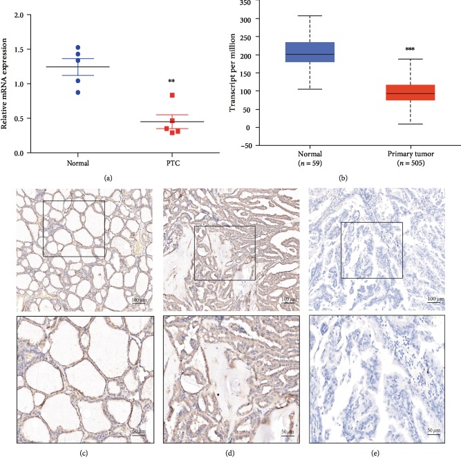
The results of qPCR showed that the expression of WBP5 in the adjacent normal tissues was significantly higher than that in PTC (Figure 1(a)). Consistently, the TCGA-thyroid carcinoma data also suggested that the expression of WBP5 was decreased in PTC (Figure 1(b)). Moreover, WBP5 detected by IHC exhibited high expression in 45.45% of the adjacent normal tissues (10/22) and 42.75% of PTC tissues (56/131), as shown in Figures 1(c)–1(d). Finally, the immunohistochemical staining intensity of WBP5 in the adjacent normal tissues was significantly higher than that in PTC tissues.
Figure 1.
Expression of WBP5 in papillary thyroid cancer (PTC) and the paired adjacent normal tissue samples. (a) qPCR was used to detect the mRNA level of WBP5 expression in PTC and the adjacent normal tissues. ∗∗P ≤ 0.01, two-sided paired t-test. (b) The TCGA-thyroid carcinoma data analysis of WBP5 expression level between the normal thyroid gland versus PTC. ∗∗∗P ≤ 0.001, two-sided unpaired t-test. (c) Positive expression of WBP5 in the normal thyroid tissue. (d) Positive expression of WBP5 in PTC. (e) Negative expression of WBP5 in PTC.
3.2. Clinicopathological Implication of WBP5 Expression in PTC
The association between the expression of WBP5 and the clinicopathological features of patients with PTC is summarized in Table 1. Statistical analysis indicated that bilaterality (P = 0.043), capsule invasion (P = 0.042), and N-stage (P = 0.026) were significantly associated with WBP5 expression (P < 0.05). Notably, the postoperative recurrence rate of patients with high WBP5 expression was lower than that of patients with low WBP5 expression in PTC (P = 0.006). However, there was no significant correlation between WBP5 expression and age, gender, tumor number, maximal tumor diameter, and intrathyroidal dissemination.
3.3. Survival Analysis
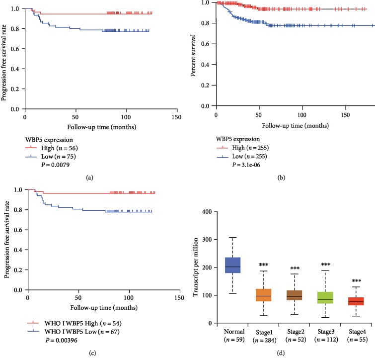
As shown in Table 2, the univariate analysis revealed that patients with PTC with high WBP5 expression had lower relapse risk than those with low WBP5 expression (hazard ratio [HR] = 0.221, 95% confidence interval [CI]: 0.065–0.753, P = 0.016). Moreover, significant correlations between relapse and bilaterality (P = 0.003), maximal tumor diameter (P = 0.015), capsule invasion (P = 0.016), total thyroidectomy (P = 0.005), lymph node dissection (P = 0.039), and iodine radiotherapy (P ≤ 0.001) were also identified using univariate analysis. The subsequent multivariate analysis confirmed that both total thyroidectomy (P = 0.039) and iodine radiotherapy (P ≤ 0.001) were predictors of DFS in patients with PTC (Table 2). However, WBP5 was not identified as an independent prognostic factor using multivariate analysis (HR = 0.746, 95% CI: 0.191–2.921, P = 0.674). Kaplan–Meier analysis indicated that patients with high WBP5 expression (P < 0.05) had a lower risk to recur than those with low expression (Figure 2(a)). Our findings were consistent with the results of the TCGA-thyroid carcinoma data, which contained 510 samples (Figure 2(b)). Furthermore, patients with high WBP5 expression (P < 0.05) had a significantly longer DFS in patients with WHO grade I PTC (Figure 2(c)), while other stages were not significantly associated with WBP5 expression. In the TCGA-thyroid carcinoma data, the expression of WBP5 significantly declined in high-grade PTC compared with that in the normal thyroid tissue (Figure 2(d)).
Table 2.
Univariate and multivariate cox regression analysis of WBP5 expression with patient prognosis.
| Variable | Univariate analysis | Multivariate analysis | ||
|---|---|---|---|---|
| HR (95% CI) | P-value | HR (95% CI) | P-value | |
| Age (years) | 0.969 (0.933–1.007) | 0.106 | ||
| Gender | 1.642 (0.631–4.272) | 0.310 | ||
| Histological variants | 0.048 (0.000–9.588) | 0.262 | ||
| Bilaterality | 3.834 (1.593–9.226) | 0.003∗ | 2.704 (0.555–13.183) | 0.218 |
| Tumor number | 1.555 (0.635–3.803) | 0.334 | ||
| Maximal tumor diameter | 2.735 (1.214–6.161) | 0.015∗ | 1.364 (0.458–4.058) | 0.577 |
| Capsule invasion | 1.826 (1.121–2.975) | 0.016∗ | 1.088 (0.552–2.145) | 0.808 |
| Intrathyroidal dissemination | 2.020 (0.734–5.559) | 0.173 | ||
| TNM staging (I/II VS III/IV) | 0.049 (0.000–234490.651) | 0.700 | ||
| Total thyroidectomy | 3.580 (1.482–8.650) | 0.005∗ | 0.138 (0.021–0.902) | 0.039∗ |
| Lymph node dissection | 2.297 (1.044–5.055) | 0.039∗ | 1.053 (0.469–2.362) | 0.900 |
| Iodine radiotherapy | 15.265 (5.086–45.813) | ≤0.001∗ | 26.947 (6.232–116.515) | ≤0.001∗ |
| WBP5 expression | 0.221 (0.065–0.753) | 0.016∗ | 0.746 (0.191–2.921) | 0.674 |
HR : Hazard ratio. ∗Statistical significance.
Figure 2.
Kaplan–Meier curve and bioinformatics analyses of WBP5 expression. (a) WBP5 expression was significantly correlated with DFS in PTC (P < 0.05). (b) Association between WBP5 expression level and DFS in patients with PTC based on the gene expression profiling interactive analysis (GEPIA). Patients with PTC with a high WBP5 had longer DFS than those with low WBP5 expression (P < 0.05). (c) WBP5 expression was significantly correlated with the WHO grade I prognosis in PTC (P < 0.05). (d) The expression of WBP5 at different stages of PTC in the UALCAN database. ∗∗∗P ≤ 0.001, two-sided unpaired t-test.
4. Discussion
Currently, patients with PTC usually have a favorable prognosis. However, some patients with PTC still have a high risk of recurrence; therefore, screening for novel molecular markers of PTC recurrence is important for effective treatment and follow-up. In the present study, we found that WBP5 was highly expressed in 56 of the 131 PTC tissues (42.75%) and 10 of the 22 (45.45%) adjacent normal thyroid tissue. The data analyse showed that patients with PTC with a high WBP5 expression had a lower relapse risk than that in patients with PTC with a low WBP5 expression. Therefore, WBP5 might serve as a valuable and specific prognostic biomarker in PTC.
There have been only a few studies on WBP5, and to the best of our knowledge, the association between WBP5 and PTC clinicopathological features has not been reported. In this study, we explored the correlation between WBP5 expression and clinical features of PTC by statistical analysis of specimens from 131 patients with PTC. Analysis result suggested that WBP5 might be a favorable prognostic indicator of PTC. Interestingly, in our study, the expression of WBP5 was higher in patients with unilateral tumorigenesis, no membrane invasion, and no lymph node metastasis, suggesting that WBP5 expression is an indicator of less aggressive tumors. Nevertheless, Tang et al. [15] revealed that the expression of WBP5 might be higher in patients at the advanced disease stage than at the initial disease stage, suggesting that WBP5 can be a marker for predicting short survival time in patients with SCLC. Although it has been suggested that WBP5 might be an oncogene in human colorectal cancer with microsatellite instability [16], a recent study by Suh et al. [17] indicated that WBP5 is a possible tumor suppressor gene in gastric carcinogenesis. WBP5 (pp21 homolog) is homologous to TCEAL7 [21, 22], which is regarded as a possible tumor suppressor gene in ovarian cancer [23]. In addition, studies have reported that pp21 homolog can inhibit the activation of Rous sarcoma virus in chicken embryo fibroblasts [24]. The reason for the inconsistency in the function of WBP5 might be that WBP5 plays different roles in different tumors; there are only a few detailed studies on WBP5. In order to improve the accuracy and reliability of the usefulness of WBP5 in predicting clinical behavior of PTC, a larger sample size and extended follow-up time are needed. In the future, it is necessary to explore other functions of WBP5 in thyroid cancer, as the function of WBP5 in PTC has not been fully revealed.
5. Conclusions
In summary, our results suggested that WBP5 could predict favorable DFS in patients with PTC. WBP5 might serve as a valuable and specific prognostic biomarker for PTC, and WBP5 up-regulation might provide a therapeutic method for improving DFS in patients with PTC.
Acknowledgments
The authors are thankful to the staff of the Pathology Department of Zhejiang Cancer Hospital for the supply and histological analysis of the tumors. They are also indebted to the patients for sharing their medical records.
Data Availability
The data that support the findings of this study are available from the corresponding author, Minghua Ge, upon reasonable request.
Conflicts of Interest
The authors declare that they have no conflicts of interest.
Funding
This study was supported by National Natural Science Foundation of China (Grant Nos. 81872170, 81672642, and 81602348) and the Key research and development project in Zhejiang Province (Grant No. 2015C03G1360022).
References
- 1. Mcleod D. S., Sawka A. M., Cooper D. S. Controversies in primary treatment of low-risk papillary thyroid cancer. Lancet. 2013;381:1046–1057. doi: 10.1016/s0140-6736(12)62205-3. [DOI] [PubMed] [Google Scholar]
- 2.Zheng R., Zeng H., Zhang S., Chen T., Chen W. National estimates of cancer 3. Prevalence in China, 2011. Cancer Letters. 2016;370:33–38. doi: 10.1016/j.canlet.2015.10.003. [DOI] [PubMed] [Google Scholar]
- 3.La Vecchia C., Malvezzi M., Bosetti C., et al. Thyroid cancer mortality and incidence: a global overview. International Journal of Cancer. 2015;136(9):2187–2195. doi: 10.1002/ijc.29251. [DOI] [PubMed] [Google Scholar]
- 4.Siegel R. L., Miller K. D., Jemal A. Cancer statistics, 2018. CA: A Cancer Journal for Clinicians. 2018;68(1):7–30. doi: 10.3322/caac.21442. [DOI] [PubMed] [Google Scholar]
- 5.de Melo T. G., Zantut-Wittmann D. E., Ficher E., da Assumpção L. V. M. Factors related to mortality in patients with papillary and follicular thyroid cancer in long-term follow-up. Journal of Endocrinological Investigation. 2014;37(12):1195–1200. doi: 10.1007/s40618-014-0131-4. [DOI] [PubMed] [Google Scholar]
- 6.Brassard M., Borget I., Edet-Sanson A., et al. Long-term follow-up of patients with papillary and follicular thyroid cancer: a prospective study on 715 patients. The Journal of Clinical Endocrinology & Metabolism. 2011;96(5):1352–1359. doi: 10.1210/jc.2010-2708. [DOI] [PubMed] [Google Scholar]
- 7.Lin K. L., Wang O. C., Zhang X. H., Dai X. X., Hu X. Q., Qu J. M. The BRAF mutation is predictive of aggressive clinicopathological characteristics in papillary thyroid microcarcinoma. Annals of Surgical Oncology. 2010;17(12):3294–3300. doi: 10.1245/s10434-010-1129-6. [DOI] [PubMed] [Google Scholar]
- 8.Bastos A. U., Oler G., Nozima B. H. N., Moysés R. A., Cerutti J. M. BRAF V600E and decreased NIS and TPO expression are associated with aggressiveness of a subgroup of papillary thyroid microcarcinoma. European Journal of Endocrinology. 2015;173(4):525–540. doi: 10.1530/EJE-15-0254. [DOI] [PubMed] [Google Scholar]
- 9.Howell G. M., Nikiforova M. N., Carty S. E., et al. BRAF V600E mutation independently predicts central compartment lymph node metastasis in patients with papillary thyroid cancer. Annals of Surgical Oncology. 2013;20:47–52. doi: 10.1530/eje-15-0254. [DOI] [PubMed] [Google Scholar]
- 10.Lee S. E., Hwang T. S., Choi Y. L., et al. Molecular profiling of papillary thyroid carcinoma in Korea with a high prevalence of BRAFV600E. Thyroid. 2017;27:802–810. doi: 10.1089/thy.2016.0547. [DOI] [PubMed] [Google Scholar]
- 11.Sudol M., Chen H. I., Bougeret C., Einbond A., Bork P. Characterization of a novel protein-binding module—the WW domain. FEBS Letters. 1995;369(1):67–71. doi: 10.1016/0014-5793(95)00550-S. [DOI] [PubMed] [Google Scholar]
- 12.Bedford M. T., Chan D. C., Leder P. FBP WW domains and the Abl SH3 domain bind to a specific class of proline-rich ligands. EMBO Journal. 1997;16(9):2376–2383. doi: 10.1093/emboj/16.9.2376. [DOI] [PMC free article] [PubMed] [Google Scholar]
- 13.Liu W., Wu J., Xiao L., et al. Regulation of neuronal cell death by c-Abl-Hippo/MST2 signaling pathway. PLoS One. 2012;7(5):p. e36562. doi: 10.1371/journal.pone.0036562. [DOI] [PMC free article] [PubMed] [Google Scholar]
- 14.Guo L., Liu Y., Bai Y., Sun Y., Xiao F., Guo Y. Gene expression profiling of drug-resistant small cell lung cancer cells by combining microRNA and cDNA expression analysis. European Journal of Cancer. 2010;46(9):1692–1702. doi: 10.1016/j.ejca.2010.02.043. [DOI] [PubMed] [Google Scholar]
- 15.Tang R., Lei Y., Hu B., et al. WW domain binding protein 5 induces multidrug resistance of small cell lung cancer under the regulation of miR-335 through the hippo pathway. British Journal of Cancer. 2016;115(2):243–251. doi: 10.1038/bjc.2016.186. [DOI] [PMC free article] [PubMed] [Google Scholar]
- 16.Gylfe A. E., Kondelin J., Turunen M., et al. Identification of candidate oncogenes in human colorectal cancers with microsatellite instability. Gastroenterology. 2013;145(3):540–543.e22. doi: 10.1053/j.gastro.2013.05.015. [DOI] [PubMed] [Google Scholar]
- 17.Suh Y. S., Yu J., Kim B. C., et al. Overexpression of plasminogen activator inhibitor-1 in advanced gastric cancer with aggressive lymph node metastasis. Cancer Research and Treatment. 2015;47(4):718–726. doi: 10.4143/crt.2014.064. [DOI] [PMC free article] [PubMed] [Google Scholar]
- 18.Amin M. B., Greene F. L. Edge SB AJCC Cancer Staging Manual. 8th. New York: Springer; 2016. [Google Scholar]
- 19.Lanczky A., Nagy A., Bottai G., et al. miRpower: a web-tool to validate survival-associated miRNAs utilizing expression data from 2178 breast cancer patients. Breast Cancer Research and Treatment. 2016;160(3):439–446. doi: 10.1007/s10549-016-4013-7. [DOI] [PubMed] [Google Scholar]
- 20.Tang Z., Li C., Kang B., Gao G., Li C., Zhang Z. GEPIA: a web server for cancer and normal gene expression profiling and interactive analyses. Nucleic Acids Research. 2017;45(W1):W98–W102. doi: 10.1093/nar/gkx247. [DOI] [PMC free article] [PubMed] [Google Scholar]
- 21.Mukai J., Hachiya T., Shoji-Hoshino S., et al. NADE, a p75NTR-associated cell death executor, is involved in signal transduction mediated by the common neurotrophin receptor p75NTR. Journal of Biological Chemistry. 2000;275(23):17566–17570. doi: 10.1074/jbc.C000140200. [DOI] [PubMed] [Google Scholar]
- 22.Mukai J., Kimura T., Sano H., et al. Structure-function analysis of NADE: identification of regions that mediate nerve growth factor-induced apoptosis. Journal of Biological Chemistry. 2002;277(16):13973–13982. doi: 10.1074/jbc.M106342200. [DOI] [PubMed] [Google Scholar]
- 23.Rattan R., Narita K., Chien J., et al. TCEAL7, a putative tumor suppressor gene, negatively regulates NF-B pathway. Oncogene. 2009;29:1362–1373. doi: 10.1038/onc.2009.431. [DOI] [PubMed] [Google Scholar]
- 24.Yeh C. H., Shatkin A. J. Down-regulation of rous sarcoma virus long terminal repeat promoter activity by a HeLa cell basic protein. Proceedings of the National Academy of Sciences. 1994;91(23):11002–11006. doi: 10.1073/pnas.91.23.11002. [DOI] [PMC free article] [PubMed] [Google Scholar]
Associated Data
This section collects any data citations, data availability statements, or supplementary materials included in this article.
Data Availability Statement
The data that support the findings of this study are available from the corresponding author, Minghua Ge, upon reasonable request.


