Abstract
We present a case of an unusually large, circumferential tubulovillous adenoma involving the terminal ileum and the caecum with ileocaecal valve consumption, presenting as intussusception in an otherwise healthy 90-year-old woman. The patient presented with several months of chronic symptoms of weight loss and diarrhoea. Clinical examination revealed a right-sided mass. Investigations revealed a large right-sided lesion suspicious of intussusception. The patient underwent a right-sided hemicolectomy where the intussusception was resected. Histology of the resected mass revealed a tubulovillous adenoma with focal invasive adenocarcinoma.
Keywords: colon cancer, pathology, cancer intervention, surgical oncology, general surgery
Background
Adult intussusception is a rare clinical phenomenon with paediatric cases accounting for the majority, 95%, of cases.1 Adult intussusception also presents a clinical challenge as, unlike paediatric cases, it does not present in a specific manner. Tumours have been shown to be the most common cause of adult intussusception, where they act as lead points for the formation of the intussusception.2 In our case, the patient presented with chronic abdominal pain secondary to intussusception with an unusually large underlying tubulovillous adenoma. The chronicity of the patient’s symptoms highlights the importance for physicians to always consider intussusception in chronic abdominal pain and investigate for potential underlying malignancy.
Case presentation
A normally fit and well 90-year-old woman was referred to colorectal clinic by her general practitioner for several months of change in bowel habit. She described weight loss, diarrhoea, bloating and 1 month of bleeding per rectum which she was not concerned about as this had stopped some months ago after she stopped a trial of non-vitamin K oral anticoagulants for atrial fibrillation (AF). Her medical history was significant only for AF and atrial flutter. There was no family history of bowel cancer.
On examination, there was a palpable abdominal mass in the right abdomen but no evidence of acute abdomen or peritonitis. Blood biochemistry and full blood count were unremarkable. CT pneumocolon (CT-VC) showed a right-sided abdominal mass suspicious for intussusception. The tumour was excised laparoscopically. The mass was resected without reduction. Histology confirmed the mass to be a large circumferential tubulovillous adenoma with predominantly low-grade and focal high-grade dysplasia. Adenocarcinoma was shown to be present within the tubulovillous adenoma but only invaded as far as the lamina propria making this a stage pT1 tumour. Examination of 21 mesenteric lymph nodes showed no evidence of spread.
Despite the extensive size of the lesion causing the intussusception, the patient did not present with significant obstructing symptoms. Instead, she presented with more chronic symptoms of weight loss, diarrhoea, bloating and 1 month of bleeding per rectum. This may be attributed to the fact that the carpeting ileocaecal lesion of the tubulovillous adenoma was destroying the ileocaecal valve allowing for the bowel contents to pass freely from the small bowel to the large bowel.
Investigations
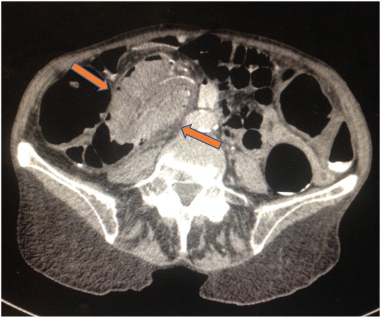
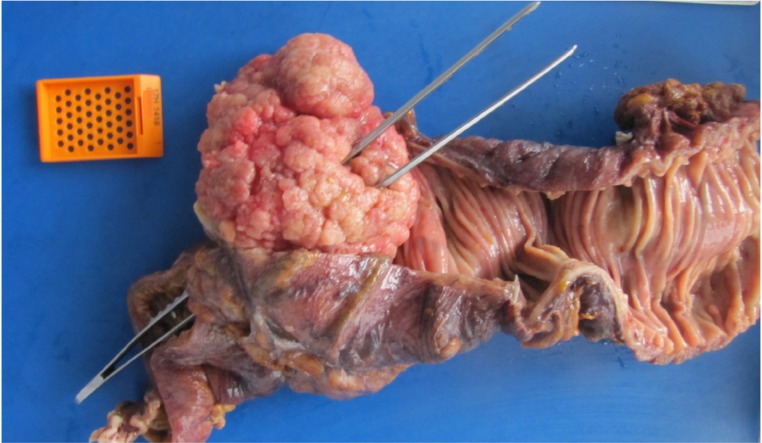
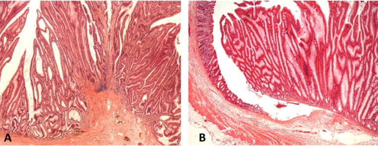
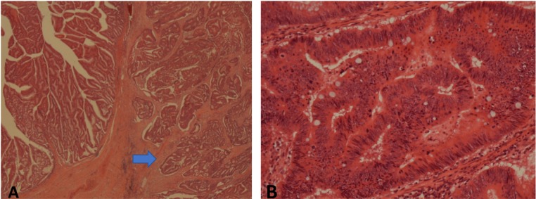
CT-VC was done which showed a caecal mass with invaginating ileocolic blood vessels suggestive of intussusception (figure 1). Radiological findings together with the clinical picture were suggestive of cancer. A CT scan of the chest was negative for metastases. She underwent a laparoscopic right hemicolectomy which showed an ileocaecal intussusception. The mass was resected (figure 2) without reduction. Histology confirmed the mass to be a large 8×6 cm circumferential tubulovillous adenoma with predominantly low-grade (figure 3) and focal high-grade dysplasia with focal invasive adenocarcinoma arsing within it (figure 4). The adenoma was involving all of the caecal lumen with ileocaecal valve consumption and growth into the terminal ileum. The bowel was almost completely obstructed.
Figure 1.
CT scan showing an elongated sausage-shaped mass (arrows) invaginating into the colon.
Figure 2.
Right-hemicolectomy specimen.
Figure 3.
(A and B) ×20 magnification of the tubulovillous adenoma showing areas of low-grade dysplasia.
Figure 4.
(A) ×20 magnification of the adenoma with adjacent invasive moderately differentiated adenocarcinoma (arrow); (B) ×200 magnification for the areas of adenoma with high-grade dysplasia.
Treatment
Laparoscopic right hemicolectomy. This was both diagnostic and curative. Intussusception was seen on laparoscopy. The diseased bowel was resected without intestinal reduction. Clear margins of healthy bowel were anastomosed.
Outcome and follow-up
The patient made an unremarkable recovery postoperatively. She was discharged back to her home a week after her procedure and followed up in colorectal clinic 2 months later with blood tests to include full blood count, liver profile, renal profile and carcinoembryonic antigen tumour marker. The results of these tests were unremarkable. She continues to do well and will be followed up as per guidelines.
Discussion
Adult intussusception is a rare clinical phenomenon with an incidence of 2 to 3 million per year.2 Diagnosis, however, presents a clinical challenge given the non-specific presentation of adult intussusception. Moreover, in adult intussusception, there is often a lead point causing the phenomenon. Indeed, large case series have revealed tumours as the most common type of lead point.1–3 Inopportunely, adult intussusception is often longstanding and presents in a non-specific manner.2 3 Given the significant likelihood of underlying malignancy, it is important for physicians to consider intussusception in chronic abdominal pain in adults and investigate accordingly. A review of the literature shows that CT scanning is currently the most accurate modality in the diagnosis of adult intussusception.3 4
In our case, a CT-VC was done, which revealed the classic target lesion of intussusception. The resected tumour was shown to be a circumferential tubulovillous adenoma involving the caecum, ileocaecal valve and extending into the ileum with total consumption of the ileocaecal valve. This is a very unusual presentation of a right-sided adenoma as these tumours do not typically grow in an annular fashion or to be so large.1 3 5
Learning points.
Consider intussusception in chronic abdominal pain.
Adult intussusception is a rare clinical phenomenon without specific symptoms but often with an identifiable cause.
Resection of intussusception is preferred in adults given the high likelihood of malignancy in large-bowel intussusception.
Despite the large size of the ileocaecal adenoma, the bowel lumen may remain patent as the ileocaecal valve is totally consumed by the lesion.
Right-sided tumours such as very large circumferential adenomas may, on rare occasions, present with features more typical of left-sided tumours.
Acknowledgments
I would like to thank the patient for all their patience and understanding throughout the whole process of writing the manuscript. Thank you to Dr Kubba for all his invaluable input in writing and submitting the manuscript.
Footnotes
Contributors: EM wrote the case report with help from PM and FK. EM assisted in ensuring the patient’s procedure went ahead as planned—there were several obstacles but managed to get it done at scheduled time. EM gathered all the necessary data and wrote out the initial draft of the report. PM performed the hemicolectomy and edited the surgical section of the case report. FK analysed the resected specimen and provided all of the histology input. EM and FK wrote the bulk of the case report—EM did much of the initial drafting and FK did most of the editing. PM provided very useful input on the direction and focus of the report.
Funding: The authors have not declared a specific grant for this research from any funding agency in the public, commercial or not-for-profit sectors.
Competing interests: None declared.
Patient consent for publication: Obtained.
Provenance and peer review: Not commissioned; externally peer reviewed.
References
- 1. Anon Case record of the Massachusetts General Hospital. Weekly clinicopathological exercises. Case 26-2002. An 87-year-old woman with abdominal pain, vomiting, bloody diarrhea, and an abdominal mass. N Engl J Med 2002;347:601–6. 10.1056/NEJMcpc020109 [DOI] [PubMed] [Google Scholar]
- 2. Yalamarthi S, Smith RC. Adult intussusception: case reports and review of literature. Postgrad Med J 2005;81:174–7. 10.1136/pgmj.2004.022749 [DOI] [PMC free article] [PubMed] [Google Scholar]
- 3. Sarma D, Prabhu R, Rodrigues G. Adult intussusception: a six-year experience at a single center. Ann Gastroenterol 2012;25:128–32. [PMC free article] [PubMed] [Google Scholar]
- 4. Amr MA, Polites SF, Alzghari M, et al. Intussusception in adults and the role of evolving computed tomography technology. Am J Surg 2015;209:580–3. 10.1016/j.amjsurg.2014.10.019 [DOI] [PMC free article] [PubMed] [Google Scholar]
- 5. Gujar A, Ambardekar R, Rane N, et al. Intussusception due to caecal carcinoma in a young man: unusual cause of presentation a case report. Int J Res Med Sci 2015;3:3901–3. 10.18203/2320-6012.ijrms20151467 [DOI] [Google Scholar]


