ABSTRACT
In this supplement, we build on work previously published under the Human Connectome Project. Specifically, we seek to show a comprehensive anatomic atlas of the human cerebrum demonstrating all 180 distinct regions comprising the cerebral cortex. The location, functional connectivity, and structural connectivity of these regions are outlined, and where possible a discussion is included of the functional significance of these areas. In part 7, we specifically address regions relevant to the lateral parietal lobe.
Keywords: Anatomy, Cerebrum, Connectivity, DTI, Functional connectivity, Human, Parcellations
ABBREVIATIONS
- AIP
anterior intraparietal
- HCP
Human Connectome Project
- IFOF
inferior fronto-occipital fasciculus
- ILF
inferior longitudinal fasciculus
- IP0
intraparietal 0
- IP1
intraparietal 1
- IP2
intraparietal 2
- IPS1
intraparietal sulcus 1
- LIPd
lateral intraparietal, dorsal
- LIPv
lateral intraparietal, ventral
- MdLF
middle longitudinal fasciculus
- MIP
medial intraparietal
- MR
magnetic resonance
- PFop
parietal area F, operculum
- PF
parietal area F
- PFm
parietal area F, part m
- PFt
parietal area F, part t
- PGi
parietal area G inferior
- PGp
parietal area G, posterior
- PGs
parietal area G superior
- SLF
superior longitudinal fasciculus
- TPOJ
temporo-parieto-occipital junction
- VIP
ventral intraparietal
It has long been recognized that the parietal lobe is an important part of the cerebrum, particularly the inferior parietal lobule. Despite this, our understanding of the exact localization of parietal functions leaves much to be desired. While most neurosurgeons can locate the parts of the cerebrum responsible for motor and speech function within a gyrus, the neglect and praxis centers are not easily localized and are much less familiar. We would argue that lacking a familiar nomenclature to describe the lateral parietal lobe, as well as the complexity of this part of the cerebrum have led to incomplete efforts to localize these functions accurately. In this section, we discuss the lateral surface of the parietal lobe and highlight the substantially different anatomic subdivisions outlined under the Human Connectome Project (HCP).
BASIC ORGANIZATION OF THE PARIETAL AREAS
The lateral surface of the parietal lobe is made up of two gross anatomic regions: the superior surface of the superior parietal lobule and the inferior parietal lobule, which is itself made up of the angular and supramarginal gyri. These lobules are separated by the deep intraparietal sulcus, which is slanted across the parietal surface, angled inferiorly as one follows it from posterior to anterior where it joins the postcentral sulcus. The temporo-parieto-occipital junction (TPOJ) is a vague boundary running along the inferior portion of the parietal lobules.
The naming scheme for these regions is at a first glance somewhat confusing, but a basic understanding of the evolution of the parietal lobe is helpful for making sense of how these areas are named. Dutiful students of neuroscience will note that much of the work with areas 5 and 7 has been performed in macaques, and that these animals have a rudimentary inferior parietal lobule, primarily containing area 7.1 The intraparietal sulcus is also well known to possess several areas involved in spatial orientation, named for their position in the macaque equivalent, including anterior intraparietal (AIP), lateral intraparietal, medial intraparietal (MIP), and so on.1 These areas similarly exist in humans, and wherever possible the macaque naming scheme has been utilized in the human nomenclature. Complicating this is the expansion of the human intraparietal lobule, which contains areas without obvious analogs in macaques.2,3 The most obvious explanation for this is that these areas involve semantic tasks that monkeys have a limited ability to perform.4,5 The result is that the entirety of the macaque parietal lobe seems to have been pushed into the superior parietal lobule, which contains all of areas 5 and 7, as well as the intraparietal sulcus areas.6 The result is that the inferior parietal lobule in humans requires its own naming scheme, and that the superior parietal lobule nomenclature does not always make spatial sense, especially the intraparietal sulcus regions, which are all pushed onto the superior bank of the sulcus.
The Inferior Parietal Lobule and TPOJ Junction Areas
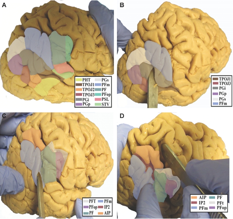
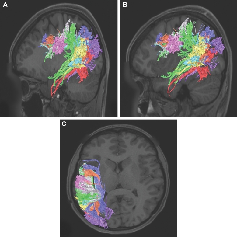
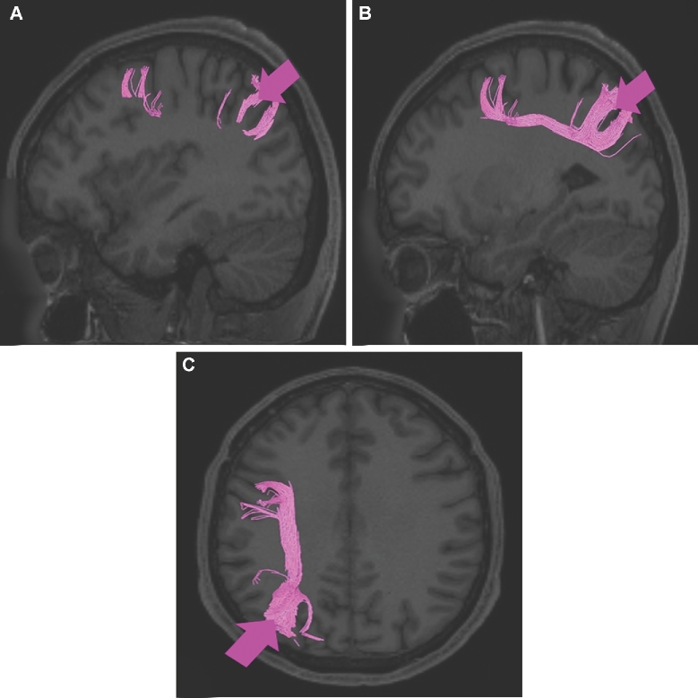
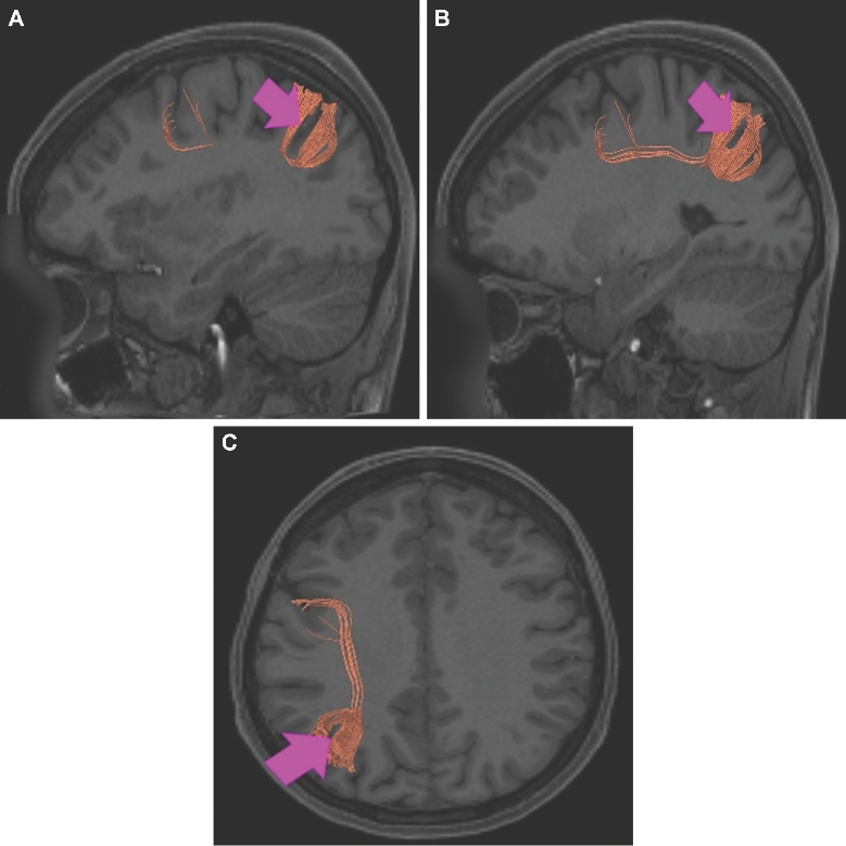
The parcellations that comprise the inferior parietal lobule and TPOJ junction areas include parietal area F, operculum (PFop), parietal area F, part t (PFt), parietal area F (PF), parietal area F, part m (PFm), parietal area G superior (PGs), parietal area G inferior (PGi), parietal area G, posterior (PGp), TPOJ1, TPOJ2, and TPOJ3. The anatomic location of these parcellations is shown on a cadaver brain in Figure 1. The combined tractography of these parcellations is shown in Figure 2.
FIGURE 1.
Anatomic view of the inferior parietal lobule and TPOJ. Parcellations are shown on the right hemisphere of a cadaver brain. A, Lateral view of the inferior parietal lobule. B, Lateral view of the inferior parietal lobule with widening of the superior temporal sulcus at the junction of the angular gyrus. C, Lateral view of the inferior parietal lobule with widening of the postcentral sulcus to show the extent of PFm and PF. D, Lateral view of the inferior parietal lobule with widening of the intraparietal sulcus.
FIGURE 2.
Combined structural connectivity of the parcellations comprising the inferior parietal lobule and the TPOJ, shown on T1-weighted magnetic resonance (MR) images. A and B, Sagittal views and C, axial view.
Area PFop
Where is it?
Area PFop is found on the anterior inferior parietal lobule where it joins the postcentral gyrus anteroinferiorly.
What are its borders?
Area PFop borders area 1 anteriorly and area PF posteriorly. Its superior border is made of PFt, and area 2, and its inferior border is made of OP4, OP1, and PFcm.
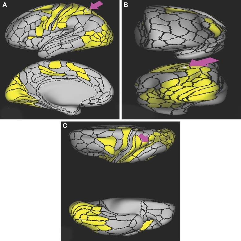
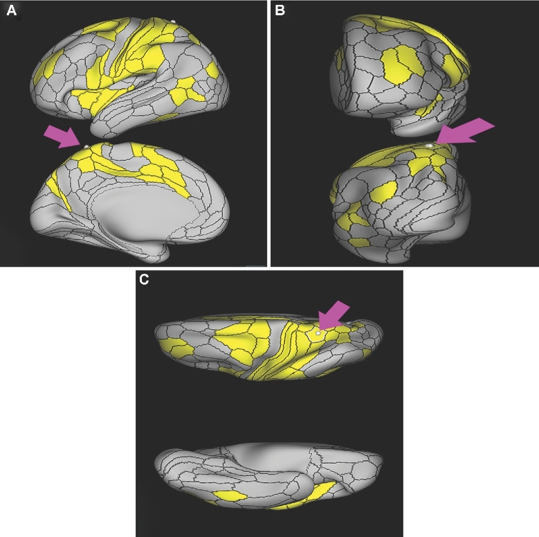
What is its functional connectivity?
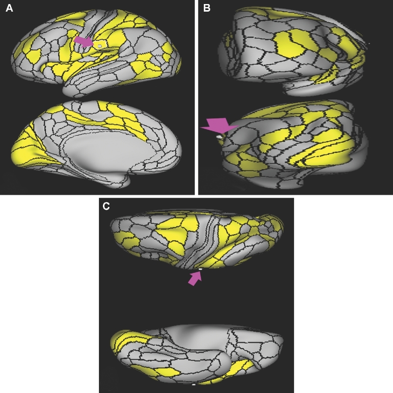
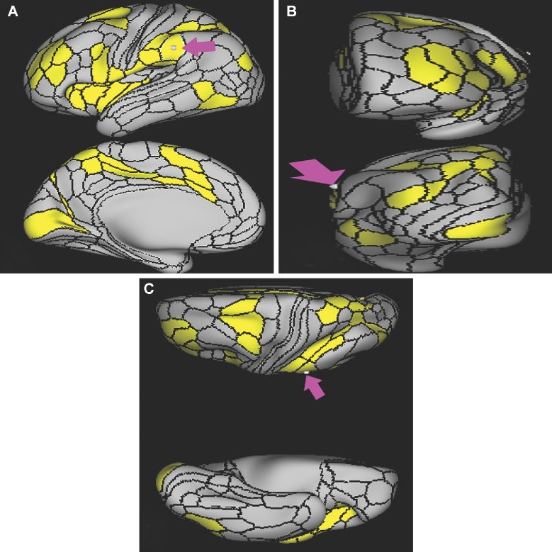
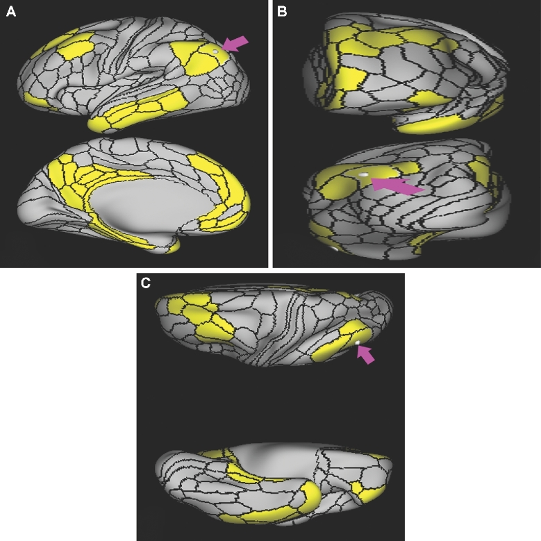
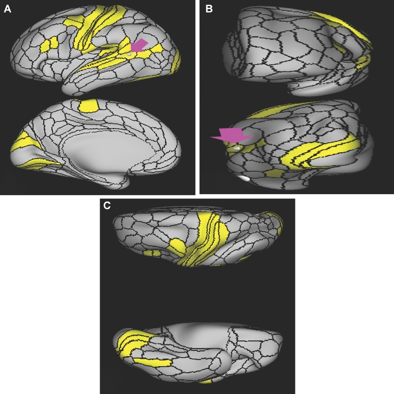
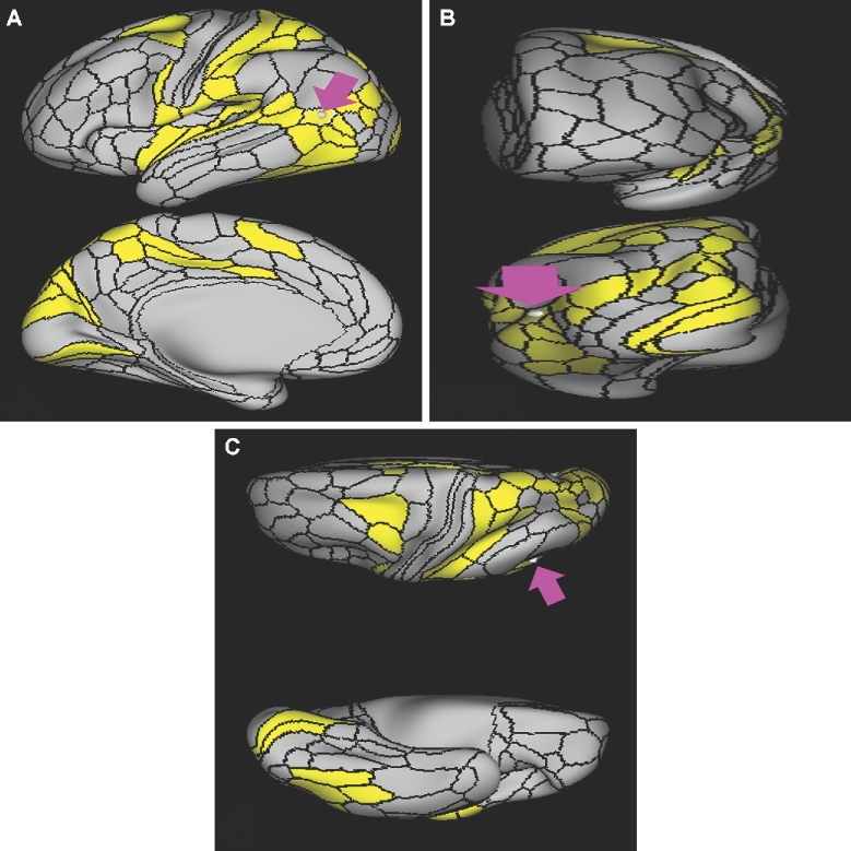
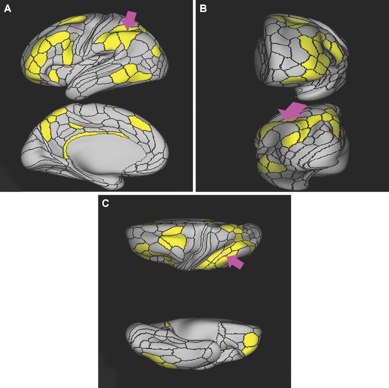
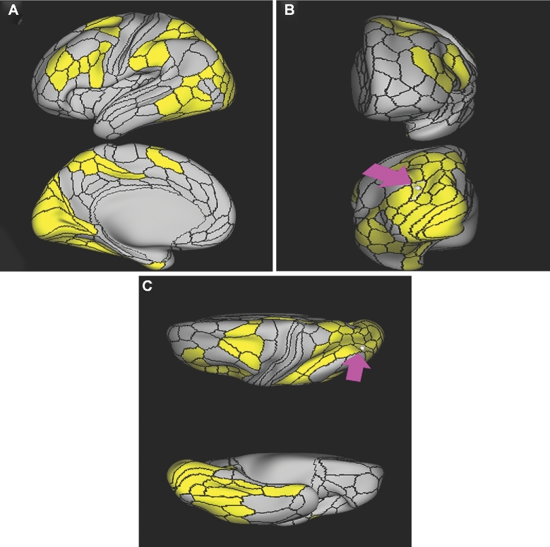
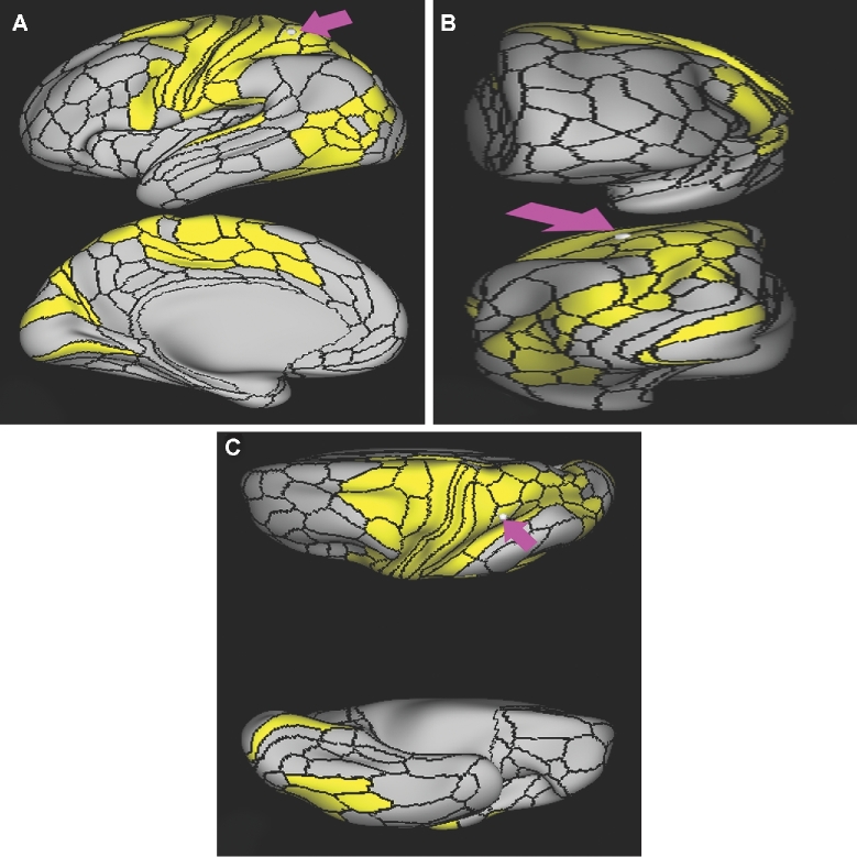
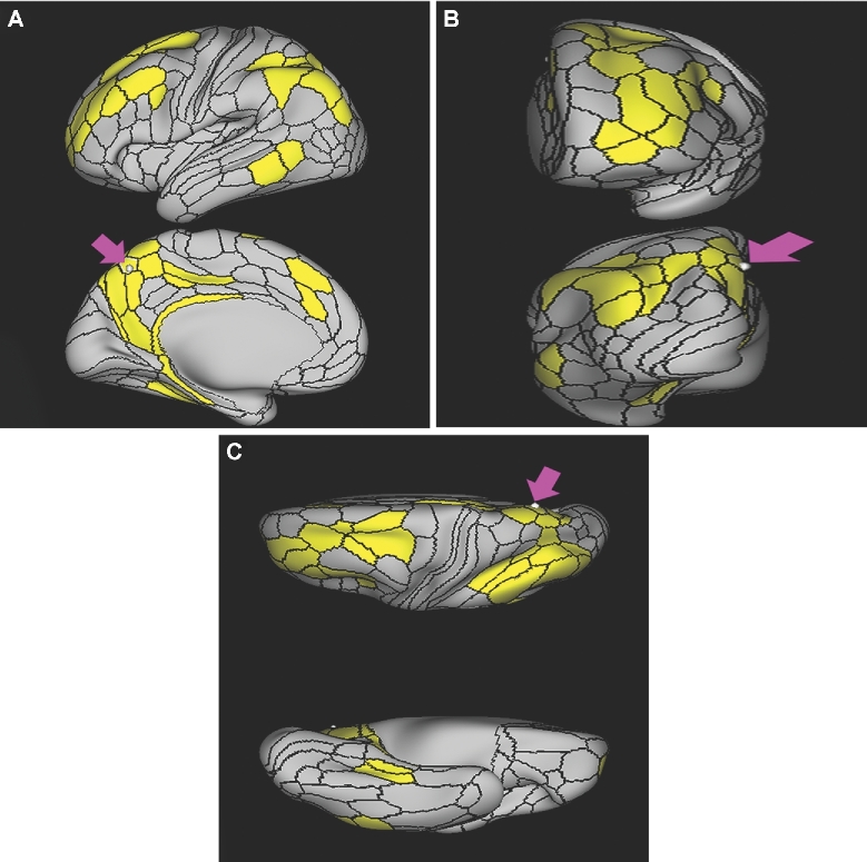
Area PFop demonstrates functional connectivity to area 2 in the sensory strip, areas SCEF, FEF, PEF, 6ma, 6mp, 6a, 6r, and 6v in the premotor regions, areas a24prime, p24prime, p32prime, 24dv, 5mv, and 23c in the middle cingulate regions, areas IFSa, 46, and 9-46d in the lateral frontal lobe, areas OP4, OP2-3, OP1, PFcm, FOP1, FOP3, FOP4, and FOP5 in the superior insula opercular regions, areas LBelt, PBelt, PI, A4, MI, 52, RI, PoI1, and PoI2 in the lower opercula and Heschl's gyrus regions, area TE2p and PHT in the temporal lobe, areas PFt, PF, PGp, intraparietal 0 (IP0), AIP, MIP, lateral intraparietal, ventral (LIPv), lateral intraparietal, dorsal (LIPd), 7PC, 7PL, and 7AL in the lateral parietal lobe, areas 7AM and DVT in the medial parietal lobe, areas V1, V2, and V3 in the medial occipital lobe, areas V3a and V6 of the dorsal visual stream, areas V8 and FFC of the ventral visual stream, and areas LO3, TPOJ2, PH, and FST of the lateral occipital lobe (Figure 3).
FIGURE 3.
Functional connectivity of PFop demonstrated on an inflated left hemisphere. A, Lateral and medial views. B, Rostral and caudal views. C, Dorsal and ventral views. Parcellations with the strongest functional connectivity are shown in yellow. Pink arrows designate the parcellation of interest.
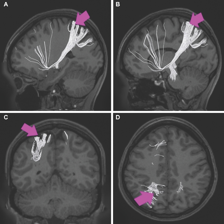
What are its white matter connections?
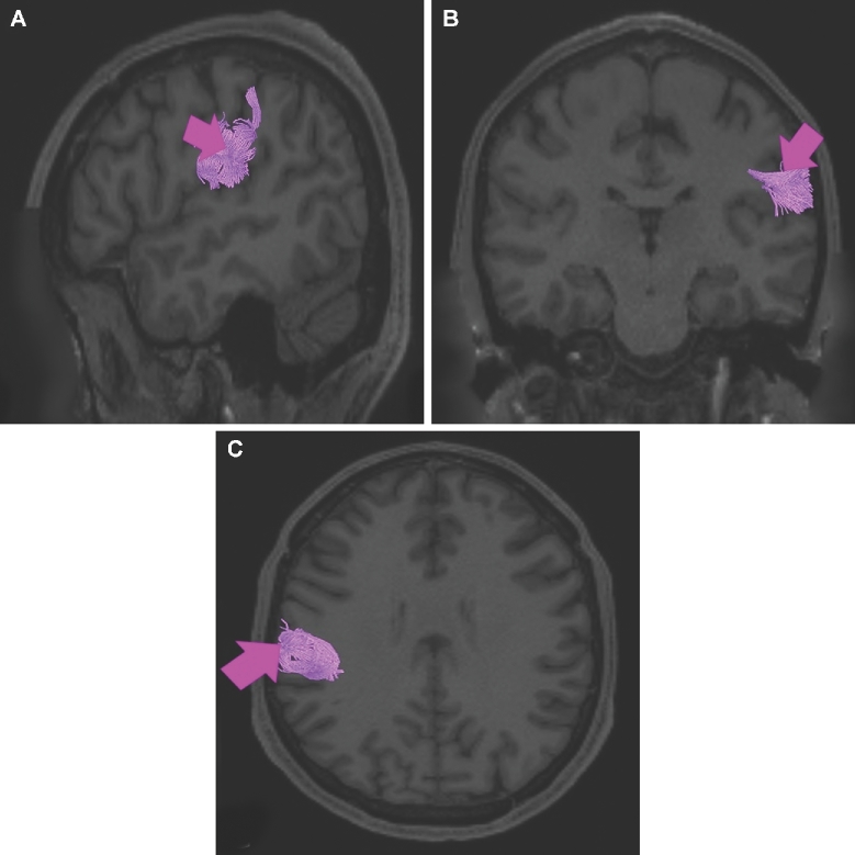
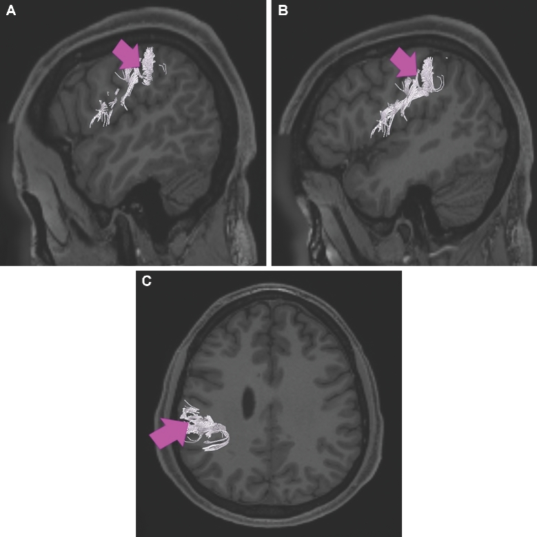
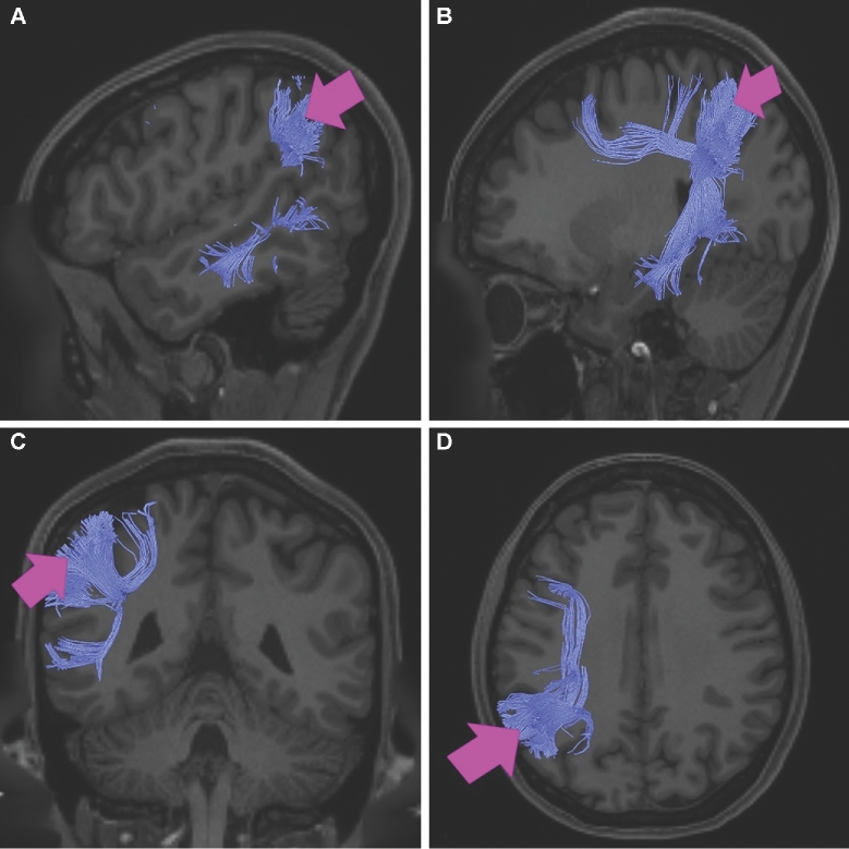
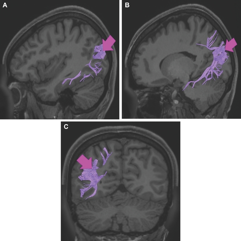
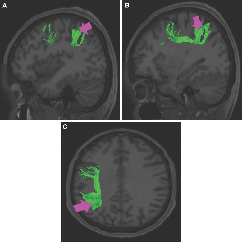
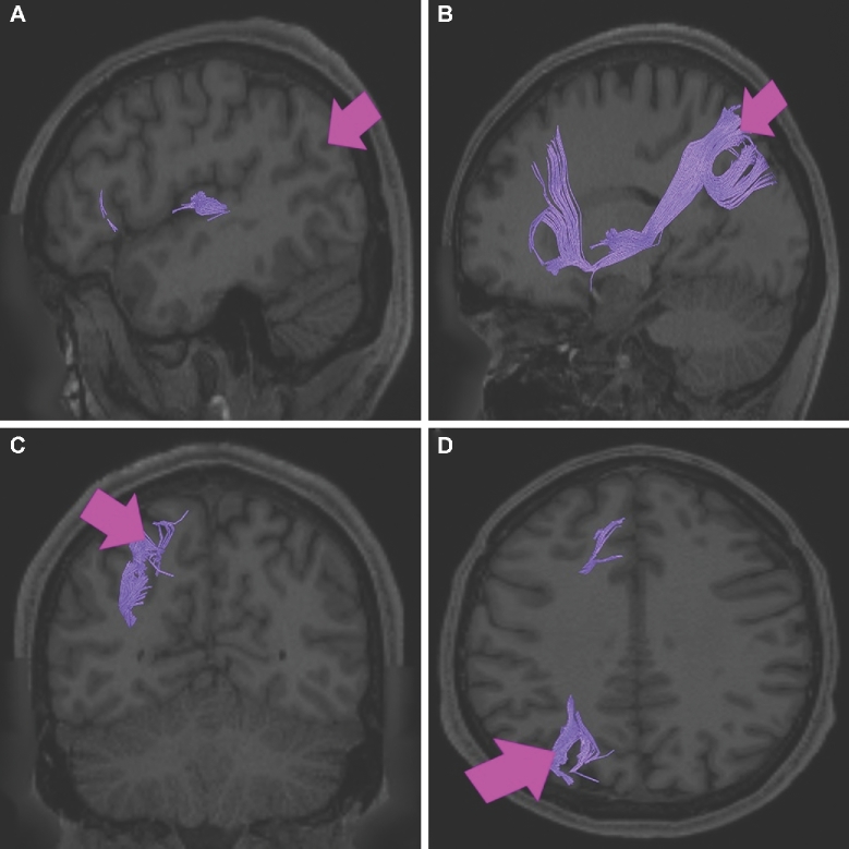
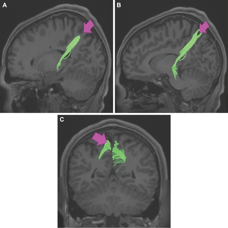
Area PFop is structurally connected to local parcellations. Local short association fibers connect with PF, PFcm, PFt, 4, OP1, and OP4 (Figure 4).
FIGURE 4.
Structural connectivity of PFop in the left hemisphere, shown on T1-weighted MR images. A, Sagittal view, B, coronal view, and C, axial view showing projections to local parcellations. Light purple: white matter tracts of PFop demonstrating connections with local parcellations. Pink arrows indicate the location of the parcellation of interest.
What is known about its function?
This region is a part of the rostral inferior parietal lobule that is involved in motor planning and action-related functions.7 For example, it is activated in humans when they observe tools being used.7 PFop has also been described as part of a mirror neuron system in which actions or behaviors are learned by observing the actions of others.8 The rostral inferior parietal lobule has also been implicated in the task-positive network.6
Area PFt
Where is it?
Area PFt is found on the posterior bank of the postcentral sulcus, on the anterosuperior edge of the inferior parietal lobule. It lies on the inferior edge of the intraparietal sulcus, just where it meets the postcentral sulcus.
What are its borders?
Area PFt borders AIP superiorly, and PFop inferiorly. Area 2 is its anterior neighbor, and area PF is its posterior neighbor.
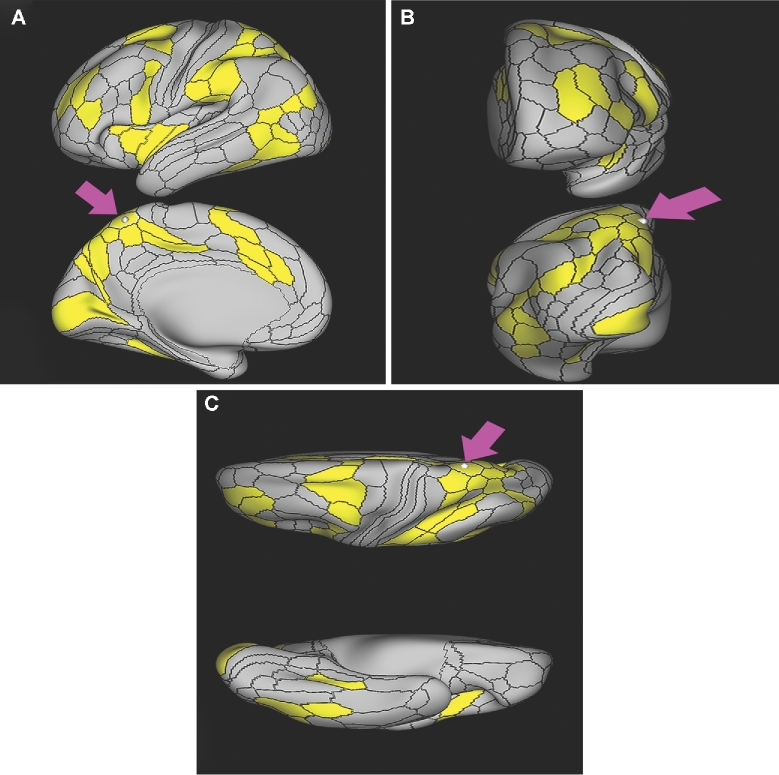
What is its functional connectivity?
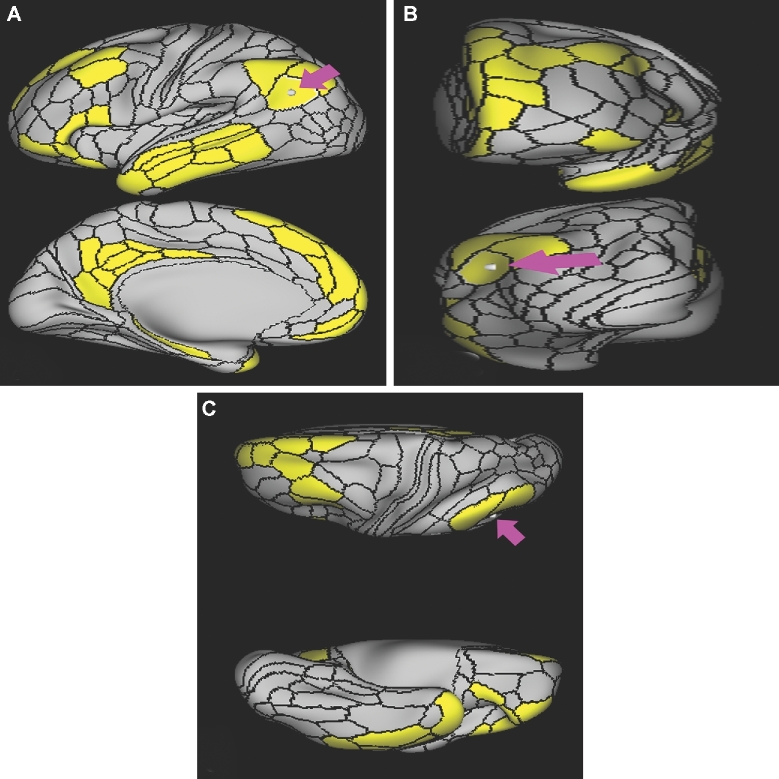
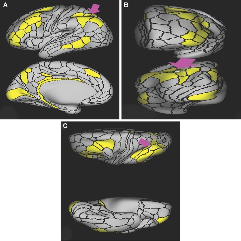
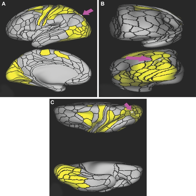
Area PFt demonstrates functional connectivity to areas 1 and 2 in the sensory strip, areas SCEF, FEF, PEF, 6ma, 6mp, 6a, 6d, 6r, and 6v in the premotor regions, areas p32prime, 5mv, and 23c in the middle cingulate regions, areas IFSa, IFJp, and 46 in the lateral frontal lobe, areas OP4, OP1, PFcm, FOP2, and FOP4 in the superior insula opercular regions, areas MI, PoI1, and PoI2 in the lower opercula and Heschl's gyrus regions, area PHA3 and PHT in the temporal lobe, areas PFop, PF, PGp, intraparietal 2 (IP2), IP0, intraparietal sulcus 1 (IPS1), AIP, ventral intraparietal (VIP), MIP, LIPv, LIPd, 7PC, 7PL, and 7AL in the lateral parietal lobe, area 7AM in the medial parietal lobe, area FFC in the ventral visual stream, and areas PH and FST of the lateral occipital lobe (Figure 5).
FIGURE 5.
Functional connectivity of PFt demonstrated on an inflated left hemisphere. A, Lateral and medial views. B, Rostral and caudal views. C, Dorsal and ventral views. Parcellations with the strongest functional connectivity are shown in yellow. Pink arrows designate the parcellation of interest.
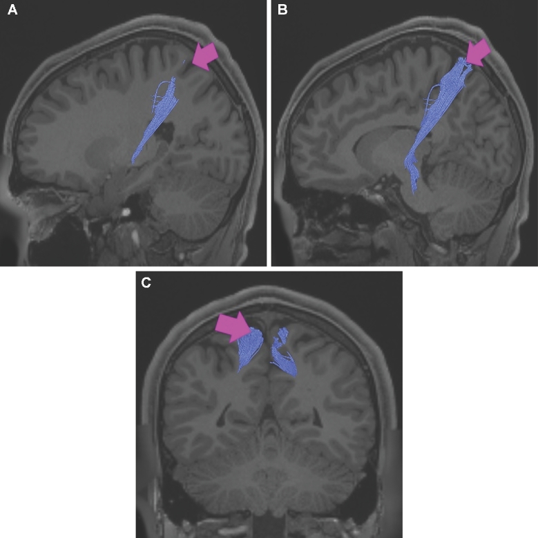
What are its white matter connections?
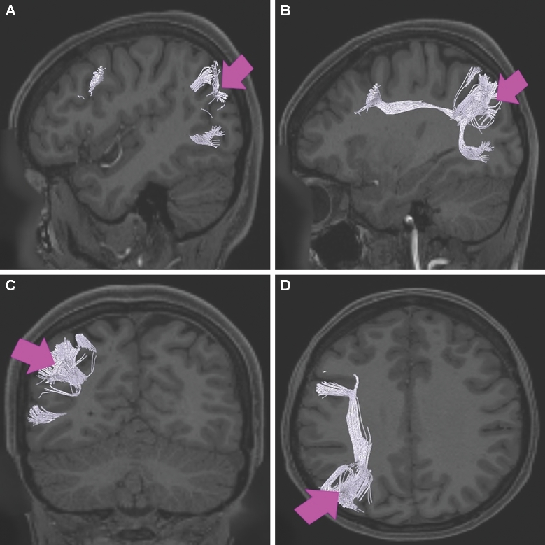
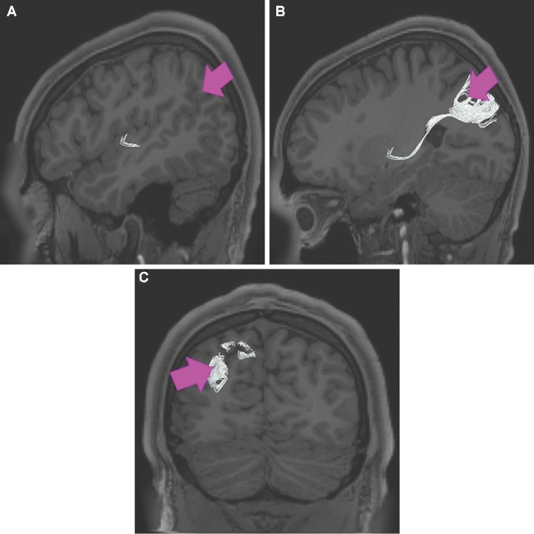
Area PFt is structurally connected to local parcellations, the inferior parietal lobule and opercular parcellations through the arcuate/superior longitudinal fasciculus (SLF). Connections from PFt project diagonally to the inferior parietal lobule and anterior inferior to the operculum. Operculum projections end at OP4, 43, 6r, and 4. Inferior parietal projections end at 2 and AIP. Local short association bundles are PF and PFop (Figure 6).
FIGURE 6.
Structural connectivity of PFt in the left hemisphere, shown on T1-weighted MR images. A and B, Sagittal and C, axial views showing projections to local parcellations and the arcuate/SLF. White: white matter tracts of PFt demonstrating connections with local parcellations and the arcuate/SLF. Pink arrows indicate the location of the parcellation of interest.
What is known about its function?
Area PFt is important in the action observation and imitation network as well as in grasping objects under visual guidance.7 This region is also implicated in the mirror neuron system described above.7 PFt is also activated when individuals observe the use of tools to move objects.9
Area PF
Where is it?
Area PF is found on the lateral surface of the superior portion of the supramarginal gyrus.
What are its borders?
Area PF borders PSL and PFcm inferiorly, PFop and PFt anteriorly, IP2 superiorly, and PFM posteriorly.
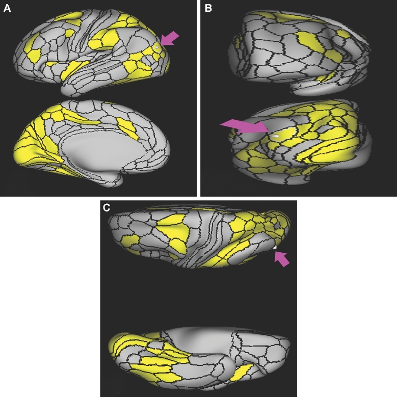
What is its functional connectivity?
Area PF demonstrates functional connectivity to areas SCEF, FEF, 6ma, 6a, and 6r in the premotor regions, areas a24prime, p24prime, p32prime, 5mv, and 23c in the middle cingulate regions, areas IFSa, IFJp, a9-46v, p9-46v, 46, and 9-46d in the lateral frontal lobe, areas OP4, PFcm, FOP1, FOP3, and FOP5 in the superior insula opercular regions, areas AVI, MI, 52, PoI1, and PoI2 in the lower opercula and Heschl's gyrus regions, area PHT in the temporal lobe, areas PFt, PFop, PGp, IP0, IP2, AIP, MIP, LIPd, 7PL, and 7AL in the lateral parietal lobe, areas 7AM, PCV, and DVT in the medial parietal lobe, and area V1 in the medial occipital lobe (Figure 7).
FIGURE 7.
Functional connectivity of PF demonstrated on an inflated left hemisphere. A, Lateral and medial views. B, Rostral and caudal views. C, Dorsal and ventral views. Parcellations with the strongest functional connectivity are shown in yellow. Pink arrows designate the parcellation of interest.
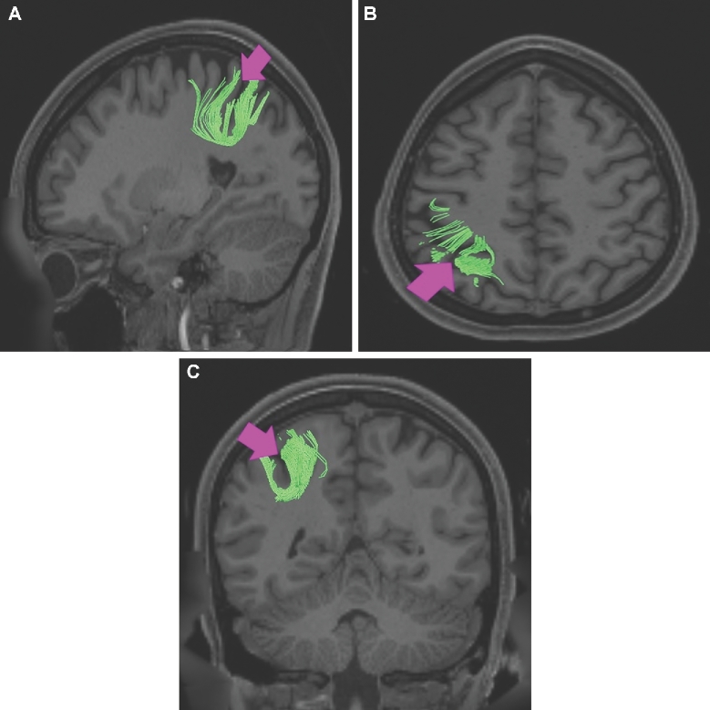
What are its white matter connections?
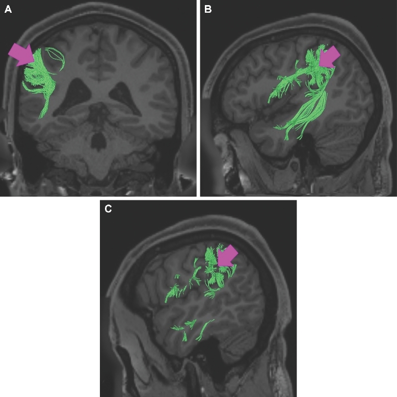
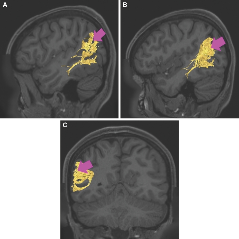
Area PF is structurally connected to the arcuate/SLF and local parcellations. Arcuate/SLF connections course anteriorly from PF to somatosensory areas 1, 3a, 4, 43, and OP4, and inferiorly to middle and inferior temporal gyrus parcellations TE1a, STSva, and TE2a. Local short association bundles connect with AIP, PFcm, PFm, PFop, PFt, PSL, and STV (Figure 8).
FIGURE 8.
Structural connectivity of PF in the left hemisphere, shown on T1-weighted MR images. A, Coronal view, B, medial sagittal view and C, lateral sagittal view showing projections to local parcellations and the arcuate/SLF. Green: white matter tracts of PF demonstrating connections with local parcellations and the arcuate/SLF. Pink arrows indicate the location of the parcellation of interest.
What is known about its function?
PF has functions similar to those localized to area PFt. It is important in the action observation and imitation network and has been implicated in the mirror neuron system described above.7 It is also activated when individuals observe the use of tools to move objects.9
Area PFm
Where is it?
Area PFm is found on the anterior superior surface of the angular gyrus, and straddles the sulcus to lie on the posterior superior bank of the supramarginal gyrus.
What are its borders?
Area PFm borders IP2 and intraparietal 1 (IP1) superiorly, PF anteriorly, PSL and STV inferiorly, and PGI and PGS posteriorly.
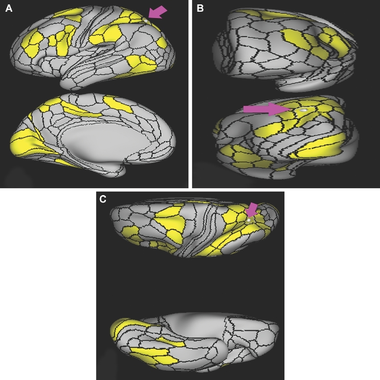
What is its functional connectivity?
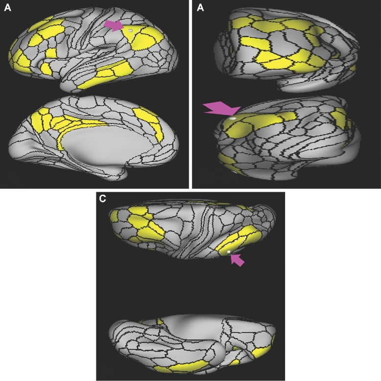
Area PFm demonstrates functional connectivity to areas 8AV, 8AD, 8BL, 8C, s6-8, i6-8, a47r, p47r, a10p, p10p, 9a, a9-46v, p9-46v, and area 44 in the lateral frontal lobe, area d32 in the medial frontal lobe, area AVI in the insula, areas STSvp, TE1m, TE1p, and TE2a, in the temporal lobe, areas PGs, PGi, IP2, and IP1 in the lateral parietal lobe, and areas 7m, 7pm, POS2, 31a, 31pv, d23ab, 23d, and RSC in the medial parietal lobe (Figure 9).
FIGURE 9.
Functional connectivity of PFm demonstrated on an inflated left hemisphere. A, Lateral and medial views. B, Rostral and caudal views. C, Dorsal and ventral views. Parcellations with the strongest functional connectivity are shown in yellow. Pink arrows designate the parcellation of interest.
What are its white matter connections?
Area PFm is structurally connected to the arcuate/SLF. Arcuate/SLF connections course anteriorly from PFm to 8C and 8BM, and inferiorly to middle temporal gyrus parcellations TE1a, TE1m, TE1p, STSva, STSvp, and PHT. Local short association bundles connect with AIP, 7PC, IP1, IP2, LIPd, LIPv, PGi, PGs, 2, and 1 (Figure 10).
FIGURE 10.
Structural connectivity of PFm in the left hemisphere, shown on T1-weighted MR images. A and B, Sagittal views, C, coronal view, and D, axial view showing projections to local parcellations and the arcuate/SLF. Blue: white matter tracts of PFm demonstrating connections with local parcellations and arcuate/SLF. Pink arrows indicate the location of the parcellation of interest.
What is known about its function?
PFm shows activation in nonspatial attention tasks, decision making when switching choices, rule change during visually guided attention, and reorientation.10 Intermediate regions of the inferior parietal lobule also provide syntactical components to language processing.4 This area also plays a role in attentional processing,11 and is activated in working memory, motor cue, and risk-related tasks.6
Area PGs
Where is it?
Area PGs is found on the superior surface of the angular gyrus.
What are its borders?
Area PGs borders PFm anteriorly, PGi inferiorly, PGp posteriorly, and IP1 and IP0 superiorly.
What is its functional connectivity?
Area PGs demonstrates functional connectivity to areas 8AD, 8AV, 8BL, 8C, s6-8, i6-8, a47r, 47m, 10d, and 9p in the lateral frontal lobe, areas 9m, 10v, 10r, 8BM, a24, d32, and s32 in the medial frontal lobe, areas PHA1, PHA2, PreS, EC, the hippocampus, STSva, STSvp, TGd, TE1a, TE1m, TE1p, and TE2a in the temporal lobe, areas PFm, PGi, and IP1 in the lateral parietal lobe, and areas 7m, 7pm, POS1, POS2, 31a, 31pd, 31pv, d23ab, v23ab, 23d, and RSC in the medial parietal lobe (Figure 11).
FIGURE 11.
Functional connectivity of PGs demonstrated on an inflated left hemisphere. A, Lateral and medial views. B, Rostral and caudal views. C, Dorsal and ventral views. Parcellations with the strongest functional connectivity are shown in yellow. Pink arrows designate the parcellation of interest.
What are its white matter connections?
Area PGs is structurally connected to the arucate/SLF. Arcuate/SLF connections course anteriorly from PGs to inferior frontal sulcus parcellations IFJa, IFJp, and IFSp, and inferiorly to occipitotemporal junction areas PHT, FST, and TPOJ2. Local short association bundles connect with PFm, PGi, PGp, IP0, IP1, IP2, and MIP (Figure 12).
FIGURE 12.
Structural connectivity of PGs in the left hemisphere, shown on T1-weighted MR images. A and B, Sagittal views, C, coronal view, and D, axial view showing projections to local parcellations and the arcuate/SLF. White: white matter tracts of PGs demonstrating connections with local parcellations and the arcuate/SLF. Pink arrows indicate the location of the parcellation of interest.
What is known about its function?
Area PGs shows activity when individuals change their visuospatial attention from one area to another.7 Specifically, PGs is involved in the response to biological motion.7 It is a major node in the task-negative network, which mostly functions to redirect attention towards relevant stimuli.6 This region is also relevant in number processing.11 Area PGs is superior to area PGi, but these regions are not differentiated well within the literature.
Area PGi
Where is it?
Area PGi is found on the inferior surface of the angular gyrus.
What are its borders?
Area PGi borders PGs superiorly, and PFm and STV anteriorly. Its inferior and its short posterior border are made up of TPOJ1, TPOJ2, and TPOJ3.
What is its functional connectivity?
Area PGi demonstrates functional connectivity to areas 8AD, 8AV, 8BL, 8C, 47s, 47l, a47r, 44, 45, 10d, 9a, and 9p in the lateral frontal lobe, areas SFL, 9m, 10v, 10r, 8BM, and d32 in the medial frontal lobe, areas STSda, STSdp, STSva, STSvp, the hippocampus, TGd, TE1a, TE1m, TE1p, and TE2a in the temporal lobe, areas PFm and PGs in the lateral parietal lobe, and areas 7m, POS1, 31a, 31pd, 31pv, d23ab, and v23ab in the medial parietal lobe (Figure 13).
FIGURE 13.
Functional connectivity of PGi demonstrated on an inflated left hemisphere. A, Lateral and medial views. B, Rostral and caudal views. C, Dorsal and ventral views. Parcellations with the strongest functional connectivity are shown in yellow. Pink arrows designate the parcellation of interest.
What are its white matter connections?
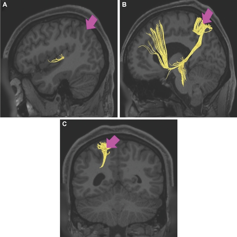
Area PGi is structurally connected to inferior parcellations of the occipitotemporal junction. Some individuals have connections to the arcuate/SLF with anterior projections to the premotor areas but this is inconsistent. Local short association bundles connect to the occipitotemporal junction at parcellations PHT, TE1p, STSvp, STSdp, TPOJ1, and TE1m (Figure 14).
FIGURE 14.
Structural connectivity of PGi in the left hemisphere, shown on T1-weighted MR images. A and B, Sagittal views and C, coronal view showing projections to local parcellations and the arcuate/SLF. Yellow: white matter tracts of PGi demonstrating connections with local parcellations and the arcuate/SLF. Pink arrows indicate the location of the parcellation of interest.
What is known about its function?
Area PGi is involved in many of the same functions as area PGs. This region shows activity when individuals change their visuospatial attention from one area to another,7 and is a major node in the task-negative network, which mostly functions to redirect attention towards relevant stimuli.6 Relative to PGs, PGi is more active when processing faces compared to a body, is more active when listening to a story compared to unrelated words, and is more active when listening to a story vs answering arithmetic problems.6
Area PGp
Where is it?
Area PGp is found on the posterior most portion of the inferior parietal lobule.
What are its borders?
Area PGp borders PGs anterosuperiorly. Its anteroinferior border is made up of TPOJ3 and LO3 (lateral occipital area 3). IP0 is its posterosuperior boundary. Its posterior boundary is completed by V3cd (visual area 3, part cd).
What is its functional connectivity?
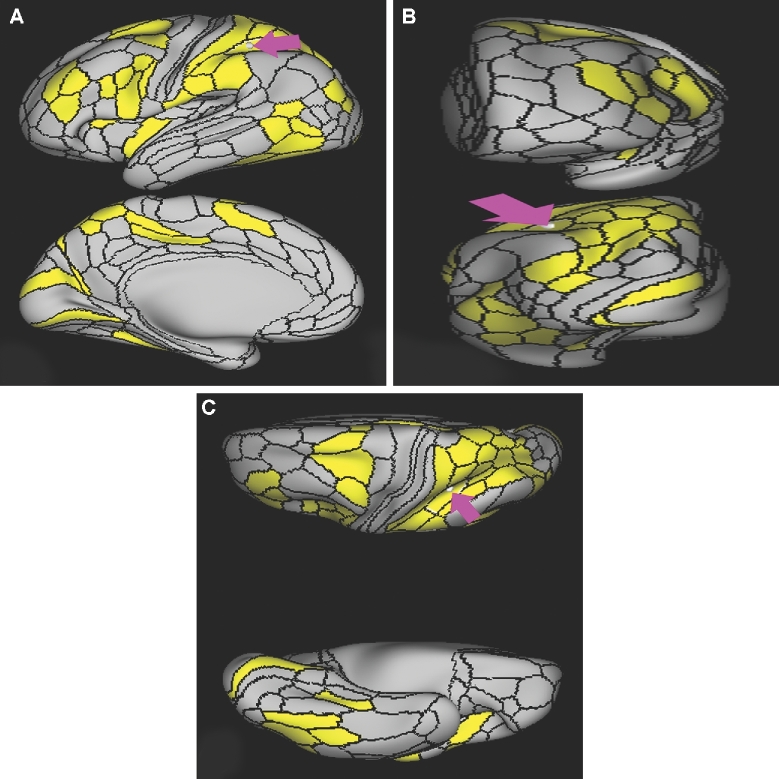
Area PGp demonstrates functional connectivity to areas FEF, PEF, 6a, and 6ma in the premotor regions, areas a24prime, p32prime, 5mv, and 23c in the middle cingulate regions, areas IFSa and 46 in the lateral frontal lobe, areas PFcm and FOP4 in the superior insula opercular regions, areas PoI1 and PoI2 in the lower opercula and Heschl's gyrus regions, areas PHA1, PHA2, PHA3, TE2p, and PHT in the temporal lobe, areas AIP, MIP, VIP, LIPd, LIPv, PFop, PF, PFt, IP2, IP0, IPS1, 7AL,7PL, and 7PC in the lateral parietal lobe, areas 7AM, 7pm, POS1, POS2, DVT, and PCV in the medial parietal lobe, areas ProS, V1, V2, and V3 in the medial occipital lobe, areas V3a, V3b, V6, and V6a of the dorsal visual stream, areas FFC, VVC, and VMV2 of the ventral visual stream, and areas V3cd, LO3, PH, TPOJ2, TPOJ3, and FST of the lateral occipital lobe (Figure 15).
FIGURE 15.
Functional connectivity of PGp demonstrated on an inflated left hemisphere. A, Lateral and medial views. B, Rostral and caudal views. C, Dorsal and ventral views. Parcellations with the strongest functional connectivity are shown in yellow. Pink arrows designate the parcellation of interest.
What are its white matter connections?
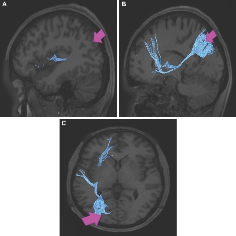
Area PGp is structurally connected with the inferior longitudinal fasciculus (ILF) and the superior parietal lobule. Connections with the ILF project from PGp to the basal surface of the occipitotemporal junction and fusiform gyrus, ending at parcellations PH, FFC, and TF. Superior parietal lobe fibers connect with 7PL and IPS1. Local short association fibers connect with surrounding parcellations IP0, PGs, TPOJ3, MT and visual processing areas LO3, LO1, V3CD, and V4 (Figure 16).
FIGURE 16.
Structural connectivity of PGp in the left hemisphere, shown on T1-weighted MR images. A and B, Sagittal views and C, coronal view showing projections to local parcellations and the inferior longitudinal fasiculus (ILF). Blue: white matter tracts of PGs demonstrating connections with local parcellations and the ILF. Pink arrows indicate the location of the parcellation of interest.
What is known about its function?
Area PGp represents a link between the occipital and parietal cortices given the similarity in their molecular expression pattern to the higher ventral extrastriate area, which has been suggested to have connection and functionality related to visuospatial processing.8 This area is also involved in moral decision-making and egocentric/allocentric perspective function.8 Caudal portions of the inferior parietal lobule determine semantic content and assist in the interpretation of meaning within information.4 This region plays a role in memory retrieval.7
Area TPOJ1
Where is it?
Area TPOJ1 (temporal-parietal-occipital junction 1) is found in the posterior superior temporal sulcus, just as it angles upward to indent the angular gyrus. It makes up both banks and the depth of this portion of the sulcus.
What are its borders?
Area TPOJ1 borders STV superiorly, and PHT inferiorly. Its anterior border is small but borders STSdp, STSvp, A4, and A5. It borders PGi posterosuperiorly and TPOJ2 posteriorly.
What is its functional connectivity?
Area TPOJ1 demonstrates functional connectivity to areas 1,2, 3a, and 3b in the sensory strip, area 4 in the motor strip, areas 55b and FEF in the premotor areas, areas IFJa and IFSp in the lateral frontal lobe, areas OP4, PFcm, RI, STV, PSL, A4, A5, PBelt, and LBelt in the insula opercular region, area STSdp in the temporal lobe, areas V2, V3, and V4 in the medial occipital lobe, area FFC in the ventral visual stream, and areas TPOJ2 and TPOJ3 in the lateral occipital lobe (Figure 17).
FIGURE 17.
Functional connectivity of TPOJ1 demonstrated on an inflated left hemisphere. A, Lateral and medial views. B, Rostral and caudal views. C, Dorsal and ventral views. Parcellations with the strongest functional connectivity are shown in yellow. Pink arrows designate the parcellation of interest.
What are its white matter connections?
Area TPOJ1 is structurally connected to the arcuate/SLF and inferior parietal lobe. Arcuate/SLF connections wrap around the sylvian fissure toward the fronal lobe to end at premotor parcellations IFJa, IFJp, and 6r. From the arcuate/SLF there are also connections to the inferior parietal lobule that end at PFm, PGi, and PGs. Local short association fibers connect with TPOJ2, STSvp, and STSdp (Figure 18).
FIGURE 18.
Structural connectivity of TPOJ1 in the left hemisphere, shown on T1-weighted MR images. A and B, Sagittal views and C, coronal view showing projections to local parcellations and the arcuate/SLF. Yellow: white matter tracts of TPOJ1 demonstrating connections with local parcellations and the arcuate/SLF. Pink arrows indicate the location of the parcellation of interest.
What is known about its function?
The TPOJ participates in several processes including detecting incongruities between expected and presented stimuli, reward processing, and saliency.12 This region also shows prominence in theory of mind, and the processing and detection of incongruous concepts and their subsequent resolution.12 The TPOJ region has specifically been shown to be activated during self-related processing, and receives input from different sensory afferent neurons.12 The TPOJ is not finely parcellated in the current literature, thus task-based functional magnetic resonance imaging differences are the only way to delineate their individual functions.6
Area TPOJ2
Where is it?
Area TPOJ2 (temporal-parietal-occipital junction 2) is found on the anterior, superior part of the lateral occipital cortex. It straddles the sulcus and is on the posteroinferior bank of the angular gyrus.
What are its borders?
Area TPOJ2 borders PGi superiorly, TPOJ1 anteriorly, PHT, FST, and MST inferiorly, and TPOJ3 posteriorly.
What is its functional connectivity?
Area TPOJ2 demonstrates functional connectivity to area 2 in the sensory strip, areas 6a, 6v, SCEF, and FEF in the premotor areas, areas 5mv, 23c, and p24prime in the cingulate regions, areas OP4, 43, PFcm, 52, RI, STV, A4, PoI1, PoI2, and PBelt in the insula opercular region, areas PHT and TE2p in the temporal lobe, areas PFop, PGp, IPS1, IP0, AIP, LIPv, 7AL, 7PL, and 7PC in the lateral parietal lobe, areas 7AM, PCV, and DVT in the medial parietal lobe, areas V2 and V3 in the medial occipital lobe, areas V3a, V6, and V6a in the dorsal visual stream, area FFC in the ventral visual stream, and areas FST, PH, LO3, MST, TPOJ1, and TPOJ3 in the lateral occipital lobe (Figure 19).
FIGURE 19.
Functional connectivity of TPOJ2 demonstrated on an inflated left hemisphere. A, Lateral and medial views. B, Rostral and caudal views. C, Dorsal and ventral views. Parcellations with the strongest functional connectivity are shown in yellow. Pink arrows designate the parcellation of interest.
What are its white matter connections?
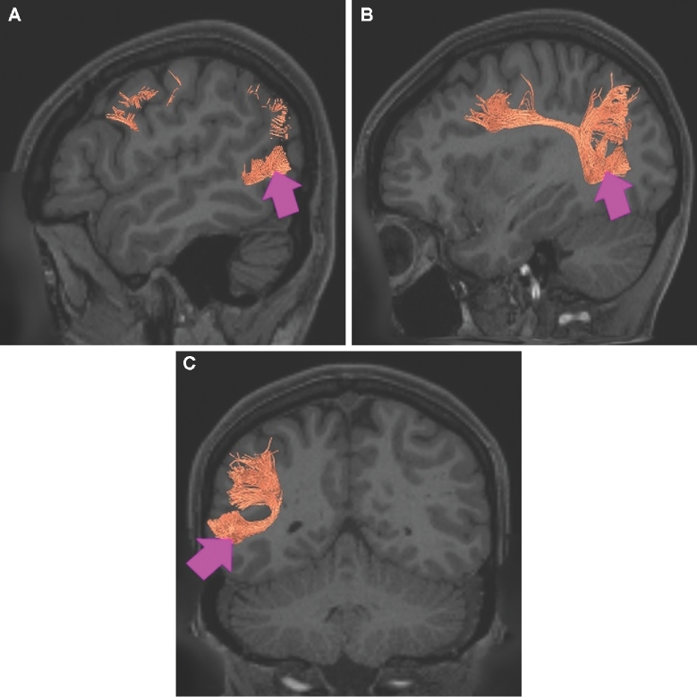
Area TPOJ2 is structurally connected to the arcuate/SLF and inferior parietal lobule. Arcuate/SLF connections wrap around the sylvian fissure toward the fronal lobe to end at premotor parcellations IFJa, IFJp, 6v, and 6r. From the arcuate/SLF there are also connections to the inferior parietal lobule that end at PFm, PGi, and PGs. Local short association fibers connect with TPOJ1 (Figure 20).
FIGURE 20.
Structural connectivity of TPOJ2 in the left hemisphere, shown on T1-weighted MR images. A and B, Sagittal views and C, coronal view showing projections to local parcellations and the arcuate/SLF. Orange: white matter tracts of TPOJ2 demonstrating connections with local parcellations and the SLF/arcuate. Pink arrows indicate the location of the parcellation of interest.
What is known about its function?
Relative to its anterior neighbor TPOJ1, area TPOJ2 shows greater activity when individuals are viewing an image of a body compared to an average image of places, tools, and faces.6 TPOJ2 is deactivated when viewing place images and when listening to a story vs unrelated words.6
Area TPOJ3
Where is it?
Area TPOJ3 (temporal-parietal-occipital junction 3) is found on the posterior inferior portion of the inferior parietal lobule.
What are its borders?
Area TPOJ3 borders PGI superiorly, TPOJ2 anteriorly, MST, MT, and LO3 inferiorly, and PGp posteriorly.
What is its functional connectivity?
Area TPOJ3 demonstrates functional connectivity to area 2 in the sensory strip, areas 6a and FEF in the premotor areas, areas 5mv and 23c in the cingulate regions, areas OP4, PFcm, and STV in the insula opercular region, areas PHA2, PHA3, and TE2p in the temporal lobe, areas PGp, IP0, and 7PC in the lateral parietal lobe, areas 7AM, PCV, and DVT in the medial parietal lobe, area V2 in the medial occipital lobe, area V6 in the dorsal visual stream, and areas FST, PH, LO3, MST, TPOJ1, and TPOJ2 in the lateral occipital lobe (Figure 21).
FIGURE 21.
Functional connectivity of TPOJ3 demonstrated on an inflated left hemisphere. A, Lateral and medial views. B, Rostral and caudal views. C, Dorsal and ventral views. Parcellations with the strongest functional connectivity are shown in yellow. Pink arrows designate the parcellation of interest.
What are its white matter connections?
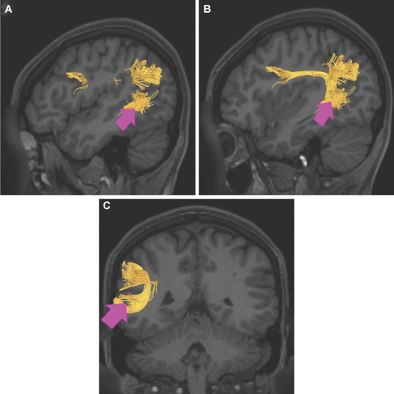
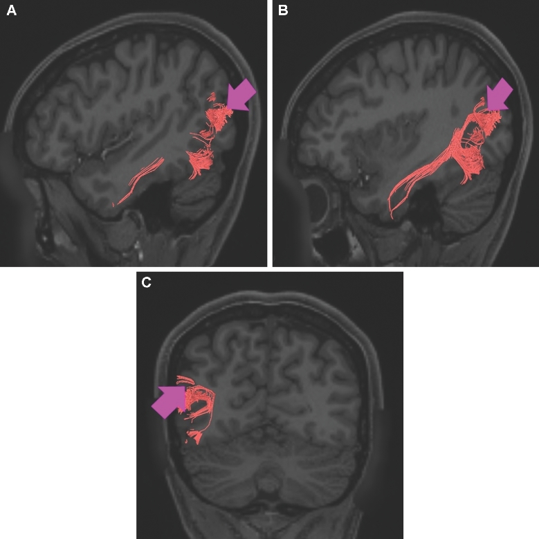
Area TPOJ3 is structurally connected with local parcellations and the ILF. Connections with the ILF project through the inferior temporal lobe to end at TF, TGv, and PeEc areas. There are also projections to the premotor cortex but this is inconsistent across individuals. Local short association bundles connect with MT, MST, LO3, FST, TPOJ3, TE2p, PH, and PGi (Figure 22).
FIGURE 22.
Structural connectivity of TPOJ3 in the left hemisphere, shown on T1-weighted MR images. A and B, Sagittal views and C, coronal view showing projections to local parcellations and the ILF. Red: white matter tracts of TPOJ2 demonstrating connections with local parcellations and the ILF. Pink arrows indicate the location of the parcellation of interest.
What is known about its function?
Area TPOJ3 is strongly activated when viewing faces compared to when viewing shapes.6 Relative to its anterior neighbor TPOJ2, area TPOJ3 is thinner, more active during working memory tasks, and less active during motor cue and shape recognition tasks.6
Intraparietal Sulcus Areas
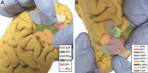
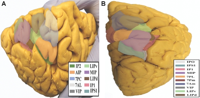
The parcellations comprised of the intraparietal sulcus areas include IP2, IP1, IP0, IPS1, AIP, LIPd, LIPv, and MIP. The anatomic location of these parcellations is shown on a cadaver brain in Figure 23. The combined tractography of these parcellations is shown in Figure 24.
FIGURE 23.
Anatomic view of the intraparietal sulcus parcellations shown on the right hemisphere of a cadaver brain. A, Superior caudal view of the intraparietal sulcus. B, Lateral view of the intraparietal sulcus with widening of the sulcus to show extent the parcellations.
FIGURE 24.
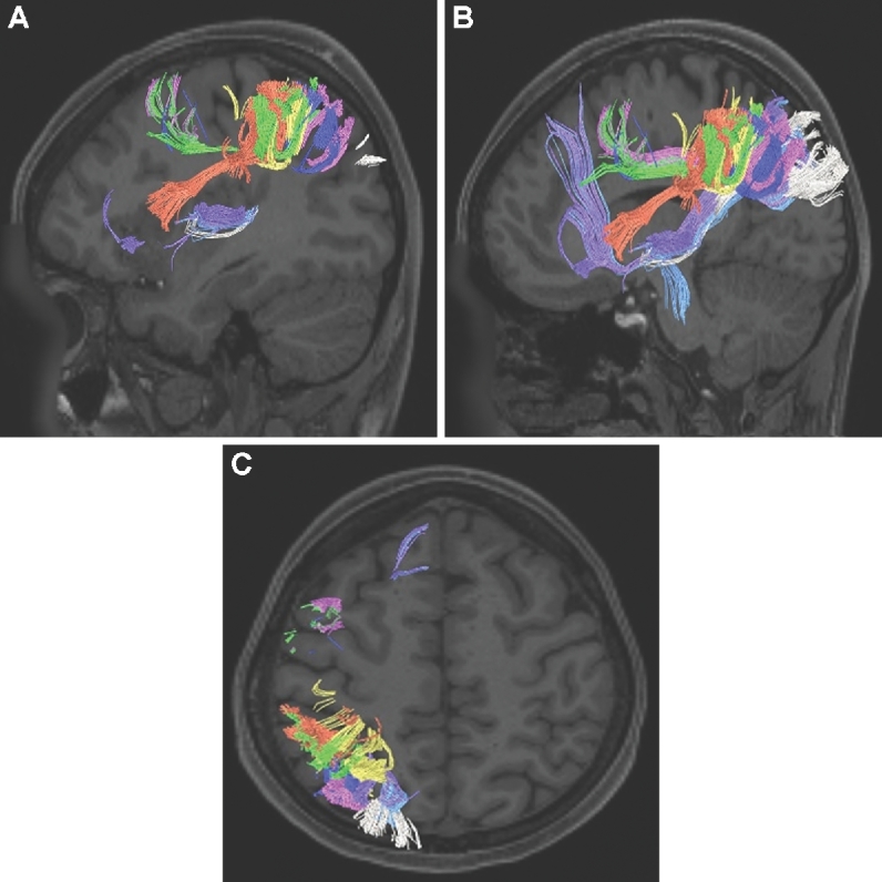
Combined structural connectivity of the parcellations comprising the intraparietal sulcus shown on T1-weighted MR images. Sagittal views, A, lateral and B, medial. C, Axial view.
Area IP2
Where is it?
Area IP2 is found on the anterior most portion of the inferior bank of the intraparietal sulcus.
What are its borders?
IP2 borders PF anteriorly, PFm inferiorly, IP1 posteriorly, and areas AIP and LIPv superiorly.
What is its functional connectivity?
Area IP2 demonstrates functional connectivity to areas 6ma, 6a, and 6r in the premotor regions, areas 33prime and 8BM in the middle cingulate regions, areas IFSa, IFJp, a9-46v, p9-46v, 46, 11L, 8C, i6-8, a47r, and p47r in the lateral frontal lobe, areas AVI in the insula regions, area PHT in the temporal lobe, areas TE1p and PHT in the temporal lobe, areas PFt, PF, PFm, PGp, IP0, IP1, AIP, MIP, LIPd, and 7PL in the lateral parietal lobe, areas 7AM, 7pm, 31a, POS2, and RSC in the medial parietal lobe, and area PH in the occipital lobe (Figure 25).
FIGURE 25.
Functional connectivity of IP2 demonstrated on an inflated left hemisphere. A, Lateral and medial views. B, Rostral and caudal views. C, Dorsal and ventral views. Parcellations with the strongest functional connectivity are shown in yellow. Pink arrows designate the parcellation of interest.
What are its white matter connections?
Area IP2 is structurally connected with the SLF. Connections with the SLF project anteriorly to the premotor cortex to end at 55b and PEF. Local short association fibers connect with PFm, LIPd, AIP, and IP1. White matter tracts in the right hemisphere have more inferior connections with the inferior frontal gyrus (Figure 26).
FIGURE 26.
Structural connectivity of IP2 in the left hemisphere, shown on T1-weighted MR images. A and B, Sagittal views and C, axial view showing projections to local parcellations and the arcuate/SLF. Green: white matter tracts of IP2 demonstrating connections with local parcellations and the arcuate/SLF. Pink arrows indicate the location of the parcellation of interest.
What is known about its function?
Area IP2 shows significant activation during mental arithmetic activities.13 Anterior IPS areas appear to support more complex parts of numeric and mathematical information processing.14 The anterolateral bank of the IPS is involved in the modulatory sensorimotor integration processes related to fine finger movements.15
Area IP1
Where is it?
Area IP1 is found on the middle portion of the inferior bank of the intraparietal sulcus.
What are its borders?
Area IP1 borders IP2 anteriorly and IP0 posteriorly. Its inferior border is formed by PFm and PGs, and its superior border is MIP and LIPv.
What is its functional connectivity?
Area IP1 demonstrates functional connectivity to area 6a in the premotor regions, areas 33prime and 8BM in the middle cingulate regions, areas IFSa, IFSp, IFJp, a9-46v, p9-46v, 8AV, 8C, i6-8, a47r, and p47r in the lateral frontal lobe, areas TE1m, TE1p, PHT, and PreS in the temporal lobe, areas TE1p and PHT in the temporal lobe, areas PFm, PGs, IP0, IP2, AIP, MIP, and LIPd in the lateral parietal lobe, areas 7pm, 31a, d23ab, POS2, and RSC in the medial parietal lobe, and area V1 in the occipital lobe (Figure 27).
FIGURE 27.
Functional connectivity of IP1 demonstrated on an inflated left hemisphere. A, Lateral and medial views. B, Rostral and caudal views. C, Dorsal and ventral views. Parcellations with the strongest functional connectivity are shown in yellow. Pink arrows designate the parcellation of interest.
What are its white matter connections?
Area IP1 is structurally connected with the SLF. Connections with the SLF project anteriorly to the premotor cortex to end at 55b, FEF, and PEF. Local short association fibers connect with PFm, LIPd, IP0, IPS1, and PGs. White matter tracts in the right hemisphere have more inferior connections with the inferior frontal gyrus (Figure 28).
FIGURE 28.
Structural connectivity of IP1 in the left hemisphere, shown on T1-weighted MR images. A and B, Sagittal views and C, axial view showing projections to local parcellations and the arcuate/SLF. Pink: white matter tracts of IP1 demonstrating connections with local parcellations and the arcuate/SLF. Pink arrows indicate the location of the parcellation of interest.
What is known about its function?
Area IP1 shows significant activation during mental arithmetic activities, and, as part of the anterior IPS, supports more complex parts of numeric and mathematical information processing.13,14 Area IP1 shows greater activation in primary contrasts, when interpreting motor cues and when hearing a story compared to when hearing arithmetic problems.6 Relative to area IP2, area IP1 is more active when individuals are processing faces than when processing shapes, and is less deactivated when hearing a story vs unrelated words.6
Area IP0
Where is it?
Area IP0 (Intraparietal 0) is found on the posterior most portion of the inferior bank of the intraparietal sulcus.
What are its borders?
Area IP0 borders IP1 anteriorly. Its posterior border is made of V3b and V3cd. Its anteroinferior edge is largely made up of PGp and a small portion of PGs. IPS1 is its superior neighbor.
What is its functional connectivity?
Area IP0 demonstrates functional connectivity to areas SCEF, FEF, PEF, 6a, 6r, and 6ma in the premotor regions, areas 5mv and 23c in the middle cingulate regions, areas IFSa, IFSp, IFJa, IFJp, i6-8, p9-46v, and 46 in the lateral frontal lobe, areas PeEc, PHA2, PHA3, TE1p, TE2p, and PHT in the temporal lobe, areas AIP, MIP, VIP, LIPd, LIPv, PFop, PF, PFt, PGp, IP2, IP1, IPS1, 7PL, and 7PC, in the lateral parietal lobe, areas 7AM, 7pm, DVT, and PCV in the medial parietal lobe, areas ProS, V1, V2, V3, and V4 in the medial occipital lobe, areas V3a, V3b, V7, V6, and V6a of the dorsal visual stream, areas FFC, VVC, V8, VMV2, and VMV3 of the ventral visual stream, and areas V3cd, LO1, LO3, PH, TPOJ2, TPOJ3, and FST of the lateral occipital lobe (Figure 29).
FIGURE 29.
Functional connectivity of IP0 demonstrated on an inflated left hemisphere. A, Lateral and medial views. B, Rostral and caudal views. C, Dorsal and ventral views. Parcellations with the strongest functional connectivity are shown in yellow. Pink arrows designate the parcellation of interest.
What are its white matter connections?
Area IP0 is structurally connected with local parcellations and to the middle longitudinal fasciculus (MdLF). Connections with the MdLF travel deep to the parietal lobe to the superior temporal gyrus to end at A4. Local short association bundles connect with IP1, IPS1, V3B, V6A, DVT, and V6 (Figure 30).
FIGURE 30.
Structural connectivity of IP0 in the left hemisphere, shown on T1-weighted MR images. A and B, Sagittal views and C, coronal view showing projections to local parcellations and the MdLF. White: white matter tracts of IP0 demonstrating connections with local parcellations and the MdLF. Pink arrows indicate the location of the parcellation of interest.
What is known about its function?
IP0 shows significant activation during mental arithmetic activities.13 Posterior IPS regions may play a role in the transformation of symbolic/nonsymbolic numeric information into spatial and semantic representation.14 Relative to IP1, IP0 is deactivated when viewing faces than an average compilation of body, tool, and place images, and is activated vs deactivated when viewing places compared to a compilation of face, body, and tool images.6 It is less deactivated when hearing a language story vs an unrelated grouping of words.6
Area IPS1
Where is it?
Area IPS1 is found on the posterior, superior bank of the intraparietal sulcus. It should not be confused with IP1, which is on the inferior bank of the sulcus. IPS1 is the posterior most region on the superior bank of the intraparietal sulcus.
What are its borders?
Area IPS1 borders IP0 inferiorly, MIP anteriorly, DVT medially, and area 7PL anteromedially. Its posterior border is made up of the visual areas V6a, V7, and V3b, which are part of the dorsal visual stream.
What is its functional connectivity?
Area IPS1 demonstrates functional connectivity to areas 1 and 2 in the sensory strip, area 4 in the motor strip, areas SCEF and FEF in the premotor regions, areas TE2p and PHT in the temporal lobe, areas AIP, MIP, VIP, LIPd, LIPv, PGp, PFt, IP0, 7PL, and 7PC in the lateral parietal lobe, areas DVT in the medial parietal lobe, areas V1, V2, V3, and V4 in the medial occipital lobe, areas V3a, V3b, V7, V6, and V6a of the dorsal visual stream, areas V8, PIT, FFC, VVC, VMV1, VMV2, and VMV3 of the ventral visual stream, and areas V3cd, V4t, LO1, LO2, LO3, PH, TPOJ2, MT, MST, and FST of the lateral occipital lobe (Figure 31).
FIGURE 31.
Functional connectivity of IPS1 demonstrated on an inflated left hemisphere. A, Lateral and medial views. B, Rostral and caudal views. C, Dorsal and ventral views. Parcellations with the strongest functional connectivity are shown in yellow. Pink arrows designate the parcellation of interest.
What are its white matter connections?
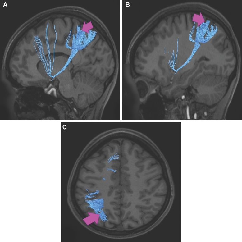
Area IPS1 is structurally connected to the inferior fronto-occipital fasciculus (IFOF), MdLF, and local parcellations. The IFOF courses from IPS1 through the posterior temporal lobe and extreme/external capsule to frontal lobe parcellations. IFOF terminations are inconsistent across individuals but the majority end at the superior temporal gyrus involving parcellations 8BL, 9a, and 9p. Connections with the MdLF travel deep to the parietal lobe to the superior temporal gyrus and planum temporale to end at A4, PBelt, and MBelt. Local short association fibers connect with MIP, V7, V6A, and V3B (Figure 32).
FIGURE 32.
Structural connectivity of IPS1 in the left hemisphere, shown on T1-weighted MR images. A and B, Sagittal views and C, axial view showing projections to local parcellations, the IFOF, and the MdLF. Light blue: white matter tracts of IPS1 demonstrating connections with local parcellations, IFOF, and MdLF. Pink arrows indicate the location of the parcellation of interest.
What is known about its function?
Area IPS1 is involved in processing object-based information.16 This suggests that this region is important in object constancy, which describes the ability of an individual to interpret object permanence.16 This region is important in mentally visualizing the process of grasping objects as well as action associations and object function.16 Area IPS1 also plays a role in the transmission of top-down spatial attention information to the visual cortex during sustained attention tasks.17
Area AIP
Where is it?
Area AIP is found on the superior bank of the intraparietal sulcus at its most anterior aspect. It extends onto the superior surface of the adjacent superior parietal lobule, and its anterior tip lies in the bank of the postcentral sulcus.
What are its borders?
Area AIP borders area 2 anteriorly and PFt anteroinferiorly. Its anterosuperior border is area 7PC, and its inferior border is with IP2 across the intraparietal sulcus. LIPv and LIPd make up its posterior boundaries.
What is its functional connectivity?
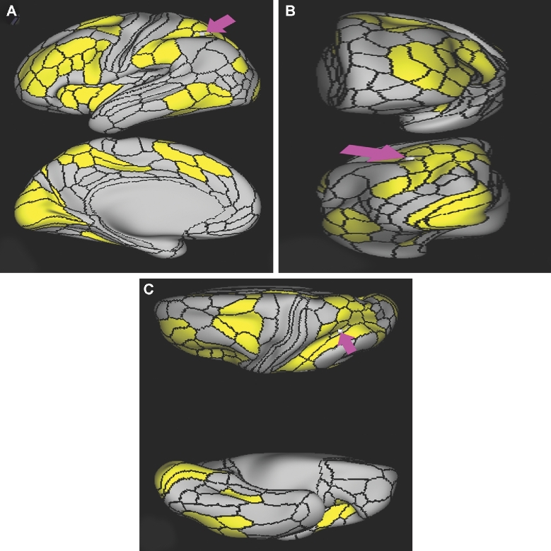
Area AIP demonstrates functional connectivity to area 2 in the sensory strip, areas SCEF, FEF, PEF 6a, 6r, and 6ma in the premotor regions, areas IFSa, IFJp, 46, and p9-46v in the lateral frontal lobe, areas 5mv, and 23c in the medial frontal lobe, areas PoI2, FOP2, FOP4, and OP4 in the insula opercular regions, areas PHA3, PHT, and TE2p, in the temporal lobe, areas 7PC, 7PL, 7AL, VIP, MIP, LIPv, LIPd, PFop, PFt, PGp, IP2, IP1, IP0, and IPS1 in the lateral parietal lobe, areas DVT, 7AM, and 7pm in the medial parietal lobe, area V2 in the medial occipital lobe, area FFC in the ventral visual stream areas, and areas V3CD, PH, TPOJ2, and FST in the lateral occipital lobe (Figure 33).
FIGURE 33.
Functional connectivity of AIP demonstrated on an inflated left hemisphere. A, Lateral and medial views. B, Rostral and caudal views. C, Dorsal and ventral views. Parcellations with the strongest functional connectivity are shown in yellow. Pink arrows designate the parcellation of interest.
What are its white matter connections?
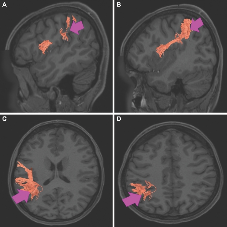
Area AIP is structurally connected to the pars opercularis and local parcellations. Connections from AIP to the pars opercularis travel anteroinferiorly to end at 43 and 6r. Local short association bundles connect with 2, PFt, PFcm, PF, IP2, and 7PC (Figure 34).
FIGURE 34.
Structural connectivity of AIP in the left hemisphere, shown on T1-weighted MR images. A and B, Sagittal views and C and D, axial views showing projections to local parcellations and the arcuate/SLF. Orange: white matter tracts of AIP demonstrating connections with local parcellations and arcuate/SLF. Pink arrows indicate the location of the parcellation of interest.
What is known about its function?
Area AIP is involved in grasping activity as well as object recognition. Neurons in this part of the cortex are oriented for grip and hand shape, and the inferior temporal cortex provides input related to object information.18 AIP also receives input from the ventral and dorsolateral visual streams.18 This region also plays a role in shaping the hand for grasping activity.19 Beyond grasping action, AIP is involved in tactile shape-processing and understanding orientation in space.20
Area LIPd
Where is it?
Area LIPd is located centrally on the superior bank of the intraparietal sulcus. Note that the nomenclature here can be confusing. LIPd is actually ventral to LIPv.
What are its borders?
Area LIPd borders AIP anteriorly, and MIP posteriorly. Its inferior border (across the intraparietal sulcus) is made up of IP2 and IP1. Its superior border is made up of LIPv and VIP.
What is its functional connectivity?
Area LIPd demonstrates functional connectivity to areas SCEF, FEF, PEF 6a, and 6r in the premotor regions, areas IFSa, IFSp, IFJa, p47r, i6-8, 8C, 9-46d, 46, a9-46v, and p9-46v in the lateral frontal lobe, areas 8BM, p32prime, 5mv, and 23c in the medial frontal lobe, areas PoI2, FOP4, FOP5, MI, AVI, and PoI2 in the insula opercular regions, areas PHA3, PHT, and TE1p in the temporal lobe, areas 7PC, 7PL, AIP, VIP, MIP, LIPv, PF, PGp, IP2, IP1, IP0, and IPS1 in the lateral parietal lobe, areas PCV, DVT, 7AM, and 7pm in the medial parietal lobe, areas V1, V2, and V3 in the medial occipital lobe, and areas PH and FST in the lateral occipital lobe (Figure 35).
FIGURE 35.
Functional connectivity of LIPd demonstrated on an inflated left hemisphere. A, Lateral and medial views. B, Rostral and caudal views. C, Dorsal and ventral views. Parcellations with the strongest functional connectivity are shown in yellow. Pink arrows designate the parcellation of interest.
What are its white matter connections?
Area LIPd is structurally connected to the premotor region and local parcellations. In some individuals the anterior projections from LIPd end at the motor cortex and do not extend to premotor areas. Premotor connections end at 55b and PEF. Local short association bundles connect with PGs, AIP, IP1, IP2, and PGs (Figure 36).
FIGURE 36.
Structural connectivity of LIPd in the left hemisphere, shown on T1-weighted MR images. A and B, Sagittal views and C, axial view showing projections to local parcellations and the arcuate/SLF. Red: white matter tracts of LIPd demonstrating connections with local parcellations and arcuate/SLF. Pink arrows indicate the location of the parcellation of interest.
What is known about its function?
LIPd is implicated in control of attention and eye movements.20 This region has specifically been implicated in saccade coordination and mapping of contralateral spaces.20
Area LIPv
Where is it?
Area LIPv is found on the inferior edge of the superior parietal sulcus. Note that it does not enter the banks of the intraparietal sulcus, but instead is located on the upper surface of the superior parietal lobule.
What are its borders?
Area LIPv borders AIP and area 7PC anteriorly, VIP superiorly, LIPv laterally, and MIP posteriorly.
What is its functional connectivity?
Area LIPv demonstrates functional connectivity to areas 1 and 2 in the sensory strip, area 4 in the motor strip, areas SCEF, FEF, PEF 6a, 6r, and 6v in the premotor regions, areas p32prime, 5mv, and 23c in the medial frontal lobe, area FOP4 in the insula opercular regions, areas PHT and TE2p in the temporal lobe, areas 7PC, 7PL, AIP, VIP, MIP, LIPv, PFop, PFt, PGp, IP0, and IPS1 in the lateral parietal lobe, area DVT in the medial parietal lobe, areas V1, V2, V3, and V4 in the medial occipital lobe, areas V3a, V3b, V7, V6a, and V6 in the dorsal visual stream areas, area V8, PIT, FFC, VVC, VMV1, VMV2, and VMV3 in the ventral visual stream areas, and areas V3CD, V4t, PH, LO1, LO2, LO3, TPOJ2, MT, MST, and FST in the lateral occipital lobe (Figure 37).
FIGURE 37.
Functional connectivity of LIPv demonstrated on an inflated left hemisphere. A, Lateral and medial views. B, Rostral and caudal views. C, Dorsal and ventral views. Parcellations with the strongest functional connectivity are shown in yellow. Pink arrows designate the parcellation of interest.
What are its white matter connections?
Area LIPv is structurally connected to local parcellations. Local short association bundles connect with 2, AIP, 7PC, IP2, LIPd, LIPv, and MIP (Figure 38). White matter tracts from LIPv in the right hemisphere have more consistent connections with the IFOF. However, this tract is not present in all individuals.
FIGURE 38.
Structural connectivity of LIPv in the left hemisphere, shown on T1-weighted MR images. A, Sagittal, B, axial, and C, coronal views showing projections to local parcellations. Light green: white matter tracts of LIPv demonstrating connections with local parcellations. Pink arrows indicate the location of the parcellation of interest.
What is known about its function?
LIPv is implicated in control of attention and eye movements,20 and is important during visually guided reaching and pointing, hand movements, and change in visuomotor contingencies.7
Area MIP
Where is it?
Area MIP is found in the posterior portion of the superior bank of the intraparietal sulcus. It extends onto the superior surface of the adjacent portion of the superior parietal lobule.
What are its borders?
Area MIP borders IPS1 posteriorly, area 7PL and VIP medially, IP1 and IP0 inferiorly, and LIPv and LIPd anteriorly.
What is its functional connectivity?
Area MIP demonstrates functional connectivity to areas SCEF, FEF, PEF 6a, 6v, 6r, and 6ma in the premotor regions, areas IFSa, IFJa, IFJp, 46, and p9-46v in the lateral frontal lobe, areas 5mv and 23c in the medial frontal lobe, areas PHA3, PHT, and TE2p in the temporal lobe, areas 7PC, 7PL, 7AL, AIP, VIP, LIPv, LIPd, PF, PFt, PGp, IP2, IP1, IP0, and IPS1 in the lateral parietal lobe, areas DVT, 7AM, and 7pm in the medial parietal lobe, areas V1 and V2 in the medial occipital lobe, area FFC in the ventral visual stream areas, and areas PH and FST in the lateral occipital lobe (Figure 39).
FIGURE 39.
Functional connectivity of MIP demonstrated on an inflated left hemisphere. A, Lateral and medial views. B, Rostral and caudal views. C, Dorsal and ventral views. Parcellations with the strongest functional connectivity are shown in yellow. Pink arrows designate the parcellation of interest.
What are its white matter connections?
Area MIP is structurally connected to the IFOF, MdLF, and local parcellations. IFOF connections travel from MIP through the posterior temporal lobe and extreme/external capsule to the frontal lobe to end at parcellations 8BL, 9p, and 45. Connections with MdLF travel deep to the parietal lobe to the planum temporale to end at PBelt and MBelt. The majority of local short association bundles project inferior to 7PL, IP0, IP1, and IPS1 (Figure 40).
FIGURE 40.
Structural connectivity of MIP in the left hemisphere, shown on T1-weighted MR images. A and B, Sagittal views, C, coronal view, and D, axial view showing projections to local parcellations, the IFOF, and the MdLF. Purple: white matter tracts of MIP demonstrating connections with local parcellations, IFOF, and MdLF. Pink arrows indicate the location of the parcellation of interest.
What is known about its function?
This visuomotor area is presumed to be involved in the control of arm-reaching movements as well as prehension.21 This region is important for reception of proprioceptive signals.22 Area MIP neurons are modulated by reaching activity as well as visual and somatosensory stimulation.20,23 Furthermore, this region is important for the adjustment of reaching movements and correction of movement error.20 This shows this region's importance in transformation of visual information into motor action for precise movement.20
Superior Parietal Lobule Areas
The parcellations comprised by the superior parietal lobule include 7PC, 7AL, 7AM, 7PL, 7PM, and VIP. The anatomic location of these parcellations is shown on a cadaver brain in Figure 41. The combined tractography of these parcellations is shown in Figure 42.
FIGURE 41.
Anatomic view of the superior parietal lobule parcellations shown on the right hemisphere of a cadaver brain. A, Dorsolateral view of the superior parietal lobule. B, Caudal view of the superior parietal lobule.
FIGURE 42.

Combined structural connectivity of the parcellations comprising the superior parietal lobule shown on T1-weighted MR images. A-C, Sagittal views, from A, most medial to B and C, most lateral. D, Coronal view.
Area 7PC
Where is it?
Area 7PC (7 postcentral) is found in the anterior, inferior superior parietal lobule. It extends into the adjacent posterior bank of the postcentral sulcus.
What are its borders?
Area 7PC borders Area 2 anteriorly and AIP inferiorly. Its posterior border is made up of LIPv and VIP. Area 7AL is its medial (superior) neighbor.
What is its functional connectivity?
Area 7PC demonstrates functional connectivity to areas 1, 2, 3a, and 3b in the sensory strip, area 4 in the motor strip, areas SCEF, FEF, PEF, 6ma, 6mp, 6a, 6d, 6r, and 6v in the premotor regions, areas 24dd, 24dv, p32prime, 5L, 5mv, and 23c in the medial frontal lobe, areas A4, PBelt, PFcm, FOP2, OP4, and OP1 in the insula opercular regions, areas PHT and TE2p in the temporal lobe, areas 7PL, AIP, VIP, MIP, LIPv, LIPd, PFop, PFt, PGp, IP0, and IPS1 in the lateral parietal lobe, areas 7AM and DVT in the medial parietal lobe, area V2 in the medial occipital lobe, areas V3b, V6a, and V6 in the dorsal visual stream areas, area FFC in the ventral visual stream areas, and areas V3CD, V4t, PH, LO3, TPOJ2, TPOJ3, MST, and FST in the lateral occipital lobe (Figure 43).
FIGURE 43.
Functional connectivity of 7PC demonstrated on an inflated left hemisphere. A, Lateral and medial views. B, Rostral and caudal views. C, Dorsal and ventral views. Parcellations with the strongest functional connectivity are shown in yellow. Pink arrows designate the parcellation of interest.
What are its white matter connections?
Area 7PC is structurally connected to local parcellations and the IFOF. Connections from the IFOF course through the posterior temporal gyrus and extreme/external capsule to the frontal lobe to 8BL, 6a, 6ma, and SFL. The majority of individuals have IFOF projections but this tract is not present in everyone. Local short association bundles are abundant and connect with LIPd, LIPv, MIP, 1, and 2 (Figure 44). White matter connections from 7PC in the right hemisphere have more consistent connections with the motor and somatosensory cortex.
FIGURE 44.
Structural connectivity of 7PC in the left hemisphere, shown on T1-weighted MR images. A and B, Sagittal views, C, axial view showing projections to local parcellations and the IFOF. Light blue: white matter tracts of 7PC demonstrating connections with local parcellations and IFOF. Pink arrows indicate the location of the parcellation of interest.
What is known about its function?
Area 7PC is involved in vision motion, observation, space, and execution.24 The left hemispheric portion of region 7PC is associated with imagination, and the right region is associated with vision shape, language comprehension, sexuality, and working memory.7 This region is involved in visual and somatosensory stimulation,7 and shows strong connection to somatosensory areas.6
Area 7AL
Where is it?
Area 7AL (7 anterior-lateral) is found on the anterior superior portion of the superior parietal lobule. It extends to the interhemispheric midline medially and the postcentral sulcus anteriorly.
What are its borders?
Area 7AL borders area 2 anteriorly area 7PC inferiorly, and VIP posteriorly. Areas 5L and 7AM form its medial boundary as these areas extend onto the interhemispheric medial surface.
What is its functional connectivity?
Area 7AL demonstrates functional connectivity to areas 1, 2, 3a, and 3b in the sensory strip, areas SCEF, FEF, 6ma, 6a, 6d, and 6r in the premotor regions, areas 46 and 9-46d in the lateral frontal lobe, areas 24dv, a24prime, p24prime, p32prime, 5L, 5mv, and 23c in the medial frontal lobe, areas 43, PFcm, FOP1, FOP2, FOP3, FOP4, FOP5, 52, MI, PoI1, PoI2, OP4, and OP1 in the insula opercular regions, areas PHT and TE2p in the temporal lobe, areas 7PC, 7PL, AIP, VIP, MIP, PFop, PF, PFt, and PGp in the lateral parietal lobe, areas 7AM, PCV, and DVT in the medial parietal lobe, area V6 visual stream areas, and areas TPOJ2 and FST in the lateral occipital lobe (Figure 45).
FIGURE 45.
Functional connectivity of 7AL demonstrated on an inflated left hemisphere. A, Lateral and medial views. B, Rostral and caudal views. C, Dorsal and ventral views. Parcellations with the strongest functional connectivity are shown in yellow. Pink arrows designate the parcellation of interest.
What are its white matter connections?
Area 7AL is structurally connected to the contralateral hemisphere, IFOF, thalamus and local parcellations. Connections with the IFOF course through the posterior temporal lobe and extreme/external capsule to the frontal lobe to parcellations 9a, 9p, 6a, and 6ma. Thalamic connections project inferior through the posterior thalamus and to the brainstem and superior colliculus. Contralateral connections end at 7AL and 7AM after coursing through the corpus callosum. The majority of local association bundles project posterior to 7PL, IP0, IP1, IPS1, LIPd, MIP; bundles are also connected with 2 and 5L (Figure 46). White matter connections from 7AL in the right hemisphere do not have as consistent connections with the superior frontal gyrus via the IFOF, when IFOF connections are present the tract terminates at the lateral frontal lobe.
FIGURE 46.
Structural connectivity of 7AL in the left hemisphere, shown on T1-weighted MR images. A and B, Sagittal views, C, coronal view, and D, axial view showing projections to local parcellations, the IFOF, and the ipsilateral thalamus and contralateral hemishere. White: white matter tracts of 7PC demonstrating connections with local parcellations, IFOF, thalamus, and contralateral hemisphere. Pink arrows indicate the location of the parcellation of interest.
What is known about its function?
Area 7AL is involved in several types of information processing including space, vision shape and motion, working memory, and execution.24 The anterior portion of 7AL is involved in self-centered mental imagery and attentional processes.25 Area 7AL also demonstrates functional connectivity to the premotor cortex.6 Relative to its medial neighbor 7AM, area 7AL shows greater activity when processing an average compilation of motor functions and is more deactivated when viewing socially interacting geometric objects.6
Area 7AM
Where is it?
Area 7AM (7 anterior-medial) is found on the anterosuperior surface of the medial face of the superior parietal lobule.
What are its borders?
Area 7AM borders the area 7AL superior-laterally, VIP posterolaterally, and areas 7PL and 7PM posteriorly. On its interhemispheric face, its anterior border is made up of areas 5L and 5MV. Its inferior border is made up of PCV (precuneus visual area), and its posterior border is made up of area 7PM.
What is its functional connectivity?
Area 7AM demonstrates functional connectivity to areas SCEF, FEF, PEF, 6ma, 6a, and 6r in the premotor regions, areas IFSa, 46, and 9-46d areas in the lateral frontal lobe, areas a24prime, a32prime, p32prime, 5mv, and 23c in the medial frontal lobe, areas MI, PoI1, PoI2, PFcm, and FOP4 in the insula opercular regions, areas PHA3, PHT, and TE2p in the temporal lobe, areas 7PC, 7AL, 7PL, AIP, VIP, MIP, LIPd, PFop, PFt, PF, PGp, IP2, and IP0 in the lateral parietal lobe, areas 7pm, PCV, POS2, and DVT in the medial parietal lobe, area V1 in the medial occipital lobe, areas V6 in the dorsal visual stream areas, area FFC in the ventral visual stream areas, and areas PH, TPOJ2, TPOJ3, and FST in the lateral occipital lobe (Figure 47).
FIGURE 47.
Functional connectivity of 7AM demonstrated on an inflated left hemisphere. A, Lateral and medial views. B, Rostral and caudal views. C, Dorsal and ventral views. Parcellations with the strongest functional connectivity are shown in yellow. Pink arrows designate the parcellation of interest.
What are its white matter connections?
Area 7AM is structurally connected to the contralateral hemisphere and thalamus. Some individuals have IFOF connections but these tracts are inconsistent. Contralateral connections course through the corpus callosum to end at 7AM and 7Pm. Thalamic connections project inferior through the posterolateral thalamus to the brainstem and superior colliculus. Local association bundles connect with VIP and 7PL (Figure 48).
FIGURE 48.
Structural connectivity of 7AM in the left hemisphere, shown on T1-weighted MR images. A and B, Sagittal views and C, coronal view showing projections to local parcellations, the ipsilateral thalamus, and the contralateral hemisphere. Blue: white matter tracts of 7AM demonstrating connections with local parcellations, thalamus, and the contralateral hemisphere. Pink arrows indicate the location of the parcellation of interest.
What is known about its function?
Area 7AM is involved in several types of information processing including space, vision shape and motion, working memory, and execution.24 The anterior portion of 7AM is involved in self-centered mental imagery and attentional processes.25 Relative to its posterolateral neighbor 7PL, area 7AM is less activated during working memory and auditory story tasks.6 Relative to its posteromedial neighbor 7PM, area 7AM is less activated during working memory and shape recognition tasks.6
Area 7PL
Where is it?
Area 7PL (7 posterior-lateral) is found on the posterior superior surface of the superior parietal lobule.
What are its borders?
Area 7PL borders MIP laterally, VIP anteriorly, and areas 7AM and 7PM medially. Its posterior border is made up of small boundaries with IPS1, DVT, and POS2.
What is its functional connectivity?
Area 7PL demonstrates functional connectivity to areas SCEF, FEF, PEF, 6ma, 6a, and 6r in the premotor regions, areas IFSa, IFJp, p9-46v, 46, and 9-46d in the lateral frontal lobe, areas p32prime, 5mv, and 23c in the medial frontal lobe, area FOP4 in the insula opercular regions, areas PHA3, PHT, and TE2p in the temporal lobe, areas 7PC, 7AL, AIP, VIP, MIP, LIPd, LIPv, PFop, PFt, PF, IPS1, IP2, and IP0 in the lateral parietal lobe, areas 7AM, 7pm, PCV, and DVT in the medial parietal lobe, area V1 in the medial occipital lobe, areas V6 in the dorsal visual stream areas, area FFC in the ventral visual stream areas, and areas PH, TPOJ2, and FST in the lateral occipital lobe (Figure 49).
FIGURE 49.
Functional connectivity of 7PL demonstrated on an inflated left hemisphere. A, Lateral and medial views. B, Rostral and caudal views. C, Dorsal and ventral views. Parcellations with the strongest functional connectivity are shown in yellow. Pink arrows designate the parcellation of interest.
What are its white matter connections?
Area 7PL is structurally connected to the IFOF, thalamus, MdLF and local parcellations. IFOF connections course through the posterior temporal lobe and extreme/external capsule to superior frontal gyrus parcellations 9p, 8BL, and SFL. The MdLF terminates near the planum temporale at parcellation MBelt. Thalamic connections project inferior through the posterolateral thalamus to the brainstem and superior colliculus. The majority of local short association bundles project posterior to V3A, V7, IP0, MIP, PGp, and IPS1, there are also connections to VIP, 7PL, and 7AM (Figure 50).
FIGURE 50.
Structural connectivity of 7PL in the left hemisphere, shown on T1-weighted MR images. A and B, Sagittal views and C, coronal view showing projections to local parcellations, the IFOF, the MdLF, and the ipsilateral thalamus. Orange: white matter tracts of 7PL demonstrating connections with local parcellations, IFOF, MdLF, and thalamus. Pink arrows indicate the location of the parcellation of interest.
What is known about its function?
The function of area 7PL in the left and right hemispheres is distinct. In the left hemisphere, this region is involved in vision motion, space, vision shape, attention, and working memory.24 In the right hemisphere, this region is involved in vision motion, space, vision shape, working memory, motor learning, execution, and attention.24 Area 7PL also plays a role in episodic memory retrieval and saccade-related activity.25 Relative to its inferomedial neighbor 7PM, area 7PL is activated vs deactivated when viewing body images vs a compilation of tool, face and place images.6 7PL demonstrates greater functional activity in both emotional and social cue tasks compared to area 7PM.6
Area 7PM
Where is it?
Area 7PM (7 posterior-medial) occupies the posterior superior parietal lobule at the angle where the convexity surface of the SPL turns inferior to form its interhemispheric surface. 7PM occupies portions of both surfaces.
What are its borders?
Area 7PM borders area 7AM anteriorly and area 7PL laterally on its superior surface. On its interhemispheric surface, it borders PCV anteroinferiorly, area 7M inferiorly, and POS2 (parieto-occipital sulcus 2) posteroinferiorly.
What is its functional connectivity?
Area 7PM demonstrates functional connectivity to areas i6-8, s6-8, 8AD, 8BM, 8C, a10p, p10p, 46, 9-46d, a9-46v, p9-46v, a32prime, 23c, and IFJp in the frontal lobe, areas 6a and 6ma in the premotor areas, areas PHA2, PreS, PHT, and TE1p in the temporal lobe, areas PGp, PGs, PFm, IP0, IP1, IP2, AIP, MIP, LIPd, and 7PL in the lateral parietal lobe, and areas 7m, 7AM, PCV, DVT, 31a, POS1, POS2, and RSC in the medial parietal lobe (Figure 51).
FIGURE 51.
Functional connectivity of 7Pm demonstrated on an inflated left hemisphere. A, Lateral and medial views. B, Rostral and caudal views. C, Dorsal and ventral views. Parcellations with the strongest functional connectivity are shown in yellow. Pink arrows designate the parcellation of interest.
What are its white matter connections?
Area 7PM is structurally connected to the contralateral side and thalamus. Some individuals have IFOF connections but these are inconsistent. Contralateral connections course through the corpus callosum to end at parcellations 7Pm, POS2, and PCV. Thalamic connections project inferior through the posterolateral thalamus to the brainstem and superior colliculus. Local short association bundles connect with POS2, PCV, and 7AM (Figure 52).
FIGURE 52.
Structural connectivity of 7Pm in the left hemisphere, shown on T1-weighted MR images. A and B, Sagittal views and C, coronal view showing projections to local parcellations, the ipsilateral thalamus, and the contralateral hemisphere. Light green: white matter tracts of 7Pm demonstrating connections with local parcellations, thalamus, and the contralateral hemisphere. Pink arrows indicate the location of the parcellation of interest.
What is known about its function?
The function of area 7PM in the left and right hemispheres is distinct. In the left hemisphere, this region is involved in vision motion, space, vision shape, attention, and working memory.24 In the right hemisphere, this region is involved in vision motion, space, vision shape, working memory, motor learning, execution, and attention.24 Area 7PM also plays a role in episodic memory retrieval and saccade-related activity.25 Relative to its superolateral neighbor 7PL, area 7PM is deactivated vs activated when viewing body images vs a compilation of tool, face and place images.6 Area 7PM is also less activated during emotional and social cue tasks compared to 7PL.6
Area VIP
Where is it?
Area VIP is found on the central portion of superior surface of the superior parietal lobule. It does not extend to the intraparietal sulcus, though it does approach the interhemispheric fissure.
What are its borders?
Area VIP borders LIPv and MIP laterally, areas 7AL and 7PC anteriorly, area 7AM medially, and area 7PL posteriorly.
What is its functional connectivity?
Area VIP demonstrates functional connectivity to areas 1 and 2 in the sensory strip, area 4 in the motor strip, areas SCEF, FEF, 6a, and 6v in the premotor regions, area FOP4 in the insula opercular regions, area PHT in the temporal lobe, areas 7PC, 7PL, 7AL, AIP, VIP, MIP, LIPv, LIPd, PGp, IP0, and IPS1 in the lateral parietal lobe, area DVT in the medial parietal lobe, areas V2, V3, and V4 in the medial occipital lobe, areas V3a, V3b, V7, V6a, and V6 in the dorsal visual stream areas, area V8, PIT, FFC, VVC, VMV1, VMV2, and VMV3 in the ventral visual stream areas, and areas V3CD, V4t, PH, LO1, LO2, LO3, MT, MST, and FST in the lateral occipital lobe (Figure 53).
FIGURE 53.
Functional connectivity of VIP demonstrated on an inflated left hemisphere. A, Lateral and medial views. B, Rostral and caudal views. C, Dorsal and ventral views. Parcellations with the strongest functional connectivity are shown in yellow. Pink arrows designate the parcellation of interest.
What are its white matter connections?
Area VIP is structurally connected to the MdLF, IFOF, thalamus, and local parcellations. IFOF connections course through the posterior temporal lobe and extreme/external capsule to superior frontal gyrus parcellations 9p, 8BL, and SFL. MdLF connections project inferior, deep to the parietal lobe to the planum temporale to end at MBelt. Thalamic connections project inferior through the posterolateral thalamus to the brainstem and superior colliculus. Local short association bundles connect with 7AL, 7AM, 7PL, MIP, and LIPv (Figure 54).
FIGURE 54.
Structural connectivity of VIP in the left hemisphere, shown on T1-weighted MR images. A and B, Sagittal views and C, coronal view showing projections to local parcellations, the MdLF, the IFOF, and thalamus. Yellow: white matter tracts of 7PC demonstrating connections with local parcellations, MdLF, IFOF, and thalamus. Pink arrows indicate the location of the parcellation of interest.
What is known about its function?
Area VIP demonstrates directional selectivity.21 It is activated by optic flow and assists in encoding direction. This region is important for visual motion detection, auditory movements, and vestibular and tactile movement processing.20
DISCUSSION
Where does one start when trying to understand this area of the brain? The parietal lobe, despite its gross anatomic simplicity, is dauntingly complex. It is safe to argue that perhaps no other part of the brain requires computers, advanced neuroimaging, and connectomics more in order to define accurately the structure and function of this area. As one example of the difficulty and complexity of studying the lateral parietal lobe, we provide the following list of distinct neuropsychological, cognitive or network-based attributes that literature searches revealed while studying the cortical areas in this section:
Task positive: PFop6
Task negative: PGs, PGi6
Attention: PFm, PGs, PGi, IPS1, LIPd, 7AL, 7PM7,11,17,20,24,25
Syntax: PFm4
Tool use: PFt9
Numbers: PGs, IP2, IP011,13,14
Decision making: PGp, TPOJ1, TPOJ2, TPOJ38,12
Memory retrieval: 7PL25
Incongruity detection: TPOJ112
Finger movement: IP215
Emotional cues: 7PL6
Visual motion observation: 7PL, 7PM, VIP20,24
Imagination: 7AL, 7AM25
Face shape processing: IP16
Object representation: IPS116
Grasping, spatiomotor transfer: AIP, LIPv7,18,19
Reaching: MIP20
Sensorimotor integration: 7PC7
Visuomotor integration: 7PC, 7AL24
This list is hardly exhaustive or definitive. We are certain that more detailed analysis will find that this list is a modest assessment of what the parietal lobe does. In light of this, the discussion that follows is not exhaustive of all of the points to be made about the parietal lobe, but where possible we will summarize some of our findings in the hope of making the parietal lobe more intelligible.
The Basic Functions of the Parietal Lobe
Much of what most people think about the parietal lobe is based on the neurologic deficits caused by parietal injuries. On the left side, these syndromes involve some combination of language disturbance, dyspraxia, and Gerstmann's syndrome (finger agnosia, dyscalculia, and right-left confusion).26 On the right side, these syndromes are some form of the neglect syndrome that can be egocentric (neglect of the right side of the world centered around the individual) or allocentric (neglect of the right side of objects or scenes).26,27 Injury to either side can cause simultagnosia or optic ataxia (difficulty guiding the hand using visual cues).28,29 Despite their usual patterns, the laterality of these problems is not universal, and can double dissociate.30
Based on these observations and animal neurophysiology studies, it is generally thought that the parietal lobe's main role is to join disparate sensory modalities to create a common view of the world.31,32 For example, the perception that somatosensory, visual, and auditory sensations do not occur in separate streams but instead form a single experience, is thought to be the result of sensory binding in the parietal lobe, largely by areas that have neurons with complex, multimodal receptive fields.33
The functional connectivity of the areas comprised by the parietal lobe supports the notion that this lobe is involved in integrating information from diverse sources and sharing the integrated information with the rest of the brain. Many parietal areas have functional connectivity profiles that involve multiple lobes and areas all over the brain. Some of the most highly connected parcellations in the brain reside in the parietal lobe, and there are few minimally connected areas in the parietal lobe as we have seen in areas like the frontal pole, the temporal pole, and the inferior temporal lobe. This lobe appears to be a part of multiple large-scale, resting-state networks, such as the dorsal attention network, the frontoparietal network, the task-positive or central executive network, and the task-negative or default mode network.
Connection Patterns of the Parietal Lobe
The parietal lobe white matter is an amalgam of multiple, crisscrossing fiber pathways making it difficult to identify and differentiate the connections of the parietal cortex. Understanding the connections in light of the 24 parcellations in the parietal lobe makes learning this anatomy quite challenging. As with much of white matter anatomy, some simplifying methods allow us to learn the anatomy in parts. From our observation of the patterns of connections, we propose a scheme that seems to explain the patterns reasonably well.
Looking at the parietal lobe from the side, begin at the inferior most portion of the postcentral sulcus (which terminates in region PFop) and trace a line upward towards the midline. Then trace the intraparietal sulcus from the midpoint of this line posteriorly. Finally, return to the posterior portion of the superior temporal sulcus and trace it posterosuperiorly into the angular gyrus. This line should be taken back to bisect the angular gyrus at its posterior extent with the goal of uniting with the line in the intraparietal sulcus. When joined, these lines will have roughly traced a lowercase “h” in the left hemisphere and a backwards “h” in the right hemisphere. The rules of thumb for identifying the basic long-range connectional patterns using this lowercase “h” are as follows:
If a parcellation is mostly or completely on this “h,” then its connections traverse anteriorly towards the frontal lobe and sensorimotor regions via parts of the SLF. This includes IPS1, PFt, PFop, 7PC, AIP, LIPd, MIP, IP2, and IP1. Note that PGp and the TPOJ regions should not be on the “h” if it is drawn correctly.
If a parcellation is superior to the “h” then its connections travel inferiorly via the IFOF or to the brainstem. This includes 7AL, 7AM, VIP, 7PM, and 7PL.
If a parcellation is inside the “h” then its connections are in the arcuate, and thus usually project both to the frontal lobe and towards the temporal lobe in that complex. This includes PF, PFm, PGs, PGI, TPOJ1, and TPOJ2.
If a parcellation is below the “h” then its connections traverse inferiorly into the ILF or the occipitotemporal system. This includes PGp and TPOJ3.
These observations are not perfect. For example, TPOJ1 overlays the “h” to some extent. This description also ignores the fact that most cortical areas are connected to their neighbors through u-fibers. Nevertheless, this approach attempts to simply the connections so they can be more readily studied.
The Intraparietal Sulcus and the Default Mode Network
Much of the parietal lobe seems to share a functional connectivity pattern and act as a network, despite having different connectional patterns and having different functional roles. The common denominators in this functional connectivity pattern are the parcellations in the banks of the intraparietal sulcus, both the superior and inferior sides of the sulcus. Ultimately, most parietal areas functionally connect to all or almost all of the intraparietal sulcus areas. This should not be surprising given the known role of the intraparietal sulcus as the grid coordinate system for brain perception, and its role in helping send the motor planning system integrated sensory information.20 Many connectivity maps of parietal parcellations demonstrate connectivity to most of the parietal lobe; however, the intraparietal sulcus is part of almost all maps.
The most interesting thing to note is which parcellations do not follow this pattern. PFm, PGs, and PGi are not part of any functional connectivity map of any other area in the parietal lobe, and they do not show connectivity to any other parietal region other than each other, even their adjacent neighbors. Given their pattern of connectivity, they are almost certainly part of the default mode network, which is known to anticorrelate with the frontoparietal task positive networks, meaning the DMN is off when they are on and vice versa.34 This striking difference demonstrates the utility of parcellating the cerebrum into distinct regions, as it can highlight how different two immediately adjacent cortical areas are.
The TPOJ Regions
These regions were newly added by the HCP, and, though sharing a name, are hardly adjacent to each other and seem to associate with different networks. TPOJ1 is nearly part of the supramarginal gyrus and appears to associate with opercular areas, such as PSL and STV. In contrast, TPOJ3 is mainly linked to the lateral visual system and does not project at all to the same pathways. TPOJ2 is a hybrid between these two extremes as it has connectivity to some degree with both groups as well as having connectivity to putative hub regions such as PHT and FST described in Chapter 9 of this supplement. Interestingly, all three TPOJ areas show connectivity with each other despite being in an oddly shaped chain and not a cluster. Much more effort will be needed to decode the role of these areas in linking aspects of cognitive and sensory function.
Disclosures
Synaptive Medical assisted in the funding of all 18 chapters of this supplement. No other funding sources were utilized in the production or submission of this work.
Acknowledgments
Data were provided [in part] by the Human Connectome Project, WU Minn Consortium (Principal Investigators: David Van Essen and Kamil Ugurbil; 1U54MH091657) funded by the 16 NIH Institutes and Centers that support the NIH Blueprint for Neuroscience Research; and by the McDonnell Center for Systems Neuroscience at Washington University. We would also like to thank Brad Fernald, Haley Harris, and Alicia McNeely of Synaptive Medical for their assistance in constructing the network figures for Chapter 18 and for coordinating the completion and submission of this supplement.
REFERENCES
- 1. Seelke AM, Padberg JJ, Disbrow E, Purnell SM, Recanzone G, Krubitzer L. Topographic maps within Brodmann's area 5 of macaque monkeys. Cereb Cortex. 2012;22(8):1834-1850. [DOI] [PMC free article] [PubMed] [Google Scholar]
- 2. Kaas JH. The evolution of brains from early mammals to humans. WIREs Cogn Sci. 2013;4(1):33-45. [DOI] [PMC free article] [PubMed] [Google Scholar]
- 3. Kaas JH, Stepniewska I. Evolution of posterior parietal cortex and parietal-frontal networks for specific actions in primates. J Comp Neurol. 2016;524(3):595-608. [DOI] [PMC free article] [PubMed] [Google Scholar]
- 4. Shalom DB, Poeppel D. Functional anatomic models of language: assembling the pieces. Neuroscientist. 2008;14(1):119-127. [DOI] [PubMed] [Google Scholar]
- 5. Ishibashi R, Lambon Ralph MA, Saito S, Pobric G. Different roles of lateral anterior temporal lobe and inferior parietal lobule in coding function and manipulation tool knowledge: Evidence from an rTMS study. Neuropsychologia. 2011;49(5):1128-1135. [DOI] [PubMed] [Google Scholar]
- 6. Glasser MF, Coalson TS, Robinson ECet al. A multi-modal parcellation of human cerebral cortex. Nature. 2016;536(7615):171-178. [DOI] [PMC free article] [PubMed] [Google Scholar]
- 7. Mars RB, Jbabdi S, Sallet Jet al. Diffusion-weighted imaging tractography-based parcellation of the human parietal cortex and comparison with human and macaque resting-state functional connectivity. J Neurosci. 2011;31(11):4087-4100. [DOI] [PMC free article] [PubMed] [Google Scholar]
- 8. Caspers S, Schleicher A, Bacha-Trams M, Palomero-Gallagher N, Amunts K, Zilles K. Organization of the human inferior parietal lobule based on receptor architectonics. Cereb Cortex. 2013;23(3):615-628. [DOI] [PMC free article] [PubMed] [Google Scholar]
- 9. Passingham RE, Chung A, Goparaju B, Cowey A, Vaina LM. Using action understanding to understand the left inferior parietal cortex in the human brain. Brain Res. 2014;1582:64-76. [DOI] [PMC free article] [PubMed] [Google Scholar]
- 10. Caspers S, Geyer S, Schleicher A, Mohlberg H, Amunts K, Zilles K. The human inferior parietal cortex: cytoarchitectonic parcellation and interindividual variability. Neuroimage. 2006;33(2):430-448. [DOI] [PubMed] [Google Scholar]
- 11. Caspers S, Eickhoff SB, Rick Tet al. Probabilistic fibre tract analysis of cytoarchitectonically defined human inferior parietal lobule areas reveals similarities to macaques. Neuroimage. 2011;58(2):362-380. [DOI] [PMC free article] [PubMed] [Google Scholar]
- 12. Vrticka P, Black JM, Reiss AL. The neural basis of humour processing. Nat Rev Neurosci. 2013;14(12):860-868. [DOI] [PubMed] [Google Scholar]
- 13. Wu SS, Chang TT, Majid A, Caspers S, Eickhoff SB, Menon V. Functional heterogeneity of inferior parietal cortex during mathematical cognition assessed with cytoarchitectonic probability maps. Cereb Cortex. 2009;19(12):2930-2945. [DOI] [PMC free article] [PubMed] [Google Scholar]
- 14. Uddin LQ, Supekar K, Amin Het al. Dissociable connectivity within human angular gyrus and intraparietal sulcus: evidence from functional and structural connectivity. Cereb Cortex. 2010;20(11):2636-2646. [DOI] [PMC free article] [PubMed] [Google Scholar]
- 15. Binkofski F, Dohle C, Posse Set al. Human anterior intraparietal area subserves prehension: a combined lesion and functional MRI activation study. Neurology. 1998;50(5):1253-1259. [DOI] [PubMed] [Google Scholar]
- 16. Mruczek RE, von Loga IS, Kastner S. The representation of tool and non-tool object information in the human intraparietal sulcus. J Neurophysiol. 2013;109(12):2883-2896. [DOI] [PMC free article] [PubMed] [Google Scholar]
- 17. Lauritzen TZ, D’Esposito M, Heeger DJ, Silver MA. Top-down flow of visual spatial attention signals from parietal to occipital cortex. J Vision. 2009;9(13):18-18. [DOI] [PMC free article] [PubMed] [Google Scholar]
- 18. Galletti C, Fattori P. The dorsal visual stream revisited: Stable circuits or dynamic pathways? Cortex. 2018;98:203-217. [DOI] [PubMed] [Google Scholar]
- 19. Fogassi L, Gallese V, Buccino G, Craighero L, Fadiga L, Rizzolatti G. Cortical mechanism for the visual guidance of hand grasping movements in the monkey: A reversible inactivation study. Brain J Neurol. 2001;124(3):571-586. [DOI] [PubMed] [Google Scholar]
- 20. Grefkes C, Fink GR. Review: The functional organization of the intraparietal sulcus in humans and monkeys. J Anat. 2005;207(1):3-17. [DOI] [PMC free article] [PubMed] [Google Scholar]
- 21. Galletti C, Fattori P. The dorsal visual stream revisited: Stable circuits or dynamic pathways? Cortex. 2018;98:203-217. [DOI] [PubMed] [Google Scholar]
- 22. Prevosto V, Graf W, Ugolini G. Posterior parietal cortex areas MIP and LIPv receive eye position and velocity inputs via ascending preposito-thalamo-cortical pathways. Eur J Neurosci. 2009;30(6):1151-1161. [DOI] [PubMed] [Google Scholar]
- 23. Colby CL, Duhamel JR. Heterogeneity of extrastriate visual areas and multiple parietal areas in the macaque monkey. Neuropsychologia. 1991;29(6):517-537. [DOI] [PubMed] [Google Scholar]
- 24. Wang J, Yang Y, Fan Let al. Convergent functional architecture of the superior parietal lobule unraveled with multimodal neuroimaging approaches. Hum Brain Mapp. 2015;36(1):238-257. [DOI] [PMC free article] [PubMed] [Google Scholar]
- 25. Scheperjans F, Eickhoff SB, Homke Let al. Probabilistic maps, morphometry, and variability of cytoarchitectonic areas in the human superior parietal cortex. Cereb Cortex. 2008;18(9):2141-2157. [DOI] [PMC free article] [PubMed] [Google Scholar]
- 26. Vallar G. Spatial neglect, balint-homes' and gerstmann's syndrome, and other spatial disorders. CNS Spectr. 2007;12(07):527-536. [DOI] [PubMed] [Google Scholar]
- 27. Kenzie JM, Girgulis KA, Semrau JA, Findlater SE, Desai JA, Dukelow SP. Lesion sites associated with allocentric and egocentric visuospatial neglect in acute stroke. Brain Connect. 2015;5(7):413-422. [DOI] [PubMed] [Google Scholar]
- 28. Clark U. Simultanagnosia. In: Kreutzer JS, DeLuca J, Caplan B, eds. Encyclopedia of Clinical Neuropsychology. New York, NY: Springer New York; 2011:2299-2301. [Google Scholar]
- 29. Martin JA, Karnath HO, Himmelbach M. Revisiting the cortical system for peripheral reaching at the parieto-occipital junction. Cortex. 2015;64:363-379. [DOI] [PubMed] [Google Scholar]
- 30. Papagno C, Della Sala S, Basso A. Ideomotor apraxia without aphasia and aphasia without apraxia: the anatomical support for a double dissociation. J Neurol Neurosurg Psychiatry. 1993;56(3):286-289. [DOI] [PMC free article] [PubMed] [Google Scholar]
- 31. Whitlock JR. Posterior parietal cortex. Curr Biol. 2017;27(14):R691-R695. [DOI] [PubMed] [Google Scholar]
- 32. Borra E, Luppino G. Functional anatomy of the macaque temporo-parieto-frontal connectivity. Cortex. 2017;97:306-326. [DOI] [PubMed] [Google Scholar]
- 33. Berlucchi G, Vallar G. The history of the neurophysiology and neurology of the parietal lobe. Handb Clin Neurol. 2018;151:3-30. [DOI] [PubMed] [Google Scholar]
- 34. Chen AC, Oathes DJ, Chang Cet al. Causal interactions between fronto-parietal central executive and default-mode networks in humans. Proc Natl Acad Sci U S A. 2013;110(49):19944-19949. [DOI] [PMC free article] [PubMed] [Google Scholar]


