ABSTRACT
In this supplement, we build on work previously published under the Human Connectome Project. Specifically, we seek to show a comprehensive anatomic atlas of the human cerebrum demonstrating all 180 distinct regions comprising the cerebral cortex. The location, functional connectivity, and structural connectivity of these regions are outlined, and where possible a discussion is included of the functional significance of these areas. In part 8, we specifically address regions relevant to the posterior cingulate cortex, medial parietal lobe, and the parieto-occipital sulcus.
Keywords: Anatomy, Cerebrum, Connectivity, DTI, Functional connectivity, Human, Parcellations
ABBREVIATIONS
- DMN
default mode network
- Dpcc
dorsal posterior cingulate cortex
- DVT
dorsal visual transitional area
- fMRI
functional magnetic resonance imagings
- IFOF
inferior fronto-occipital fasciculus
- MdLF
middle longitudinal fasciculus
- MR
magnetic resonance
- PCV
precuneus visual area
- POS1
parieto-occipital sulcus 1
- POS2
parieto-occipital sulcus 2
- RSC
retrosplenial cortex
- VMV1
ventromedial visual area 1
- vPCC
ventral posterior cingulate cortex
The interhemispheric face of the parietal lobe includes the posterior cingulate and paracingulate cortices, as well as the medial face of the parietal lobe. It is an understatement to note that we lack a complete understanding of what these areas of the brain do. They rarely have lesions and are relatively inaccessible to the surgeon, making direct cortical simulation difficult. Regardless, the recent elaboration of the posterior cingulate cortex as part of the default mode network (DMN) suggests that this area is even more complex than we realize.1,2
BASIC ORGANIZATION OF THIS REGION
The gross anatomic regions described in this section include the subparietal gyrus, the posterior cingulate gyrus, the precuneus, and the banks of the parieto-occipital sulcus. The areas located within these sections are subdivided as follows:
Subparietal gyrus: three area 31 subdivisions
Posterior cingulate cortex: four area 23 subdivisions plus the retrosplenial cortex (RSC)
Precuneus: precuneus visual area (PCV) and area 7M
Parieto-occipital sulcus: the two POS areas on the anterior bank, dorsal visual transitional area (DVT) on the posterior bank, and the ProS area in the prostriate area at its anteroinferior terminus.
Subparietal Areas
The parcellations comprised by the subparietal area include 31a, 31pd, and 31pv. The anatomic location of these parcellations is shown in Figure 1. This region has consistent white matter connections with local precuneus parcellations, the cingulum and the contralateral hemisphere. The combined tractography of 31a, 31pd, and 31pv is shown in Figure 2.
FIGURE 1.

Anatomic view of the subparietal parcellations 31a, 31pd, and 31pv shown on the right hemisphere of a cadaver brain.
FIGURE 2.
Combined structural connectivity of subparietal area parcellations, shown on T1-weighted magnetic resonance (MR) images. A, Lateral sagittal view. B, Axial view. C, Medial sagittal view. Orange: white matter tracts of 31a. Purple: white matter tracts of 31pd. White: white matter tracts of 31pv.
Area 31a
Where is it?
Area 31a (31 anterior) is found on the anterior half of the subparietal gyrus, directly posterior to the marginal sulcus.
What are its borders?
Area 31a borders areas 31pd and 31pv and PCV posteriorly, and areas d23ab and 23d inferiorly. It has a long anterior and superior border with area 23c.
What is its functional connectivity?
Area 31a demonstrates functional connectivity to areas a9-46v, p10p, 10d, 8AD, 8AV, 8C, s6-8, and i6-8 in the lateral frontal lobe, areas 8BM, p32, and d32 in the medial frontal lobe, areas PreS and TE1p in the temporal lobe, areas PGi, PGs, IP2, and IP1 in the lateral parietal lobe, and areas 23d, v23ab, d23ab, parieto-occipital sulcus 2 (POS2), parieto-occipital sulcus 1 (POS1), PCV, RSC, 7pm, 7m, 31pv, and 31pd in the medial parietal lobe (Figure 3).
FIGURE 3.
Functional connectivity of 31a demonstrated on an inflated left hemisphere. A, Lateral and medial views. B, Rostral and caudal views. C, Dorsal and ventral views. Parcellations with the strongest functional connectivity are shown in yellow. Pink arrows designate the parcellation of interest.
What are its white matter connections?
Area 31a is structurally connected to the cingulum and local parcellations of the precuneus. There are tracts that connect the contralateral hemisphere through the corpus callosum but this is inconsistent across individuals. The cingulum fibers project anteriorly from 31a with connections to the cingulate sulcus and superior frontal gyrus ending at areas p24, d32, and a24pr. Short association bundles project posterior to connect 7m (Figure 4).
FIGURE 4.
Structural connectivity of 31a in the left hemisphere, shown on T1-weighted MR images. A, Lateral sagittal view, B, axial view, and C, medial sagittal view showing projections to the cingulum and frontal lobe. Orange: white matter tracts of 31a demonstrating connections with cingulum fibers.
What is known about its function?
Area 31a is considered a part of the dorsal posterior cingulate cortex (dPCC) that is highly active during tasks that require an external focus, especially concerning visuospatial, and body orientation. Task functional magnetic resonance imagings (fMRIs) indicate that this region is specifically involved in working memory processing of place and body images; focusing on socially interacting objects vs randomly moving geometric shapes; and recognizing emotional faces over neutral objects.3-6
Area 31pd
Where is it?
Area 31pd (31 posterior dorsal) is found on the posterior superior portion of the subparietal gyrus.
What are its borders?
Area 31pd borders area 31pv inferiorly, area 31a anteriorly, PCV superiorly, and area 7M posteriorly.
What is its functional connectivity?
Area 31pd demonstrates functional connectivity to areas a9-46v, 45, 47l, 47s, 10d, 8AD, 8AV, 8BL, 9a, and 9p in the lateral frontal lobe, areas SFL, 9m, 10r, 10v, and d32 in the medial frontal lobe, area TGd, STSva, STSvp, STSda, STSdp, TE1a, and the hippocampus in the temporal lobe, areas PGi, PGs, andIP2 in the lateral parietal lobe, and areas 23d, v23ab, d23ab, PCV, POS2, POS1, RSC, 7m, 31pv, and 31a in the medial parietal lobe (Figure 5).
FIGURE 5.
Functional connectivity of 31pd demonstrated on an inflated left hemisphere. A, Lateral and medial views. B, Rostral and caudal views. C, Dorsal and ventral views. Parcellations with the strongest functional connectivity are shown in yellow. Pink arrows designate the parcellation of interest.
What are its white matter connections?
Area 31pd is structurally connected to the cingulum, contralateral hemisphere, and local parcellations of the precuneus. The cingulum fibers project anteriorly from 31pd with variable connections along the cingulate sulcus and superior frontal gyrus. Connections project through the body of the corpus callosum to the contralateral precuneus to terminate at 31a, 7m, and 31pd. Short association bundles are connected to 7m and PCV (Figure 6).
FIGURE 6.
Structural connectivity of 31pd in the left hemisphere, shown on T1-weighted MR images. A, Lateral sagittal view, B, axial view, and C, medial sagittal view showing projections to the cingulum, frontal lobe, and the contralateral hemisphere. Purple: white matter tracts of 31pd demonstrating connections with anterior cingulum fibers.
What is known about its function?
Area 31pd is considered a part of the ventral posterior cingulate cortex (vPCC), which is active during self-relevant tasks, including retrieval of semantic and episodic memories. Task fMRI studies indicate that this region is specifically involved in working memory processing of body and face images; listening to stories vs answering arithmetic questions; and focusing on socially interacting objects vs randomly moving geometric shapes.3-6
Area 31pv
Where is it?
Area 31pv (31 posterior ventral) is found on the posterior inferior subparietal gyrus where it spills across the cingulate sulcus onto the posterior cingulate gyrus.
What are its borders?
Area 31pv borders area 31a anteriorly, and area 31pd superiorly. Its posterior border includes area 7m and area v23ab, and its inferior border is d23ab.
What is its functional connectivity?
Area 31pv demonstrates functional connectivity to areas 47l, 47s, p10p, 10d, 8AD, 8AV, 8BL, 8C, 9a, and 9p in the lateral frontal lobe, areas SFL, 9m, 10r, 10v, a24, p32, and d32 in the medial frontal lobe, area TGd, STSva, STSvp, TE1a, TE1m, PreS, and the hippocampus in the temporal lobe, areas PGi, PGs, and PFm in the lateral parietal lobe, and areas 23d v23ab, d23ab, POS2, POS1, RSC, 7m, 31a, and 31pd in the medial parietal lobe (Figure 7).
FIGURE 7.
Functional connectivity of 31pv demonstrated on an inflated left hemisphere. A, Lateral and medial views. B, Rostral and caudal views. C, Dorsal and ventral views. Parcellations with the strongest functional connectivity are shown in yellow. Pink arrows designate the parcellation of interest.
What are its white matter connections?
Area 31pv is structurally connected to the cingulum, contralateral hemisphere and local parcellations of the precuneus. The cingulum fibers project anteriorly from 31pv with variable connections along the cingulate sulcus and superior frontal gyrus. Connections project through the body of the corpus callosum to the contralateral precuneus to terminate at 31pv, 31a, and 31pd. Short association bundles are connected to 23c, 23d, 31a, 31pd, 31pv, and 7m (Figure 8).
FIGURE 8.
Structural connectivity of 31pv in the left hemisphere, shown on T1-weighted MR images. A, Lateral sagittal view, B, axial view, and C, medial sagittal view showing projections to the frontal lobe, cingulum, and the contralateral hemisphere. White: white matter tracts of 31pv demonstrating connections with anterior cingulate fibers.
What is known about its function?
Area 31pv is considered a part of the vPCC, which is active during self-relevant tasks, including retrieval of semantic and episodic memories. Task fMRI studies indicate that this region is specifically involved in working memory processing of body and face images; listening to stories over answering arithmetic questions; and recognizing emotional faces over neutral objects.3-6
Posterior Cingulate Cortex
The parcellations comprised by the posterior cingulate cortex include d23ab, v23ab, 23c, 23d, and RSC. The anatomic location of these parcellations is shown in Figure 9. This region has consistent white matter connections with the cingulum, contralateral hemisphere, and local parcellations. The combined tractography of d23ab, v23ab, 23c, 23d, and RSC is shown in Figure 10.
FIGURE 9.
Anatomic view of the posterior cingulate cortex parcellations shown on the right hemisphere of a cadaver brain. A, Medial view of the posterior cingulate cortex. B, Medial view of the posterior cingulate cortex with widening of the ascending ramus of the cingulate sulcus to show the extent of 23c. C, Medial view of the posterior cingulate cortex with widening of the cingulate sulcus to show the extent of RSC.
FIGURE 10.
Combined structural connectivity of the posterior cingulate cortex parcellations, shown on T1-weighted MR images. A, Lateral sagittal view, B, axial view, and C, medial sagittal view. Light blue: white matter tracts of d23ab. Green: white matter tracts of v23ab. Orange: white matter tracts of 23c. Dark blue: white matter tracts of 23d. Light red: white matter tracts of RSC.
Area d23ab
Where is it?
Area d23ab (dorsal 23 section a-b) is found on the posterior cingulate gyrus, just superior to the splenium of the corpus callosum.
What are its borders?
Area d23ab borders area 23d anteriorly, and v23ab posteriorly. It has areas 31a, and 31pv as its superior border, and RSC as its inferior border.
What is its functional connectivity?
Area d23ab demonstrates functional connectivity to areas a47r, p10p, i6-8, s6-8, 10d, 8AD, 8AV, 8BL, 8C, and 9p in the lateral frontal lobe, areas 8BM, 9m, 10r, a24, and d32 in the medial frontal lobe, area STSva, STSvp, TE1a, TE1m, TE1p, PreS, and the hippocampus in the temporal lobe, areas IP1, PGi, PGs, and PFm in the lateral parietal lobe, and areas 23d, v23ab, POS2, POS1, RSC, 7m, 31a, 31pv, and 31pd in the medial parietal lobe (Figure 11).
FIGURE 11.
Functional connectivity of d23ab demonstrated on an inflated left hemisphere. A, Lateral and medial views. B, Rostral and caudal views. C, Dorsal and ventral views. Parcellations with the strongest functional connectivity are shown in yellow. Pink arrows designate the parcellation of interest.
What are its white matter connections?
Area d23ab is structurally connected to the cingulum. The cingulum fibers project anteriorly from d23ab with connections to the anterior cingulate cortex and cingulate sulcus as it curves around the genu of the corpus callosum to terminate at a32pr and p24. Short association bundles project posterior to v23ab (Figure 12).
FIGURE 12.
Structural connectivity of d23ab in the left hemisphere, shown on T1-weighted MR images. A, Lateral sagittal view, B, axial view, and C, medial sagittal view showing projections to the frontal lobe and cingulum. Light blue: white matter tracts of d23ab demonstrating connections with anterior cingulum fibers.
What is known about its function?
Area d23ab is considered part of the dPCC, which is highly active during tasks that require an external focus, especially concerning visuospatial and body orientation. Task fMRI studies indicate that this region is specifically involved in working memory processing of body images; listening to stories over answering arithmetic questions; and recognizing emotional faces over neutral objects.3-6
Area v23ab
Where is it?
Area v23ab (ventral 23 section a-b) is found on posterior most portion of the posterior cingulate area near the superior portion of the cingulate isthmus.
What are its borders?
Area v23ab borders areas d23ab and 31pv anteriorly, and POS1 posteriorly. Area 7M is its superior neighbor, and RSC it its anterior-inferior border.
What is its functional connectivity?
Area v23ab demonstrates functional connectivity to a47r, p10p, s6-8, 10d, 8AD, 8AV, 8BL, and 9p in the lateral frontal lobe, areas 8BM, 9m, 10r, 10v, a24, s32, and d32 in the medial frontal lobe, area STSva, TGd, TE1a, TE1m, PreS, and the hippocampus in the temporal lobe, areas IP1, PGi, and PGs in the lateral parietal lobe, and areas d23ab, POS1, RSC, 7m, 31a, 31pv, and 31pd in the medial parietal lobe (Figure 13).
FIGURE 13.
Functional connectivity of v23ab demonstrated on an inflated left hemisphere. A, Lateral and medial views. B, Rostral and caudal views. C, Dorsal and ventral views. Parcellations with the strongest functional connectivity are shown in yellow. Pink arrows designate the parcellation of interest.
What are its white matter connections?
Area v23ab is structurally connected to the cingulum. The cingulum fibers project anteriorly from v23ab with connections to the anterior cingulate cortex and cingulate sulcus as the fibers curve around the genu of the corpus callosum, these fibers end at a24, p24, and 32pr. The cingulum fibers continue wrapping around the genu with its connections splitting to project to the anterior pole of the frontal lobe at p32 and 10r, as well as following the rostrum of the corpus callosum to end at 25. Short association bundles are connected to POS1 and 7m (Figure 14).
FIGURE 14.
Structural connectivity of v23ab in the left hemisphere, shown on T1-weighted MR images. A, Lateral sagittal view, B, axial view, and C, medial sagittal view showing projections to the frontal lobe and cingulum. Green: white matter tracts of v23ab demonstrating connections with anterior cingulum fibers.
What is known about its function?
Area v23ab is considered a part of the vPCC, which is active during self-relevant tasks, including retrieval of semantic and episodic memories. Task fMRI studies indicate that this region is specifically involved in working memory processing of body and face images; listening to stories over answering arithmetic questions; recognizing emotional faces over neutral objects; and focusing on socially interacting objects over randomly moving geometric shapes.3-6
Area 23c
Where is it?
Area 23c (23, part c) is a long, thin area that lies on the inferior bank of the posterior cingulate sulcus, and that makes up the posterior bank of the marginal ramus of this sulcus as it ascends.
What are its borders?
Area 23c borders PCV on its posterior end, and areas 24dv and p24prime at its anterior end. Its long superior border is with area 5mv posteriorly, and area 24dd anterosuperiorly. Its long inferior border is with area 31a posteriorly, and area 23d anteroinferiorly.
What is its functional connectivity?
Area 23c demonstrates functional connectivity to areas SCEF, FEF, PEF, 6r, 6a, 6ma in the premotor regions, areas 24dv, a24prime, p24prime, a32prime, p32prime, and 5mv in the middle and posterior cingulate regions, areas IFSa, 9-46d and 46 in the dorsolateral frontal lobe, areas 43, OP4, PFcm, FOP1, FOP3, FOP4, and FOP5 in the superior insula opercular regions, areas 52, PoI1, PoI2, and MI in the lower opercula and Heschl's gyrus regions, area PHA3 in the temporal lobe, areas AIP, MIP, LIPv, LIPd, IP0, PGp, PFop, PF, PFt, 7AL, and 7PC, in the lateral parietal lobe, areas 31a, POS2, RSC, 7am, 7pm, PCV, and DVT in the medial parietal lobe, areas V1, V2, V3, and V4 in the medial occipital lobe, areas V3b, V7, V6, and V6a in the dorsal visual stream areas, and areas PHT, PH, TPOJ2, TPOJ3, FST, and LO3 of the lateral occipital lobe (Figure 15).
FIGURE 15.
Functional connectivity of 23c demonstrated on an inflated left hemisphere. A, Lateral and medial views. B, Rostral and caudal views. C, Dorsal and ventral views. Parcellations with the strongest functional connectivity are shown in yellow. Pink arrows designate the parcellation of interest.
What are its white matter connections?
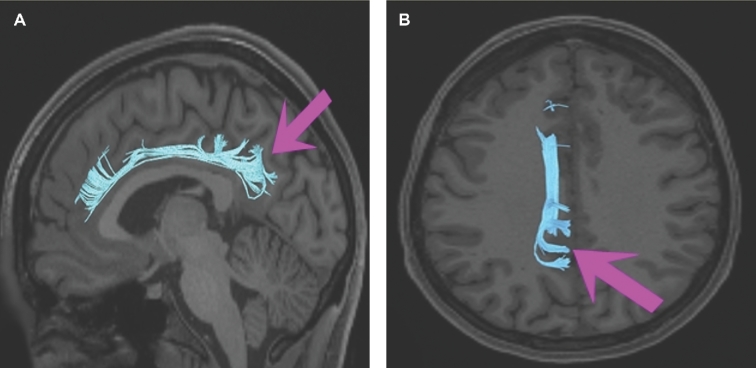
Area 23c is structurally connected to the contralateral hemisphere and local parcellations. 23c also has short projections with the cingulum but these fibers end before reaching the anterior cingulate cortex. There are consistent connections through the body of the corpus callosum to contralateral 5mv, 5m, 23c, and 5L. Short association bundles project superiorly to end at 24dd, 24dv, 5L, 5m, and 5mv (Figure 16).
FIGURE 16.
Structural connectivity of 23c in the left hemisphere, shown on T1-weighted MR images. A, Lateral sagittal view, B, axial view, and C, coronal view showing projections to the contralateral hemisphere. Orange: white matter tracts of 23c demonstrating connections with fibers running through the body of the corpus callosum to reach the contralateral hemisphere.
What is known about its function?
Area 23c is considered a part of the dPCC, which is highly active during tasks that require an external focus, especially concerning visuospatial and body orientation. Task fMRI studies indicate that this region is specifically involved in working memory processing of place and body images as well as focusing on socially interacting objects over randomly moving geometric shapes.3-6
Area 23d
Where is it?
Area 23d (23, part d) is found on the superior half of a section of the posterior cingulate gyrus. It lies just superior to the posterior part of the body of the corpus callosum.
What are its borders?
Area 23d borders area 23c superiorly, and RSC inferiorly. Its anterior border is with area p24prime, and its posterior border is with area d23ab.
What is its functional connectivity?
Area 23d demonstrates functional connectivity to a47r, p10p, s6-8, 8AV, and 9p in the lateral frontal lobe, areas 8BM, 8C 9m, a24, p24, p24prime, p32, and d32 in the medial frontal lobe, area TE1m in the temporal lobe, areas IP1, PGi, PFm, and PGs in the lateral parietal lobe, and areas d23ab, POS2, RSC, 7m, 31a, 31pv, and 31pd in the medial parietal lobe (Figure 17).
FIGURE 17.
Functional connectivity of 23d demonstrated on an inflated left hemisphere. A, Lateral and medial views. B, Rostral and caudal views. C, Dorsal and ventral views. Parcellations with the strongest functional connectivity are shown in yellow. Pink arrows designate the parcellation of interest.
What are its white matter connections?
Area 23d is structurally connected to the cingulum. The cingulum fibers project anteriorly from 23d with connections to the anterior cingulate cortex and cingulate sulcus, as the fibers curve around the genu of the corpus callosum, these fibers end at a24, p24, and a32pr. The cingulum fibers continue wrapping around the genu with its connections splitting to project anteriorly to 9m, as well as following the rostrum of the corpus callosum to end at 25. Short association bundles primarily project posteriorly to connect to d23ab and v23ab (Figure 18).
FIGURE 18.
Structural connectivity of 23d in the left hemisphere, shown on T1-weighted MR images. A, Lateral sagittal view, B, axial view, and C, medial sagittal view showing projections to the frontal lobe and cingulum. Dark blue: white matter tracts of 23d demonstrating connections with anterior cingulum fibers.
What is known about its function?
Area 23d is considered a part of the dPCC, which is highly active during tasks that require an external focus, especially concerning visuospatial and body orientation. Task fMRI studies indicate that this region is specifically involved in working memory processing of place, body, tool, and face images.3-6
Area RSC
Where is it?
Area RSC is a long thin area of posterior cingulate cortex that is immediately adjacent to the callosal sulcus wrapping around the splenium. Its length begins just superior to the midportion of the corpus callosum body, and it follows the cingulate sulcus until the bottom of the isthmus of the cingulum, where the parahippocampal gyrus begins.
What are its borders?
Area RSC borders the corpus callosum along its entire inferior border. Its anterior border is with area 33prime, and its posteroinferior border is with the presubiculum. Its long superior border includes (from anterior to posterior): area 23d, area d23ab, area v23ab, POS1, and ProS.
What is its functional connectivity?
Area RSC demonstrates functional connectivity to 9-46d, p10p, and 8AD in the lateral frontal lobe, areas 8BM, 9m, a24, p24, a32prime, p32, and d32 in the medial frontal lobe, areas IP1, IP2, PFm, and PGs in the lateral parietal lobe, and areas 23d, v23ab, d23ab, POS2, POS1, PCV, DVT, 7pm, 7m, 31a, 31pv, and 31pd in the medial parietal lobe (Figure 19).
FIGURE 19.
Functional connectivity of RSC demonstrated on an inflated left hemisphere. A, Lateral and medial views. B, Rostral and caudal views. C, Dorsal and ventral views. Parcellations with the strongest functional connectivity are shown in yellow. Pink arrows designate the parcellation of interest.
What are its white matter connections?
Area RSC is structurally connected to the cingulum. The cingulum fibers project both anteriorly and posteriorly from RSC, following the full length of the cingulate cortex. Posterior projections continue around the splenium of the corpus callosum ending at the parahippocampal gyrus at area EC. Anterior cingulum projections split near the genu of the corpus callosum to project superiorly to 10d and 9m, and inferiorly to 25. White matter tracts in the right hemisphere have more consistent connections with the precuneus when compared to left (Figure 20).
FIGURE 20.
Structural connectivity of RSC in the left hemisphere, shown on T1-weighted MR images. A to C, Three sagittal views from A, most lateral to B and C, most medial showing projections to the frontal lobe, cingulum, and parahippocampal region. Light red: white matter tracts of RSC demonstrating anterior and posterior cingulum fibers.
What is known about its function?
The RSC is primarily responsible for transitioning between allocentric (view-independent) spatial perspectives and egocentric (view-dependent) spatial perspectives.3,5-7 As such, the RSC is implicated in spatial navigation, episodic memory, future planning, and imagination. In addition, the RSC has been suspected of being involved in the retrieval of recent autobiographical information from memory.3,5-7
Precuneus Areas
The parcellations comprised by the precuneus area include 7m and PCV. The anatomic location of these parcellations is shown in Figure 21. This region has consistent white matter connections with the cingulum and contralateral hemisphere. The combined tractography of 7m and PCV is shown in Figure 22.
FIGURE 21.
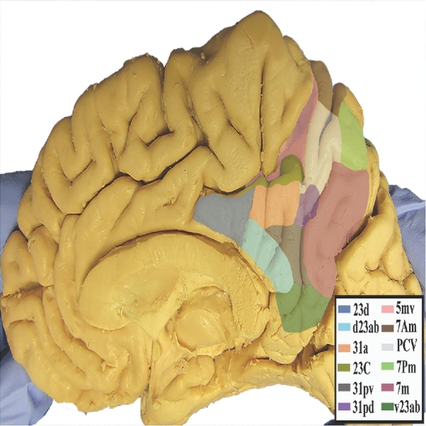
Anatomic view of the precuneus parcellations PCV and 7m shown on the medial right hemisphere of a cadaver brain.
FIGURE 22.
Combined structural connectivity of the precuneus parcellations, shown on T1-weighted MR images. A, Lateral sagittal view, B, axial view, and C, coronal view. White: white matter tracts of PCV. Red: white matter tracts of 7m.
Area PCV
Where is it?
Area PCV is found in the anterior precuneus, just posterior to the marginal ramus of the cingulate sulcus.
What are its borders?
Area PCV borders areas 5mv, 23c, and 31a anteriorly, area 31pd inferiorly, areas 7M and 7pm posteriorly, and area 7am superiorly.
What is its functional connectivity?
Area PCV demonstrates functional connectivity to 46, 9-46d, 8AD, and in the lateral frontal lobe, areas a24prime, 5mv, 23c, and s32 in the medial frontal lobe, areas FEF, 6ma, and 6a in the premotor region, area STV in the insula opercular regions, areas PHA2, PHA3, and PHT in the temporal lobe, areas 7AL, 7PL, IP0, LIPd, PF, and PGp in the lateral parietal lobe, areas 23d, POS2, POS1, RSC, DVT, 7am, 7pm, 7m, 31a, and 31pd in the medial parietal lobe, areas V1, V2, in the medial occipital lobe, area V6 in the dorsal visual stream areas, and areas TPOJ2 and TPOJ3 in the lateral occipital lobe (Figure 23).
FIGURE 23.
Functional connectivity of PCV demonstrated on an inflated left hemisphere. A, Lateral and medial views. B, Rostral and caudal views. C, Dorsal and ventral views. Parcellations with the strongest functional connectivity are shown in yellow. Pink arrows designate the parcellation of interest.
What are its white matter connections?
Area PCV is structurally connected to local parcellations and the contralateral hemisphere. Connections through the splenium of the corpus callosum terminate at contralateral 5m, 7am, PCV, and 7AL. Short association bundles project superiorly to connect to 7am, 7pm, and 5m (Figure 24).
FIGURE 24.
Structural connectivity of PCV in the left hemisphere, shown on T1-weighted MR images. A, Lateral sagittal view, B, axial view, and C, coronal view showing projections to the contralateral hemisphere. White: white matter tracts of PCV demonstrating white matter connections through the splenium of the corpus callosum to reach the contralateral hemisphere.
What is known about its function?
Area PCV is a part of the precuneus, which is involved in visual-spatial perception (including spatial reflection, visual motion perception, and spatial conflict resolution), episodic memory retrieval, self-processing, and consciousness. Task fMRI studies indicate that this region is specifically involved in working memory processing of place, body, tool, and face images and recognizing emotional faces over neutral objects.4,5,8
Area 7m
Where is it?
Area 7M (7 medial) is found in the posterior precuneus, just anterior to the parieto-occipital sulcus. It does not form the banks of this sulcus.
What are its borders?
Area 7M borders POS2 posteriorly, area v23asb and POS1 inferiorly, area 31pd anteriorly, and area 7pm and PCV superiorly.
What is its functional connectivity?
Area 7M demonstrates functional connectivity to 8AV, 8BL, 8AD, i6-8, 47s, 9a, 9p, 10d, 10v, and 10r in the lateral frontal lobe, areas 9m, a24, d32, and s32 in the medial frontal lobe, areas STSva, STSvp, TGd, TE1a, TE1m, TE1p, PreS, and the hippocampus in the temporal lobe, areas PFm, PGi, and PGs in the lateral parietal lobe, and areas 23d, v23ab, d23ab, POS2, POS1, PCV, RSC, 7pm, 31a, 31pv, and 31pd in the medial parietal lobe (Figure 25).
FIGURE 25.
Functional connectivity of 7M demonstrated on an inflated left hemisphere. A, Lateral and medial views. B, Rostral and caudal views. C, Dorsal and ventral views. Parcellations with the strongest functional connectivity are shown in yellow. Pink arrows designate the parcellation of interest.
What are its white matter connections?
Area 7M is structurally connected to the cingulum and contralateral hemisphere. Cingulum fibers project anteriorly from 7m and have connections along the midcingulate and anterior cingulate cortex to d32, a24, p24, a24pr, and p24pr. Connections through the splenium of the corpus callosum terminate at contralateral 7m and PCV. Short association bundles connect to POS1, POS2, 7pm, and PCV (Figure 26).
FIGURE 26.
Structural connectivity of 7m in the left hemisphere, shown on T1-weighted MR images. A, Sagittal view and B, axial view showing projections to the cingulum and frontal lobe. Light blue: white matter tracts of 7m demonstrating connections with anterior cingulum fibers.
What is known about its function?
Area 7m is a part of the precuneus, which is involved in visual-spatial perception (including spatial reflection, visual motion perception, and spatial conflict resolution), episodic memory retrieval, self-processing, and consciousness. Task fMRI studies indicate that this region is specifically involved in working memory processing of place, body, tool, and face images; listening to stories over answering arithmetic questions; focusing on socially interacting objects over randomly moving geometric shapes; recognizing emotional faces over neutral objects; and comparing featural dimensions of objects vs matching objects based on verbal classifications.4,5,8
Parieto-Occipital Sulcus Areas
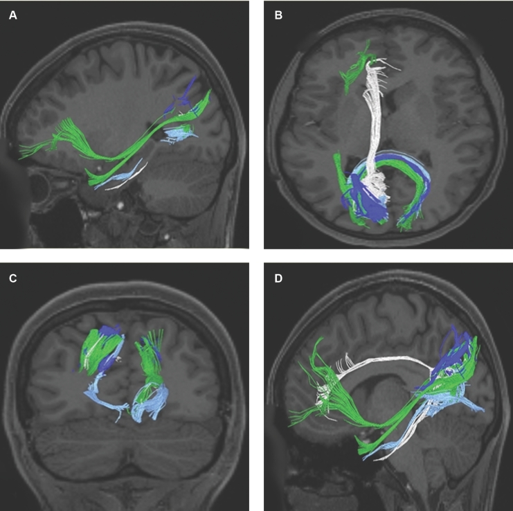
The parcellations comprised by the parieto-occipital sulcus include POS2, POS1, DVT, and ProS. The anatomic location of these parcellations is shown in Figure 27. This region has consistent white matter connections with the cingulum and contralateral hemisphere. Area DVT is unique in that it connects to the inferior fronto-occipital fasciculus (IFOF) and middle longitudinal fasciculus (MdLF). The combined tractography of POS2, POS1, DVT, and ProS is shown in Figure 28.
FIGURE 27.
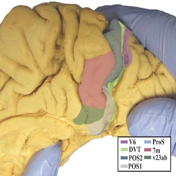
Anatomic view of the parieto-occipital sulcus parcellations POS2, POS1, DVT, and ProS shown on the medial aspect of the right hemisphere of a cadaver brain.
FIGURE 28.
Combined structural connectivity of the parieto-occipital sulcus parcellations, shown on T1-weighted MR images. A, Lateral sagittal view. B, Axial view. C, Coronal view. D, Medial sagittal view. Dark blue: white matter tracts of POS2. White: white matter tracts of POS1. Green: white matter tracts of DVT. Light blue: white matter tracts of ProS
Area POS2
Where is it?
Area POS2 is found on the anterior bank of the parieto-occipital sulcus and makes up the superior half of that bank.
What are its borders?
Area POS2 borders area 7M anteriorly and DVT posteriorly. Its superior border is made of areas 7pm and 7pl and its inferior border is made of POS1.
What is its functional connectivity?
Area POS2 demonstrates functional connectivity to 46, 9-46d, i6-8, 8C, p10p, and a10p in the lateral frontal lobe, areas 8BM, 9m, a24, p24, a32prime, p32, and d32 in the medial frontal lobe, areas IP1, IP2, PFm, PGp, and PGs in the lateral parietal lobe, and areas 23d, d23ab, POS1, PCV, RSC, DVT, 7am, 7pm, 7m, 31a, 31pv, and 31pd in the medial parietal lobe (Figure 29).
FIGURE 29.
Functional connectivity of POS2 demonstrated on an inflated left hemisphere. A, Lateral and medial views. B, Rostral and caudal views. C, Dorsal and ventral views. Parcellations with the strongest functional connectivity are shown in yellow. Pink arrows designate the parcellation of interest.
What are its white matter connections?
Area POS2 is structurally connected to local parcellations and the contralateral hemisphere. The white matter tracts from this parcellation are highly variable. Connections to the contralateral hemisphere travel with the forceps major fiber bundle to end at V1, though the termination of this tract is inconsistent. Short association bundles are connected to DVT, POS1, V2, and 7pm (Figure 30).
FIGURE 30.
Structural connectivity of POS2 in the left hemisphere, shown on T1-weighted MR images. A, Sagittal view, B, axial view, and C, coronal view showing projections to the contralateral hemisphere. Dark blue: white matter tracts of POS2 demonstrating connections with the forceps major fiber bundle to reach the contralateral hemisphere.
What is known about its function?
Area POS2 has a strong, coupled functional correlation with the RSC. Task fMRI studies indicate that this region is specifically involved in working memory processing of place, body, tool, and face images; processing of visual cues instructing movement, and comparing featural dimensions of objects vs matching objects based on verbal classifications.5
Area POS1
Where is it?
Area POS1 found on the anterior bank of the parieto-occipital sulcus and makes up the inferior half of that bank.
What are its borders?
Area POS1 borders area 7M, area d23ab, and RSC anteriorly, and DVT posteriorly. Its superior border is made of POS2, and its inferior border is made of ProS and RSC.
What is its functional connectivity?
Area POS1 demonstrates functional connectivity to 8AD, i6-8, 47m, 10d, 10v, and 10r in the lateral frontal lobe, areas 9m, a24, s32, p32, and d32 in the medial frontal lobe, areas STSva, TE1a, PHA1, PHA2, PHA3, PreS, and the hippocampus in the temporal lobe, areas PGi, PGp, and PGs in the lateral parietal lobe, and areas ProS, v23ab, d23ab, POS2, PCV, RSC, DVT, 7pm, 7m, 31a, 31pv, and 31pd in the medial parietal lobe (Figure 31).
FIGURE 31.
Functional connectivity of POS1 demonstrated on an inflated left hemisphere. A, Lateral and medial views. B, Rostral and caudal views. C, Dorsal and ventral views. Parcellations with the strongest functional connectivity are shown in yellow. Pink arrows designate the parcellation of interest.
What are its white matter connections?
Area POS1 is structurally connected to the contralateral hemisphere and to anterior and parahippocampal cingulum projections. Anterior cingulum fibers have connections to the anterior cingulate cortex at a24, a24pr, and p24. Posterior cingulum fibers curve around the splenium of the corpus callosum to end at the parahippocampal gyrus at area EC. Connections to contralateral V1 connect with FM. Short association bundles are connected to V1, V2, POS2, and V6 (Figure 32).
FIGURE 32.
Structural connectivity of POS1 in the left hemisphere, shown on T1-weighted MR images. A, Lateral sagittal view, B, axial view, and C, medial sagittal view showing projections to the frontal lobe, cingulum, parahippocampal regions, and the contralateral hemisphere. White: white matter tracts of POS1 demonstrating connections with the anterior cingulate cortex, parahippocampus, and the contralateral hemisphere.
What is known about its function?
Task fMRI studies demonstrate that area POS1 is activated during working memory processing of place images. This area also shows greater functional activity related to socially interacting objects vs randomly moving geometric shapes. It is suggested that this region is involved in scene-comprehension with the RSC.5
Area DVT
Where is it?
Area DVT (dorsal visual transitional area) is a long thin area that makes up the entire posterior bank of the parieto-occipital sulcus.
What are its borders?
Area DVT borders POS2 and POS1 anteriorly. Its superior tip wedges between V6 and POS2. Its posterior boundary is the anterior limb of V2, and its inferior boundary is with ProS.
What is its functional connectivity?
Area DVT demonstrates functional connectivity to areas SCEF, FEF, 6ma, and 6a in the premotor region, areas 9-46d and 46 in the lateral frontal lobes, areas p32prime, a24prime, 5mv, and 23c in the cingulate regions, areas FOP4, PFcm, 43, and 52 in the insula and opercular region, areas PHA1, PHA2, PHA3, and PHT in the temporal lobe, areas 7PC, 7AL, 7PL, PGp PF, PFop, AIP, VIP, LIPd, LIPv, IP0, and IPS1 in the lateral parietal lobe, areas 7am, 7pm, RSC, POS2, POS1, and PCV, in the medial occipital lobe, areas ProS, V1, V2, V3, and V4 in the medial occipital lobe, areas V3a, V3b, V7, V6, and V6a of the dorsal visual stream, areas FFC, VVC, V8, VMV1 (ventromedial visual area 1), VMV2, and VMV3 of the ventral visual stream, and areas TPOJ2, TPOJ3, V3cd, LO1, LO3, PH, and FST of the lateral occipital lobe (Figure 33).
FIGURE 33.
Functional connectivity of DVT demonstrated on an inflated left hemisphere. A, Lateral and medial views. B, Rostral and caudal views. C, Dorsal and ventral views. Parcellations with the strongest functional connectivity are shown in yellow. Pink arrows designate the parcellation of interest.
What are its white matter connections?
Area DVT is structurally connected to the IFOF, MdLF, and contralateral hemisphere. IFOF projections pass just posterior to the insula to end at the anterior pole of the frontal lobe with terminations extending from 8BL to a10p. MdLF projections course from the DVT to the superior temporal gyrus to end at STGa. Connections to contralateral V1, V2, V3, DVT, POS1, and POS2 follow the forceps major through the splenium of the corpus callosum. Short association bundles are connected to V6, DVT, POS1, and POS2 (Figure 34).
FIGURE 34.
Structural connectivity of DVT in the left hemisphere, shown on T1-weighted MR images. A, Sagittal view and B, axial view showing projections to the frontal lobe, occipital lobe, temporal lobe, and the contralateral hemisphere. Green: white matter tracts of DVT demonstrating connections with the inferior fronto-occipital fasciculus (IFOF), middle longitudinal fasciculus (MdLF), and contralateral hemisphere.
What is known about its function?
The dorsal visual transitional area is a newly defined region and is suspected to have a transitional function between the early visual cortex and posterior cingulate association cortex. It is functionally connected to the dorsal stream visual cortex, which perceives where stimuli are located, as well as the superior posterior parietal cortex, which plays an important role in planned movements, spatial reasoning, and attention, lending to its probable transitional function between these two areas. Task fMRI studies indicate that this region is specifically involved in focusing on socially interacting objects vs randomly moving geometric shapes.5
Area ProS
Where is it?
Area ProS (prostriate) is found in the prostriate cortex, which lies at the anterior most limit of the calcarine fissure. ProS lies between the anteroinferior tip of the parieto-occipital sulcus and the calcarine fissure and is just behind the isthmus of the cingulate gyrus.
What are its borders?
Area ProS borders V1 posteriorly, and PreS (presubiculum) anteriorly. Its superior border is made up of DVT, POS1, and RSC. VMV1, and the inferior limb of V2 forms its inferior border.
What is its functional connectivity?
Area ProS demonstrates functional connectivity to areas PHA1, PHA2, and PreS in the temporal lobe, areas IP0, DVT, POS1, and PGp in the parietal lobe, areas V1, V2, V3, and V4 in the medial occipital lobe, areas V3a, V3b, V7, V6, and V6a of the dorsal visual stream areas, areas VVC and V8 of the ventral visual stream, and area V3cd of the lateral occipital lobe (Figure 35).
FIGURE 35.
Functional connectivity of ProS demonstrated on an inflated left hemisphere. A, Lateral and medial views. B, Rostral and caudal views. C, Dorsal and ventral views. Parcellations with the strongest functional connectivity are shown in yellow. Pink arrows designate the parcellation of interest.
What are its white matter connections?
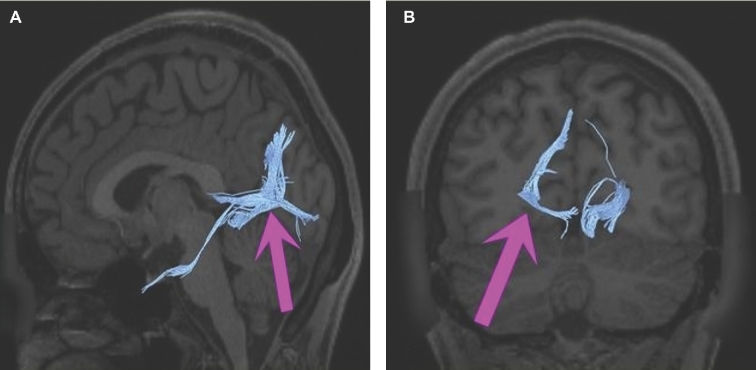
Area ProS is structurally connected to local parcellations, the contralateral hemisphere, and parahippocampal cingulum projections. Parahippocampal fibers from ProS end at PeEc. Connections to contralateral V1 course with forceps major through the splenium of the corpus callosum. Short association bundles project superiorly to V6 and POS2, and posteriorly to V1 (Figure 36).
FIGURE 36.
Structural connectivity of ProS in the left hemisphere, shown on T1-weighted MR images. A, Sagittal view and B, coronal view showing projections to the occipital lobe and parahippocampal regions of the temporal lobe. Light blue: white matter tracts of ProS demonstrating connections with parahippocampal cingulum fibers and with the forceps major bundle to reach the contralateral hemisphere.
What is known about its function?
The ProStriate area is suspected to have a transitional function between the early visual cortex and posterior cingulate association cortex like DVT, and is suspected to be primarily responsible for the coarse, rapid integration and analysis of peripheral visual stimuli. In addition, area ProS is suspected to coordinate responses and shifts in attentional focus across multiple cortical systems. It is most activated during working memory and relational functions when compared to other areas in the anterior bank of the parietal-occipital sulcus.5,9-11
DISCUSSION
The medial parietal lobe is a fascinating area. It has received little clinical attention, as damage to this area is rarely associated with a describable or obvious neuropsychological syndrome, as such the function of this is poorly understood to many surgeons. Neuroimaging research has changed this view substantially, as these areas include the posterior cingulate cortices, which have been linked to the DMN,1,2 a large scale, resting-state network, whose dysfunction has been linked to numerous brain diseases.12-17 Furthermore, graph theoretical analyses have suggested that the PCC areas are among the highest functionally connected clusters of brain regions in the cerebrum.18 Our data support this view, as the functional connectivity maps of some of these areas involve a large number of spatially and functionally distinct areas across the brain (see Figure 15 as an example), meaning the PCC is likely more important for proper brain functioning than we have realized.
The Connectional Pattern of the PCC Regions
What is truly fascinating about this is that, despite the wide variety of distinct functional regions with which PCC areas coactivate, its major white matter connections are relatively modest. The PCC is part of the DMN that classically includes the PCC, and anterior cingulate cortex and adjacent medial frontal lobe, and the lateral parietal lobe.19 The main connections support this orientation, as the primary connections of many PCC regions are via the cingulum to the anterior DMN cortices. Interestingly, while these regions are densely interconnected with each other, almost all of them seem to give long-distance projections to the anterior cingulate areas via the cingulum, which is different than a typical small world model would predict. This suggests that despite spatial distance between the two DMN clusters, regions in the ACC and PCC are technically one large cluster with hubs between the DMN and other brain networks.
Based on our work with large-scale connections, it is not entirely clear the primary way information leaves the PCC and the DMN, and which areas represent the hubs of the network. The most obvious hub-like candidate, at least in the PCC, is area 23c, which has by far the widest range of functionally connected areas as it is the only area in this section that demonstrates functional connectivity outside of the typical DMN component regions and the visual areas. Complicating this idea is our observation that 23c is mainly connected to its homolog in the other hemisphere, with some connections terminating locally. It appears to contribute minimally to the cingulum bundle, which raises the question of how it is connected functionally. Another possible candidate is an area like RSC that connects into the medial temporal lobe via the posterior cingulum. While these are all reasonable candidates, it is still possible that the true way information flows out of the PCC and, thus the DMN, is transthalamic. It is also possible that our methods are unable to resolve small or crossing fibers that are the true communication point for this region. This obviously deserves closer study, and it raises the key issue of how to preserve the DMN when its connections to other networks may be too small to resolve on imaging, thus making it difficult to establish a framework to avoid those fibers during surgery.
Works by others (Braga et al, Neuron, IN PRESS) suggest that the subregions of the DMN interdigitate anteriorly and segregate their connections in a spatially similar, interdigitated fashion. In other words, there is a spatial relationship between the site of origin of cingulum/DMN fibers and their target. Examination of Figure 2 should reinforce the idea that the connections are somewhat distinct and yet interdigitated. For example, fibers highlighted in white begin more posteroinferior and end most anteriorly, suggesting that while the cingulum bundle is compact, there is some degree of segregation of fibers.
What is This Part of the Brain Doing?
It should be obvious from this chapter that the medial parietal cortex is not a monolithic structure. Many of the more posterior precuneus areas are involved in the visual system, while the posterior cingulate areas have markedly different and more complex roles. It is interesting that these areas are tightly interdigitated in this junctional area.
Dorsal Posterior Cingulate Cortex
The parcellations that are best aligned with areas commonly viewed as dPCC are 31a, d23ab, 23c, and 23d. This division of the brain is primarily involved in visuospatial/body orientation.20 The dPCC has strong functional connectivity with DMN areas, and exhibits strong anti-correlation with the dorsal attention network during tasks that require an external focus.5 This indicates that the dPCC may be implicated in a transitional function that allows for detecting and responding to environmental events, allowing for a change in behavior. In addition, this region is involved in the integration of the dorsal visual stream contributing to its role in spatial processing.20
Ventral Posterior Cingulate Cortex
The parcellations that we believe best delinate the vPCC are 31pd, 31pv, and v23ab. This division of the brain is primarily involved with self-relevant assessments.20 The vPCC features a strong functional connectivity with the DMN during periods of rest or tasks that require internal focusing, including self-monitoring, self-reflection, retrieval of semantic and episodic memories, and planning.3-6 This region is suspected to be involved in evaluation of information from the ventral visual stream—contributing to its role in object processing in perceived and imagined stimuli.20
Default Mode Network
It is likely that both sets of regions in the PCC contribute to the DMN based on their patterns of connections, though we were unable to define clearly the targets of regions 31pv and 31pd. However, we were able to identify multiple areas that project to these regions when studying the anterior cingulate areas (see Chapter 2), and based on their patterns of functional connectivity, we have a strong suspicion they are part of this network. The DMN is an integrated system within the brain that is most active during periods of internal thought when individuals are left undisturbed.21 It is suspected to be involved during mental exercises in which the individual considers memories and future planning to construct and manipulate hypothetical scenarios,22 which provide the adaptive function of preparing for upcoming, self-relevant events.
The Precuneus Portions of the Parietal Lobe
The anatomy of these regions is quite varied and complex. As an example, see Figure 28 and note that on the banks of the parieto-occipital sulcus you have areas that are functionally visual areas dominated by local connections (POS2); areas that are members of the visual system but who project to DMN regions and the hippocampus (POS1); areas that are entirely visual and project in visual pathways (DVT); areas that are part of the visual and DMN systems functionally but only connected locally (PCV); and areas that are really part of the DMN both structurally and functionally (7M). Not surprisingly, this is a true transitional area that fits with the model of interdigitating functional network organization. This will likely frustrate efforts to define a clear function for the PCC for some time. Nevertheless, our work highlights this area as a key crossroads of higher brain function.
Disclosures
Synaptive Medical assisted in the funding of all 18 chapters of this supplement. No other funding sources were utilized in the production or submission of this work.
Acknowledgments
Data were provided [in part] by the Human Connectome Project, WU- Minn Consortium (Principal Investigators: David Van Essen and Kamil Ugurbil; 1U54MH091657) funded by the 16 NIH Institutes and Centers that support the NIH Blueprint for Neuroscience Research; and by the McDonnell Center for Systems Neuroscience at Washington University. We would also like to thank Brad Fernald, Haley Harris, and Alicia McNeely of Synaptive Medical for their assistance in constructing the network figures for Chapter 18 and for coordinating the completion and submission of this supplement.
REFERENCES
- 1. Raichle ME, MacLeod AM, Snyder AZ, Powers WJ, Gusnard DA, Shulman GL. A default mode of brain function. Proc Natl Acad Sci U S A. 2001;98(2):676-682. [DOI] [PMC free article] [PubMed] [Google Scholar]
- 2. Raichle ME. The brain's default mode network. Annu Rev Neurosci. 2015;38:433-447. [DOI] [PubMed] [Google Scholar]
- 3. Aggleton JP, Saunders RC, Wright NF, Vann SD. The origin of projections from the posterior cingulate and retrosplenial cortices to the anterior, medial dorsal and laterodorsal thalamic nuclei of macaque monkeys. Eur J Neurosci. 2014;39(1):107-123. [DOI] [PMC free article] [PubMed] [Google Scholar]
- 4. Bzdok D, Heeger A, Langner R et al. . Subspecialization in the human posterior medial cortex. Neuroimage. 2015;106:55-71. [DOI] [PMC free article] [PubMed] [Google Scholar]
- 5. Glasser MF, Coalson TS, Robinson EC et al. . A multi-modal parcellation of human cerebral cortex. Nature. 2016;536(7615):171-178. [DOI] [PMC free article] [PubMed] [Google Scholar]
- 6. Leech R, Sharp DJ. The role of the posterior cingulate cortex in cognition and disease. Brain. 2014;137(1):12-32. [DOI] [PMC free article] [PubMed] [Google Scholar]
- 7. Vann SD, Aggleton JP, Maguire EA. What does the retrosplenial cortex do? Nat Rev Neurosci. 2009;10(11):792-802. [DOI] [PubMed] [Google Scholar]
- 8. Cavanna AE, Trimble MR. The precuneus: a review of its functional anatomy and behavioural correlates. Brain. 2006;129(3):564-583. [DOI] [PubMed] [Google Scholar]
- 9. Morecraft RJ, Rockland KS, Van Hoesen GW. Localization of area prostriata and its projection to the cingulate motor cortex in the rhesus monkey. Cereb Cortex. 2000;10(2):192-203. [DOI] [PubMed] [Google Scholar]
- 10. Rockland Kathleen S. Visual system: prostriata — a visual area off the beaten path. Curr Biol. 2012;22(14):R571-R573. [DOI] [PubMed] [Google Scholar]
- 11. Yu H-H, Chaplin Tristan A, Davies Amanda J, Verma R, Rosa Marcello GP. A specialized area in limbic cortex for fast analysis of peripheral vision. Curr Biol. 2012;22(14):1351-1357. [DOI] [PubMed] [Google Scholar]
- 12. Shao J, Meng C, Tahmasian M et al. . Common and distinct changes of default mode and salience network in schizophrenia and major depression. Brain Imaging Behav. 2018. [DOI] [PubMed] [Google Scholar]
- 13. Brakowski J, Spinelli S, Dorig N et al. . Resting state brain network function in major depression - depression symptomatology, antidepressant treatment effects, future research. J Psychiatr Res. 2017;92:147-159. [DOI] [PubMed] [Google Scholar]
- 14. Stephens JA, Salorio CF, Barber AD, Risen SR, Mostofsky SH, Suskauer SJ. Preliminary findings of altered functional connectivity of the default mode network linked to functional outcomes one year after pediatric traumatic brain injury. Dev Neurorehabil. 2017:1-8. [DOI] [PMC free article] [PubMed] [Google Scholar]
- 15. Wang C, Pan Y, Liu Y et al. . Aberrant default mode network in amnestic mild cognitive impairment: a meta-analysis of independent component analysis studies. Neurol Sci. 2018;39(5):919-931. [DOI] [PubMed] [Google Scholar]
- 16. Li M, Zheng G, Zheng Y et al. . Alterations in resting-state functional connectivity of the default mode network in amnestic mild cognitive impairment: an fMRI study. BMC Med Imaging. 2017;17(1):919-931. [DOI] [PMC free article] [PubMed] [Google Scholar]
- 17. Padmanabhan A, Lynch CJ, Schaer M, Menon V. The default mode network in autism. Biol Psychiatry Cogn Neurosci Neuroimaging. 2017;2(6):476-486. [DOI] [PMC free article] [PubMed] [Google Scholar]
- 18. Hagmann P, Cammoun L, Gigandet X et al. . Mapping the structural core of human cerebral cortex. PLoS Biol. 2008;6(7):e159. [DOI] [PMC free article] [PubMed] [Google Scholar]
- 19. Harrison BJ, Pujol J, López-Solà M et al. . Consistency and functional specialization in the default mode brain network. Proc Natl Acad Sci U S A. 2008;105(28):9781-9786. [DOI] [PMC free article] [PubMed] [Google Scholar]
- 20. Vogt BA, Vogt L, Laureys S. Cytology and functionally correlated circuits of human posterior cingulate areas. Neuroimage. 2006;29(2):452-466. [DOI] [PMC free article] [PubMed] [Google Scholar]
- 21. Buckner RL, Andrews-Hanna JR, Schacter DL. The brain's default network. Ann N Y Acad Sci. 2008;1124(1):1-38. [DOI] [PubMed] [Google Scholar]
- 22. Poerio GL, Sormaz M, Wang HT, Margulies D, Jefferies E, Smallwood J. The role of the default mode network in component processes underlying the wandering mind. Soc Cogn Affect Neurosci. 2017;12(7):1047-1062. [DOI] [PMC free article] [PubMed] [Google Scholar]


