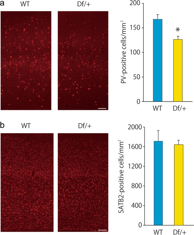
Fig. 5.

Decreased parvalbumin (PV)-positive cells in the sensory cortex in Df/+ mice. a PV immunostaining showing the decreased number of PV-positive cells in the adult Df/+ mice. Representative figures (left). Scale bar, 100 μm. Quantification of the number of PV-positive cells (each n = 3) (right). *P < 0.05, Student’s t test. b SATB2 immunostaining showing the normal number of SATB2-positive cells in adult Df/+ mice. Representative figures (left). Scale bar, 100 μm. Quantification of the number of SATB2-positive cells (each n = 3) (right). Data are presented as the mean ± s.e.m.
