ABSTRACT
In this supplement, we build on work previously published under the Human Connectome Project. Specifically, we show a comprehensive anatomic atlas of the human cerebrum demonstrating all 180 distinct regions comprising the cerebral cortex. The location, functional connectivity, and structural connectivity of these regions are outlined, and where possible a discussion is included of the functional significance of these areas. In part 3, we specifically address regions relevant to the sensorimotor cortices.
Keywords: Anatomy, Cerebrum, Connectivity, DTI, Functional connectivity, Human, Parcellations
ABBREVIATIONS
- FAT
frontal aslant tract
- HCP
Human Connectome Project
- PEF
premotor eye field
- SFL
superior frontal language
- SMA
supplementary motor area
This chapter describes the subdivisions of Brodmann areas 1 through 6 which most people will recognize as the primary sensorimotor network and premotor areas.1 Though preservation of the motor cortex has always been a goal of neurosurgeons operating in this part of the cerebrum, it is becoming increasingly clear that effective motor control requires more than just maintaining the connection between the motor strip and the spinal cord. Rather, it likely requires the maintenance of connections between the primary motor area, the sensory areas, the premotor areas, the basal ganglia, and parts of the parietal lobe. Thus, we consider many of these areas together in this section as we begin to discuss the hypothetical shape of the motor network.
PRIMARY MOTOR CORTEX
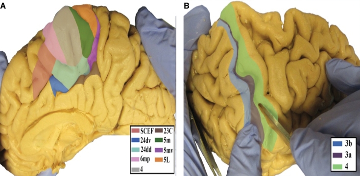
The anatomic location of the primary motor cortex (area 4) is shown in Figure 1. This region has consistent white matter connections with the pyramidal tracts, the contralateral hemisphere, and parietal lobe.
FIGURE 1.
Anatomical location of primary motor cortex parcellations shown on the right hemisphere of a cadaver brain. A, Medial view of paracentral lobule areas. B, Dorsolateral view with dilated central sulcus to show extension of parcellations into the sulcus.
Area 4
Where Is It?
Area 4 is found in the precentral gyrus. It is predominantly located on the posterior half of the gyrus, making up the anterior bank of the central sulcus. It widens medially to fill a larger proportion of the paracentral lobule, which is the leg motor region. The somatotopic organization of this area is well known; however, it is worth noting that there is an area dedicated to the eye which is adjacent to the frontal eye fields.
What Are Its Borders?
Area 4 borders area 3a along its posterior convexity border. Its inferior end contains area 43. At its superior end, on the medial surface (ie, the paracentral lobule), it borders area 5m posteriorly, area 44dd inferiorly,and area 6mp anteriorly. Its long anterior border contacts several areas, including (from superior to inferior) area 6d, FEF, area 55b, premotor eye field (PEF), and area 6v, most of which contact it on the anterior half of the precentral gyrus.
What Is Its Functional Connectivity?
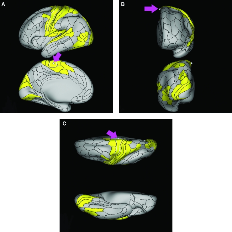
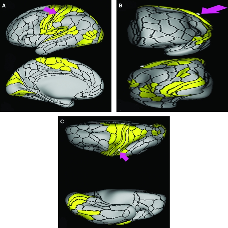
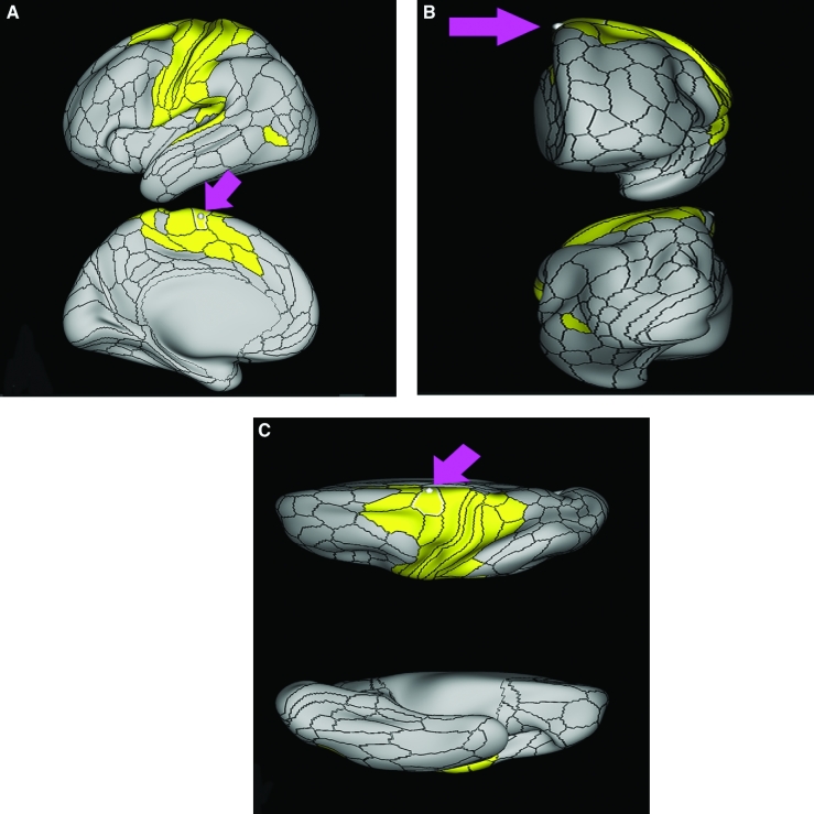
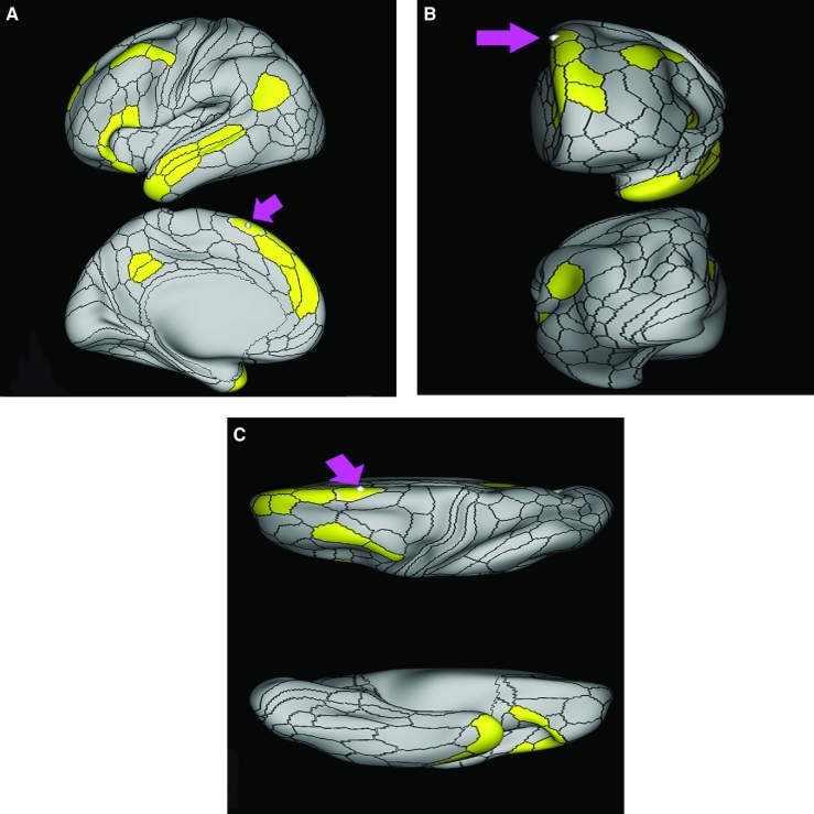
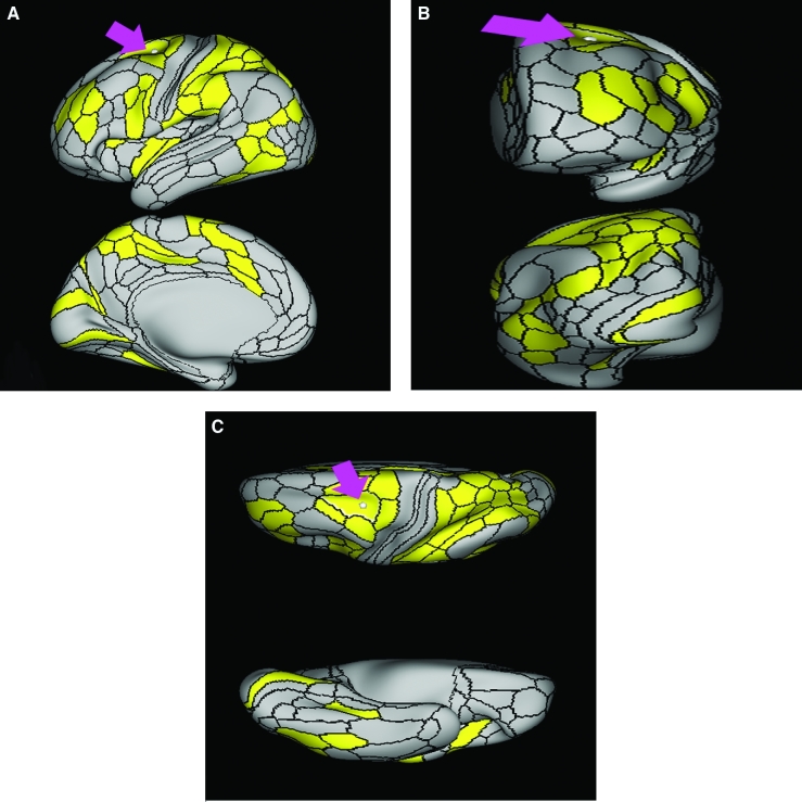
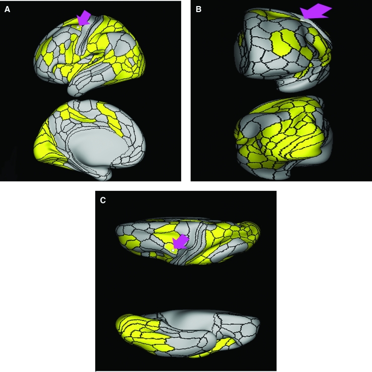
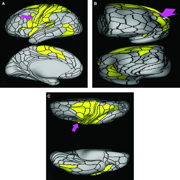
Area 4 demonstrates functional connectivity to areas 1, 2, 3a, and 3b in the sensory strip, areas SCEF, 55b, 6d, 6v, and 6mp in the premotor regions, areas 24dd, 24dv, 5m, and 5L in the middle cingulate regions, areas 43, OP1, OP2-3, OP4, IG, and FOP2 in the superior insula opercular regions, areas A4, A5, RI, PBelt, LBelt, TA2, and STV in the lower opercula and Heschl's gyrus regions, areas VIP, IPS1, LIPv, and 7PC in the parietal lobe, areas V2, V3, and V4 in the medial occipital lobe, areas V6, V6a, V3a, and V7 in the dorsal visual stream areas, area FFC of the ventral visual stream, and areas PH, TPOJ1, FST, V4t, MT, and LO3 of the lateral occipital lobe (Figure 2).
FIGURE 2.
Functional connectivity of area 4 demonstrated on an inflated left hemisphere. A, Lateral and medial views. B, Rostral and caudal views. C, Dorsal and ventral views. Parcellations with the strongest functional connectivity are shown in yellow. Pink arrows designate the parcellation of interest.
What Are Its White Matter Connections?
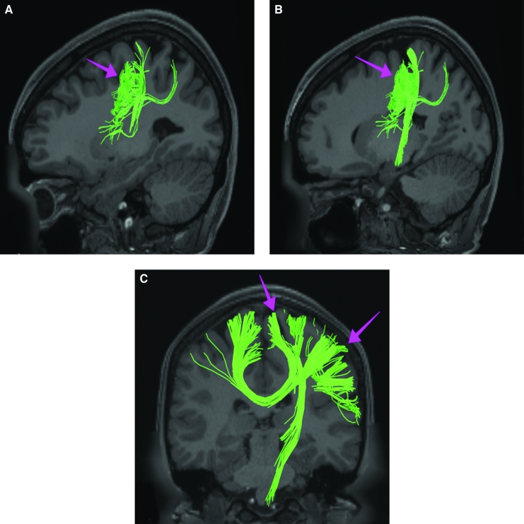
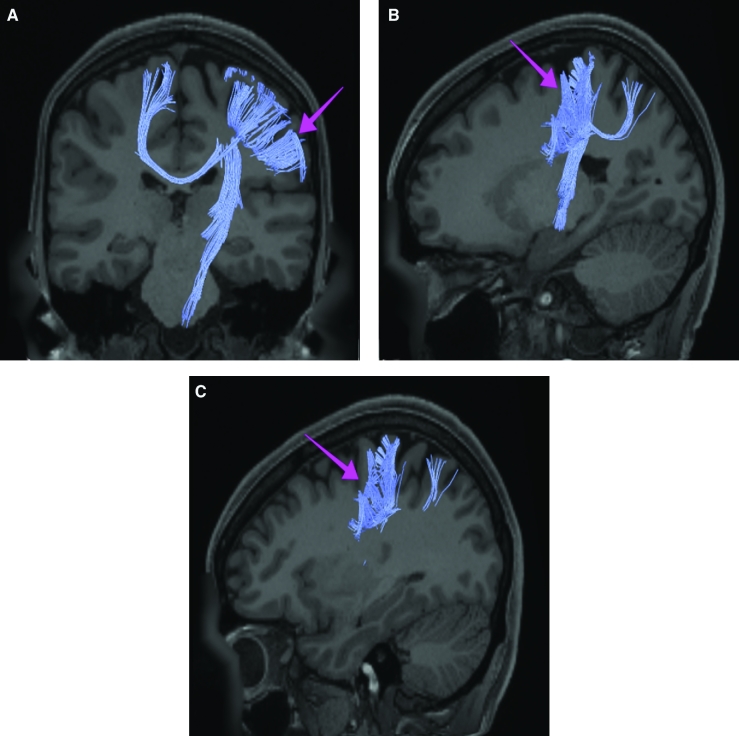
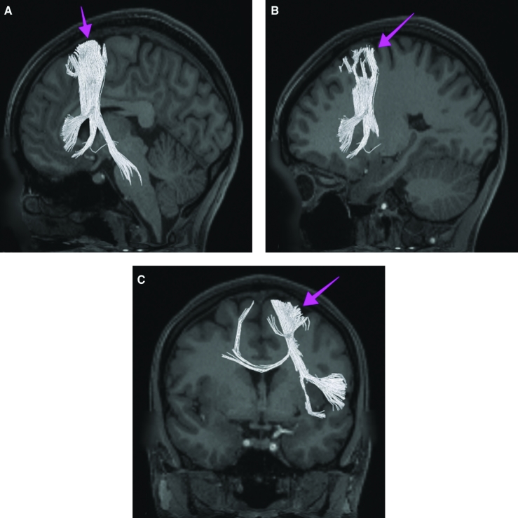
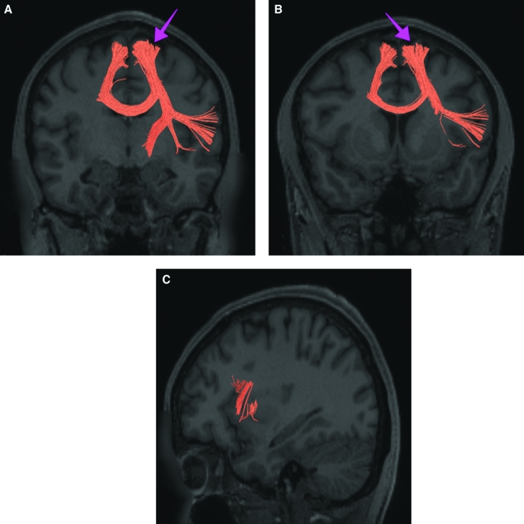
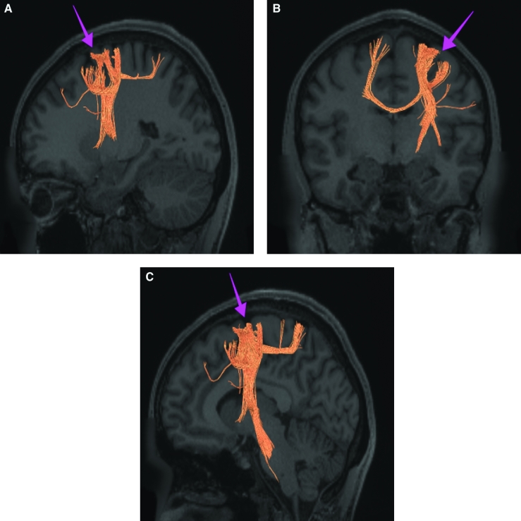
Area 4 is structurally connected to pyramidal tracts, the contralateral hemisphere, and the parietal lobule. Connections to pyramidal tracts descend through the posterior limb of the internal capsule and cerebral peduncle to the brainstem. Contralateral connections course through the body of the corpus callosum to parcellations 4, 6ma, and 6mp. Parietal projections are portions of the SLF and connect to PFm. Local short association fibers are connected with 3a, 3b, 2, 1, and 6v (Figure 3).
FIGURE 3.
Structural connectivity of area 4 in the left hemisphere, shown on T1-weighted MR images. Sagittal views of A, lateral and B, medial planes. C, Coronal view showing projections to the contralateral hemisphere. Green: white matter tracts of area 4 demonstrating connections with the pyramidal tracts, contralateral hemisphere, and the parietal lobule. Pink arrows designate the parcellation of interest.
What Is Known About Its Function?
Area 4 is known to produce fine motor movements of the distal forearm and fingers.2 Additionally, it functions in giving a muscle its tone and producing forceful muscle contractions.2 It is also thought to play a role in visual learning of motor-based skills in the early stages of life.3
PRIMARY SENSORY AREAS
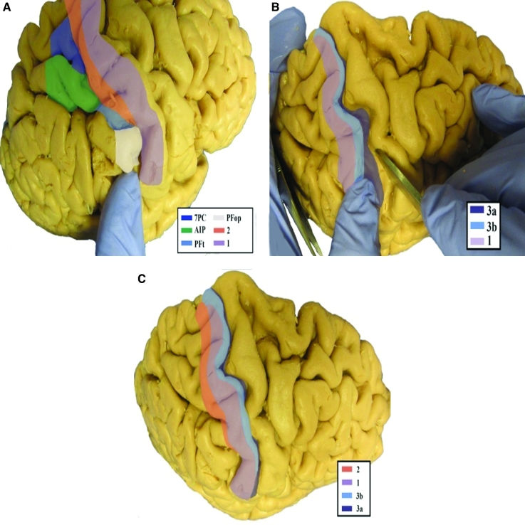
It is easy to think of these areas as simple strips which parallel each other along the postcentral gyrus from the Sylvian fissure to the midline. This is, however, not correct, as they do not perfectly overlap, and most interestingly, do not enter the paracentral lobule region at all. The anatomic location of the parcellations comprised of the primary sensory cortex is shown in Figure 4. These parcellations include 3a, 3b, 1, and 2. This region has consistent white matter connections with the pyramidal tracts, thalamocortical projections, parietal lobe, and contralateral hemisphere. The combined tractography of the parcellations is shown in Figure 5.
FIGURE 4.
Anatomical location of primary sensory area parcellations shown on the right hemisphere of a cadaver brain. A, Oblique view with dilated postcentral sulcus. B, Dorsolateral view with dilated central sulcus. C, Lateral view without dilation of the central sulcus.
FIGURE 5.
Combined structural connectivity of the primary sensory parcellations in the left hemisphere, shown on T1-weighted MR images. Sagittal views of A, medial and D, lateral planes. Coronal views of B, caudal and C, rostral planes showing projections to the contralateral hemisphere. Tracts correspond to areas 3a (green), 3b (light blue), 1 (purple), and 2 (orange).
Area 3a
Where Is It?
Area 3a is found in the depth of the central sulcus. It follows this sulcus up to the midline but does not fold onto the medial face of the hemisphere.
What Are Its Borders?
Area 3a borders area 2 along its long anterior border and area 3b along its posterior border. Inferiorly, it borders area 23 and superiorly it borders area 5m. It has a small area of posterior contact with area 2 near the Sylvian fissure.
What Is Its Functional Connectivity?
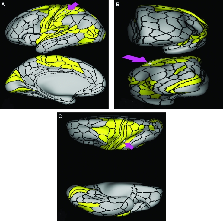
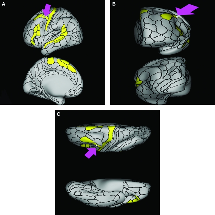
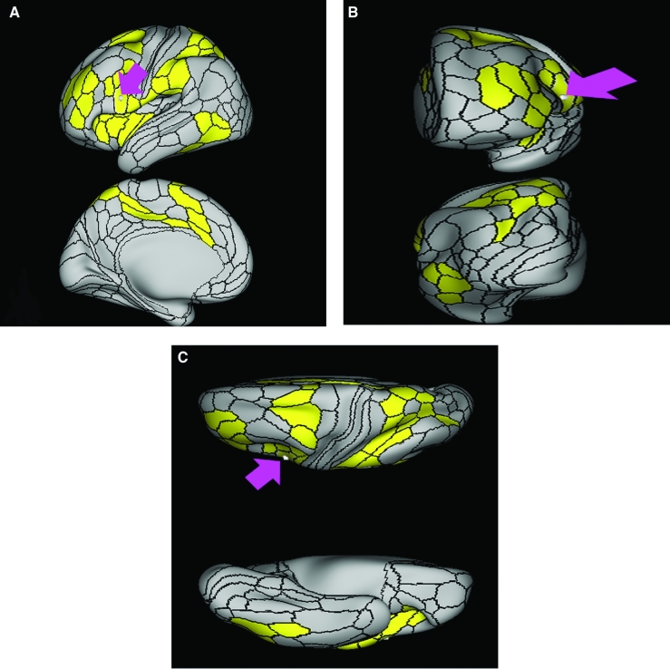
Area 3a demonstrates functional connectivity areas 1, 2, 4, 3b in the motor and sensory strips, areas SCEF, 55b, 6d, 6v, and 6mp in the premotor regions, areas 24dd, 24dv, 5m, and 5L in the middle cingulate regions, areas 43, OP1, OP2-3, OP4, IG, and FOP2 in the superior insula opercular regions, areas PoI2, 52, A4, A5, RI, A1, MBelt, PBelt, LBelt, TA2, and STV in the lower opercula and Heschl's gyrus regions, areas PFcm, 7AL, and 7PC in the parietal lobe, areas V2, V3, and V4 in the medial occipital lobe, areas V6, V6a, V3a, and V7 in the dorsal visual stream areas, area FFC of the ventral visual stream, and areas PH, TPOJ1, FST, V4t, MST, MT, and LO3 of the lateral occipital lobe (Figure 6).
FIGURE 6.
Functional connectivity of 3a demonstrated on an inflated left hemisphere. A, Lateral and medial views. B, Rostral and caudal views. C, Dorsal and ventral views. Parcellations with the strongest functional connectivity are shown in yellow. Pink arrows designate the parcellation of interest.
What Are Its White Matter Connections?
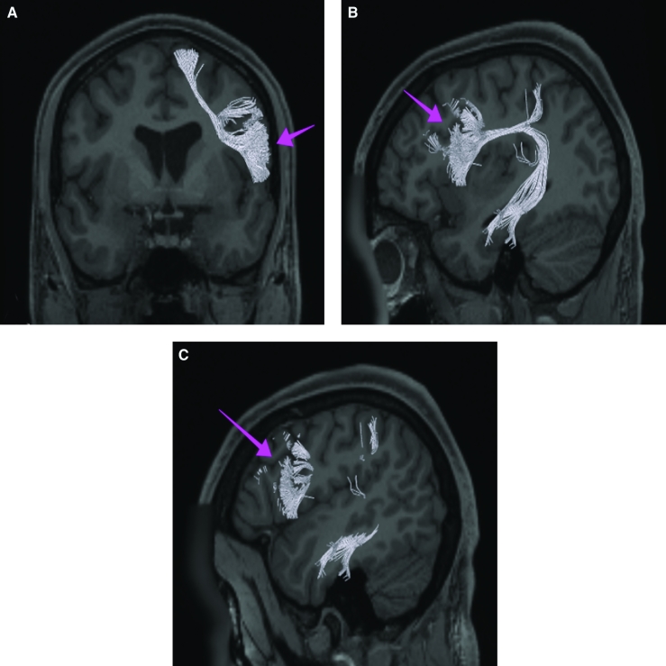
Area 3a is structurally connected to the pyramidal tracts, thalamocortical projections, contralateral hemisphere, and the parietal lobe. Connections to pyramidal tracts descend through the posterior limb of the internal capsule and cerebral peduncle to the brainstem. Thalamocortical tracts run medial to pyramidal projections to enter the thalamus. Contralateral connections course through the body of the corpus callosum to parcellations 3b and 4. Parietal projections are portions of the SLF and connect to PFm. Local short association fibers are connected with 4, 3b, 2, 1, and 6v (Figure 7).
FIGURE 7.
Structural connectivity of 3a in the left hemisphere, shown on T1-weighted MR images. A, Coronal view showing projections to the contralateral hemisphere. Sagittal view of B, medial and C, lateral planes. Green: white matter tracts of 3a demonstrating connections with the pyramidal tracts, thalamocortical projections, contralateral hemisphere, and the parietal lobe. Pink arrows designate the parcellation of interest.
What Is Known About Its Function?
Area 3a, along with area 2, is known to receive sensory information from deep body tissues.4,5 This area is also involved in the burning, chronic pain sensations that originate from deep somatic tissue.6 This suggests area 3a has relevance in many of the chronic pain ailments observed in the clinical setting.6 Area 3a is also involved in proprioception.6,7
Area 3b
Where Is It?
Area 3b is makes up the entire anterior bank of the postcentral gyrus. It does not reach the Sylvian fissure or fold onto the medial hemispheric face.
What Are Its Borders?
Area 3b borders area 3a along its anterior convexity border, and area 2 along its long posterior convexity border. Its inferior end ends superior to the Sylvian fissure and areas 3a and 2 contact each other inferior to this, forming area 3b's inferior border. Superomedially, it contacts areas 5m and 5l. It also makes a small border with area 2 posterosuperiorly.
What Is Its Functional Connectivity?
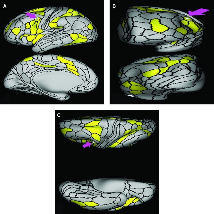
Area 3b demonstrates functional connectivity to areas 1, 2, and 3a in the sensory strip, area 4 in the motor strip, areas SCEF, 6d, and 6v in the premotor regions, areas 24dd, 24dv, 5m, and 5L in the middle cingulate regions, areas 43, OP1, OP2-3, OP4, IG, PFcm, and FOP2 in the superior insula opercular regions, areas A4, A5, RI, 52, A1, MBelt, LBelt, PBelt, TA2, and STV in the lower opercula and Heschl's gyrus regions, areas 7AL and 7PC in the parietal lobe, areas V2, V3, and V4 in the medial occipital lobe, areas V6, V6a, V3a, and V7 in the dorsal visual stream areas, area FFC of the ventral visual stream, and areas TPOJ1, FST, V4t, MST, MT, and LO3 of the lateral occipital lobe (Figure 8).
FIGURE 8.
Functional connectivity of 3b demonstrated on an inflated left hemisphere. A, Lateral and medial views. B, Rostral and caudal views. C, Dorsal and ventral views. Parcellations with the strongest functional connectivity are shown in yellow. Pink arrows designate the parcellation of interest.
What Are Its White Matter Connections?
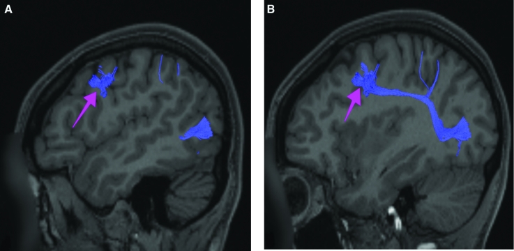
Area 3b is structurally connected to the pyramidal tracts, thalamocortical projections, contralateral hemisphere, and the parietal lobe. Connections to pyramidal tracts descend through the posterior limb of the internal capsule and cerebral peduncle to the brainstem. Thalamocortical tracts run medial to pyramidal projections to enter the thalamus. Contralateral connections course through the body of the corpus callosum to parcellations 3a, 3b, and 4. Parietal projections are portions of the SLF and connect with IP1 and IP2. Local short association fibers are connected with 1, 2, 3a, and 4 (Figure 9).
FIGURE 9.
Structural connectivity of 3b in the left hemisphere, shown on T1-weighted MR images. A, Coronal view showing projections into the contralateral hemisphere. Sagittal views of B, medial and C, lateral planes. Light blue: white matter tracts of 3b demonstrating connections with the pyramidal tracts, thalamocortical projections, contralateral hemisphere, and the parietal lobe. Pink arrows designate the parcellation of interest.
What Is Known About Its Function?
Area 3b is involved in the sensation of tactile stimuli. Specifically, this area represents the initial region of activation in tactile stimulation, followed by the activation of area 1.4 Area 3b also functions in the localization of sensation on the skin and distinguishing the features of that sensation.8 For example, activation of area 3b offers finger-specific information with respect to a tactile stimulus as opposed to areas 1 and 2 which cover a larger, more generalized receptive field.8 Additionally, area 3b receives exclusive activation in the reception of nociceptive stimuli.6
Area 1
Where Is It?
Area 1 is found on the visible surface of the postcentral gyrus. It forms the largest bulk of the postcentral operculum. It continues up to the midline, but does not fold onto the medial face.
What Are Its Borders?
Area 1 borders area 3b along its anterior convexity border, and area 2 along its posterior border. Its inferior end borders OP4 and PFop. Its superior end is surrounded by areas 3b and 2.
What Is Its Functional Connectivity?
Area 1 demonstrates functional connectivity to areas 2 and 3a in the sensory strip, area 4 in the motor strip, areas SCEF, 6mp, 6d, and 6v in the premotor regions, areas 24dd, 24dv, 5m, and 5L in the middle cingulate regions, areas 43, OP1, OP2-3, OP4, IG, PFcm, and FOP2 in the superior insula opercular regions, areas A4, A5, RI, 52, MBelt, LBelt, PBelt, TA2, and STV in the lower opercula and Heschl's gyrus regions, areas LIPv, VIP, IPS1, 7AL, and 7PC in the parietal lobe, areas V2, V3, and V4 in the medial occipital lobe, areas V6, V6a, and V7 in the dorsal visual stream areas, area FFC of the ventral visual stream, and areas PH, TPOJ1, FST, V4t, MST, MT, and LO3 of the lateral occipital lobe (Figure 10).
FIGURE 10.
Functional connectivity of 1 demonstrated on an inflated left hemisphere. A, Lateral and medial views. B, Rostral and caudal views. C, Dorsal and ventral views. Parcellations with the strongest functional connectivity are shown in yellow. Pink arrows designate the parcellation of interest.
What Are Its White Matter Connections?
Area 1 is structurally connected to the pyramidal tracts, thalamocortical projections, and the parietal lobe. Connections to pyramidal tracts descend through the posterior limb of the internal capsule and cerebral peduncle to the brainstem. Thalamocortical tracts run medial to pyramidal projections to enter the thalamus. Parietal projections are portions of the SLF and connect to PFm. Local short association fibers are connected with 1, 2, 3a, 3b, 4, and 6 (Figure 11). White matter tracts of area 1 in the right hemisphere have less consistent projections to surrounding parcellations.
FIGURE 11.
Structural connectivity of 1 in the left hemisphere, shown on T1-weighted MR images. Sagittal views of A, lateral and B, medial planes. C, Coronal view. Purple: white matter tracts of 1 demonstrating connections with the pyramidal tracts, thalamocortical projections, and the parietal lobe. Pink arrows designate the parcellation of interest.
What Is Known About Its Function?
Area 1, along with area 3b, is involved in processing tactile stimuli. Specifically, area 1 represents the secondary point of activation following the reception of a tactile sensation activating area 3b.4 Area 1 also functions along with area 2 in receiving information related to bilateral tactile stimulation of the hands.8
Area 2
Where Is It?
Area 2 is makes up most of the anterior bank of the postcentral sulcus. It neither folds onto the midline nor reaches the Sylvian fissure.
What Are Its Borders?
Area 2 borders area 1 as its main anterior border. It makes a small anterior contact with area 3b superiorly. Inferiorly, it has a small border with PFop. Its superior border is mainly made up of area 5l. It borders area 7AL posterosuperiorly. Its long posterior border contacts (from superior to inferior): area 7PC, AIP, and PFt.
What Is Its Functional Connectivity?
Area 2 demonstrates functional connectivity to areas 1, 3a, and 3b in the sensory strip, area 4 in the motor strip, areas SCEF, FEF, 6a, 6mp, 6d, and 6v in the premotor regions, areas 24dd, 24dv p32prime, 5mv, 5m, and 5L in the middle cingulate regions, areas 43, OP1, OP2-3, OP4, IG, PFcm, FOP1, and FOP2 in the superior insula opercular regions, areas PoI1, PoI2, A4, A5, RI, 52, A1, MBelt, LBelt, PBelt, TA2, and STV in the lower opercula and Heschl's gyrus regions, areas AIP, VIP, LIPv, PFop, PFt, IPS1, 7AL, and 7PC in the parietal lobe, areas V2 and V3 in the medial occipital lobe, areas V6 in the dorsal visual stream areas, area FFC of the ventral visual stream, and areas PH TPOJ1, TPOJ2, FST, V4t, MST, MT, and LO3 of the lateral occipital lobe (Figure 12).
FIGURE 12.
Functional connectivity of 2 demonstrated on an inflated left hemisphere. A, Lateral and medial views. B, Rostral and caudal views. C, Dorsal and ventral views. Parcellations with the strongest functional connectivity are shown in yellow. Pink arrows designate the parcellation of interest.
What Are Its White Matter Connections?
Area 2 is structurally connected to the pyramidal tracts, thalamocortical projections, contralateral hemisphere, and the parietal lobe. Connections to pyramidal tracts descend through the posterior limb of the internal capsule and cerebral peduncle to the brainstem. Thalamocortical tracts run medial to pyramidal projections to enter the thalamus. Contralateral connections course through the body of the corpus callosum to parcellation 7am. Parietal projections are portions of the SLF and connect to PFm, IP1, and IP2. Local short association fibers are connected with 6r, 7Pc, AIP, 1, 3a, 3b, and 4 (Figure 13).
FIGURE 13.
Structural connectivity of 2 in the left hemisphere, shown on T1-weighted MR images. A, Coronal view showing projections to the contralateral hemisphere. Sagittal view of B, lateral and C, medial planes. Orange: white matter tracts of 2 demonstrating connections with the pyramidal tracts, thalamocortical projections, contralateral hemisphere, and the parietal lobe. Pink arrows designate the parcellation of interest.
What Is Known About Its Function?
Area 2 is known to be involved in the processing of deep tissue sensations.4,5 Additionally, area 2 is activated in bilateral tactile stimulation of the hands.8
PARACENTRAL LOBULE AREAS
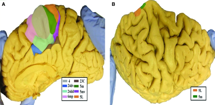
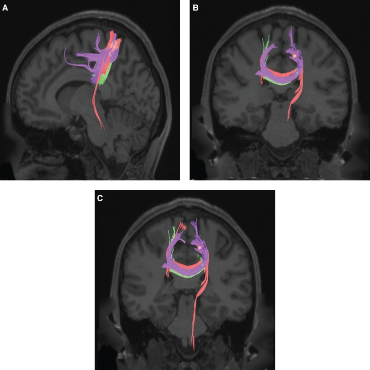
It is commonly thought that the paracentral lobule is a combined motor and sensory area, but while the anterior portion of this region contains the leg motor section of area 4, the posterior portion contains subunits of area 5 as opposed to parts of the primary sensory cortex.9 The anatomic location of the parcellations that comprise the paracentral lobule area is shown in Figure 14. These parcellations include 5l, 5m, and 5mv. This region has consistent white matter connections with the contralateral hemisphere. There are also connections with the pyramidal tracts and cingulate cortex but these white matter tracts are specific to individual parcellations. The combined tractography of the parcellations is shown in Figure 15.
FIGURE 14.
Anatomical location of paracentral lobule area parcellations shown on the right hemisphere of a cadaver brain. (left) Medial view of the paracentral lobule area. (right) Dorsolateral view of the paracentral lobule area.
FIGURE 15.
Combined structural connectivity of the paracentral lobule area parcellations in the left hemisphere, shown on T1-weighted MR images. A, Sagittal view. Coronal views of B, rostral and C, caudal planes showing projections to the contralateral hemisphere. Tracts correspond to areas 5L (red), 5m (green), and 5mv (purple).
Area 5l
Where Is It?
Area 5l (5 lateral) is located on posterior superior most portion of the postcentral gyrus. It is located at the angle where the gyrus folds onto the interhemispheric surface.
What Are Its Borders?
Area 5l borders area 2 inferiorly, and area 3b anteroinferiorly. It borders area 7AM posteriorly and area 7AL posteroinferiorly. Area 5mv is its inferior neighbor and area 5m is its anterior neighbor.
What Is Its Functional Connectivity?
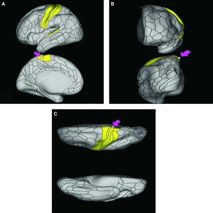
Area 5l demonstrates functional connectivity to areas 1, 2, 3a, and 3b in the sensory strip, area 4 in the motor strip, areas 6mp and 6d in the premotor regions, areas 5m and 5mv in the middle cingulate regions, areas OP1 in the superior insula opercular regions, area A4 in the lower opercula and Heschl's gyrus regions, and areas 7AL and 7PC in the parietal lobe (Figure 16).
FIGURE 16.
Functional connectivity of 5L demonstrated on an inflated left hemisphere. A, Lateral and medial views. B, Rostral and caudal views. C, Dorsal and ventral views. Parcellations with the strongest functional connectivity are shown in yellow. Pink arrows designate the parcellation of interest.
What Are Its White Matter Connections?
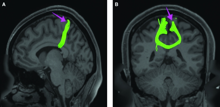
Area 5l is structurally connected to pyramidal tracts and the contralateral hemisphere. Connections to pyramidal tracts descend through the posterior limb of the internal capsule and cerebral peduncle to the brainstem. Contralateral connections course through the body of the corpus callosum to parcellations 5m, 5l, and 5mv. Local short association fibers connect with 5m and 5mv (Figure 17).
FIGURE 17.
Structural connectivity of 5L in the left hemisphere, shown on T1-weighted MR images. A, Sagittal view. Coronal views of B, caudal and C, rostral planes showing projections into the contralateral hemisphere. Red: white matter tracts of 5L demonstrating connections with the pyramidal tracts and the contralateral hemisphere. Pink arrows designate the parcellation of interest.
What Is Known About Its Function?
Area 5l functions in goal-oriented hand movement, specifically movement that is not based on visual cues.10 Recent studies suggest that area 5l, along with area 5m, is also activated in tasks that require complex coordination between the right and left hand.11
Area 5m
Where Is It?
Area 5m (5 medial) is located in the posterior superior portion of the medial face of the paracentral lobule.
What Are Its Borders?
Area 5m borders area 4 anteriorly and area 5l posteriorly. It also has a small anterior border with area 24dd. Its inferior border is area 5mv. It borders areas 3a and 3b on its lateral/superior surface.
What Is Its Functional Connectivity?
Area 5m demonstrates functional connectivity to areas 1, 2, 3a, and 3b in the sensory strip, area 4 in the motor strip, areas 6mp and 6d in the premotor regions, area 5L in the middle cingulate regions, and area A4 in the lower opercula and Heschl's gyrus regions (Figure 18).
FIGURE 18.
Functional connectivity of 5m demonstrated on an inflated left hemisphere. A, Lateral and medial views. B, Rostral and caudal views. C, Dorsal and ventral views. Parcellations with the strongest functional connectivity are shown in yellow. Pink arrows designate the parcellation of interest.
What Are Its White Matter Connections?
Area 5m is structurally connected to the contralateral hemisphere. Contralateral connections course through the body of the corpus callosum to parcellations 4 and 5l. No local short association fibers can be visualized (Figure 19).
FIGURE 19.
Structural connectivity of 5m in the left hemisphere, shown on T1-weighted MR images. A, Sagittal view. B, Coronal views showing projections to the contralateral hemisphere. Green: white matter tracts of 5m demonstrating connections with the contralateral hemisphere. Pink arrows designate the parcellation of interest.
What Is Known About Its Function?
Area 5m integrates somatosensory and visuomotor information. It participates in activities such as reaching or pointing, especially when these movements are based on somatosensory information, as opposed to visual stimuli.10 Recent studies also suggest that area 5m, along with area 5l, is activated when performing tasks that require complex coordination between the right and left hand.11
Area 5mv
Where Is It?
Area 5mv (5 medial-ventral) is located on the posterior inferior paracentral lobule, and makes up the anterior bank of the ascending ramus of the cingulate sulcus.
What Are Its Borders?
Area 5mv borders areas 5m and 5l superiorly and area 23c inferiorly across the cingulate sulcus ascending ramus. Its anterior border is with area 24dd. Area 7am and PCV make up its posterior border.
What Is Its Functional Connectivity?
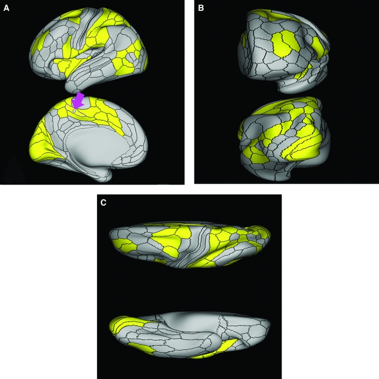
Area 5mv demonstrates functional connectivity to area 2 in the sensory strip, areas SCEF, FEF, 6r, 6a, 6mp, and 6ma in the premotor regions, areas 24dd, 24dv a24prime, p24prime, p32prime, 23c, and 5L in the middle and posterior cingulate regions, areas 9-46d and 46 in the dorsolateral frontal lobe, areas 43, OP4, PFcm, FOP1, FOP3, and FOP4 in the superior insula opercular regions, areas 52, PoI1, PoI2, and MI in the lower opercula and Heschl's gyrus regions, areas AIP, MIP, LIPv, LIPd, IP0, PGp, PFop, PF, PFt, 7AL, and 7PC in the lateral parietal lobe, areas 7am, PCV, and DVT in the medial parietal lobe, areas V1, V2, and V3 in the medial occipital lobe, areas V6 and V6a in the dorsal visual stream areas, and areas PHT, PH, TPOJ2, TPOJ3, FST, and LO3 of the lateral occipital lobe (Figure 20).
FIGURE 20.
Functional connectivity of 5mv demonstrated on an inflated left hemisphere. A, Lateral and medial views. B, Rostral and caudal views. C, Dorsal and ventral views. Parcellations with the strongest functional connectivity are shown in yellow. Pink arrows designate the parcellation of interest.
What Are Its White Matter Connections?
Area 5mv is structurally connected to the contralateral hemisphere and cingulate cortex. Contralateral connections course through the body of the corpus callosum to parcellations 5mv, 4, 5l, and 5m. Fibers to the cingulate cortex project anteriorly from 5mv to end at 24sv, p24r. No local short association fibers can be visualized (Figure 21).
FIGURE 21.
Structural connectivity of 5mv in the left hemisphere, shown on T1-weighted MR images. A, Sagittal view. B, Axial and C, coronal views showing projections to the contralateral hemisphere. Purple: white matter tracts of 5mv demonstrating connections with the contralateral hemisphere and the cingulate cortex. Pink arrows designate the parcellation of interest.
What Is Known About Its Function?
Activation of area 5mv leads to somatosensation and motor response.10 Additionally, some have found a positive correlation between activation of area 5mv and increased accuracy when a subject is imitating an upper body movement following observation of the task.12
SUPPLEMENTARY MOTOR REGION AREAS
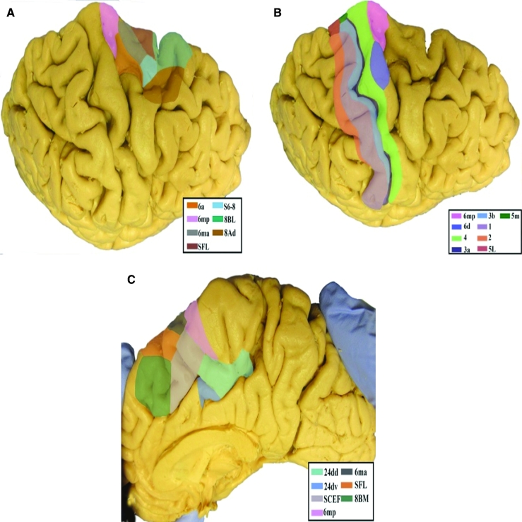
The supplementary motor area (SMA) is located on the medial bank of the posterior superior frontal gyrus and serves as a critical relay center for the imitation and production of ordered movement, eg in speech.13 Four parcellations make up the SMA. We discuss 3 of them here: 6ma, 6mp, and superior frontal language (SFL). For convenience, SCEF, the fourth part of the supplementary motor region, is discussed with the cingulate areas in the following chapter on the medial frontal lobe.
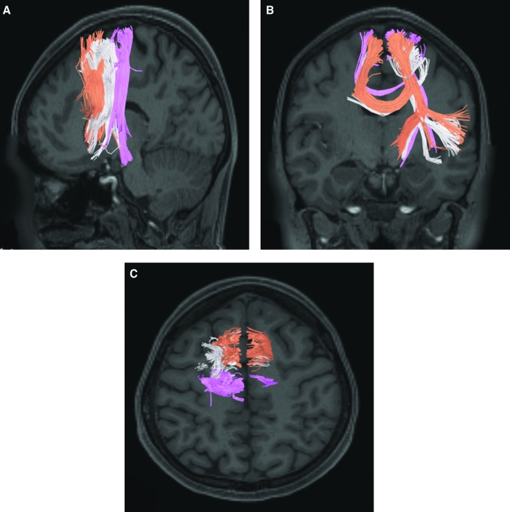
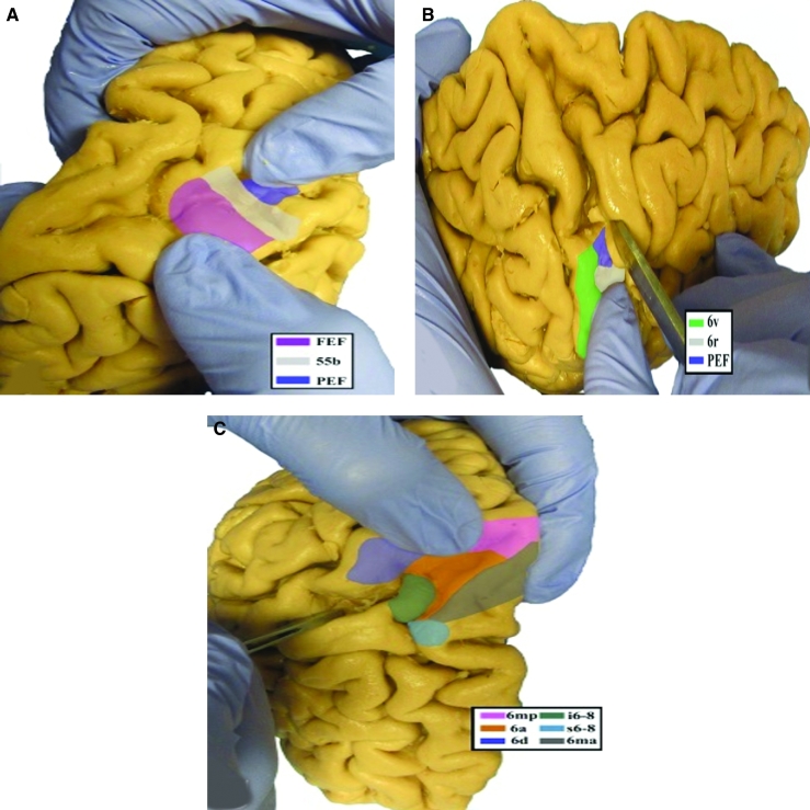
The anatomic location of the parcellations that comprise the supplementary motor region is shown in Figure 22. These parcellations include 6ma, 6mp, and SFL. This region has consistent white matter connections with the pyramidal tracts, frontal aslant tract (FAT), and contralateral hemisphere. The combined tractography of the parcellations is shown in Figure 23.
FIGURE 22.
Anatomical location of the supplementary motor area parcellations shown on the right hemisphere of a cadaver brain. A, Dorsolateral view of the supplementary motor area parcellations. B, Dorsolateral view of the primary motor cortex and corresponding parcellations. C, Medial view of the supplementary motor area.
FIGURE 23.
Combined structural connectivity of the supplementary motor area in the left hemisphere, shown on T1-weighted MR images. A, Sagittal view, B, coronal, and C, axial views showing projections to the contralateral hemisphere. Tracts correspond to areas 6ma (white), 6m (pink), and SFL (orange).
Area 6ma
Where Is It?
Area 6ma (6 medial anterior) makes up the lateral posterior portion of the superior frontal gyrus. It mainly straddles the interhemispheric angle.
What Are Its Borders?
Area 6ma borders area 6mp posteriorly, SCEF and SFL inferiorly/medially, and area 6a laterally. Its anterior neighbor is s6-8.
What Is Its Functional Connectivity?
Area 6ma demonstrates functional connectivity to areas SCEF, PEF, FEF, 6r, 6a, 6v, and 6mp in the premotor regions, areas a24prime, p24 prime, p24prime, a32prime, p32prime, 23c, and 5mv in the middle and posterior cingulate regions, areas IFSa, 9-46d a9-46v, p9-46v, and 46 in the dorsolateral frontal lobe, areas PoI1, PoI2, AVI, MI, 43, PFcm, FOP1, FOP3, FOP4, and FOP5 in the insula opercular regions, areas IP2, IP0, AIP, MIP, LIPd, PGp, PFop, PF, PFt, 7AL, 7PL, and 7PC in the lateral parietal lobe, and areas 7am, 7pm, PCV, and DVT in the medial parietal lobe (Figure 24).
FIGURE 24.
Functional connectivity of 6ma demonstrated on an inflated left hemisphere. A, Lateral and medial views. B, Rostral and caudal views. C, Dorsal and ventral views. Parcellations with the strongest functional connectivity are shown in yellow. Pink arrows designate the parcellation of interest.
What Are Its White Matter Connections?
Area 6ma is structurally connected to the pyramidal tracts, the FAT, and contralateral hemisphere. Connections to pyramidal tracts descend through the posterior limb of the internal capsule and cerebral peduncle to the brainstem. The FAT connects 6ma with the inferior frontal gyrus terminating at parcellations 44, FOP4, and AAIC. Contralateral connections course through the body of the corpus callosum to 6ma. Local short association fibers connect 6mp, SFL, 6a, i6-8, and s6-8 (Figure 25).
FIGURE 25.
Structural connectivity of 6ma in the left hemisphere, shown on T1-weighted MR images. Sagittal views of A, medial and B, lateral planes. C, Coronal view showing projections to the contralateral hemisphere. White: white matter tracts of 6ma demonstrating connections with the pyramidal tracts, the frontal aslant tract (FAT), and the contralateral hemisphere. Pink arrows designate the parcellation of interest.
What Is Known About Its Function?
Area 6ma was subdivided from adjacent parcellations due to differences in myelin thickness and functional activity.9 Specifically, 6ma shows more activation compared to areas 6mp, SFL, or s6-8 when individuals are given a visual instruction cue. Area 6ma also showed greater deactivation compared to area SFL when individuals are told a story. Area 6ma also showed less functional activity compared to area s6-8 when individuals had to match objects based on given verbal categories.9
Area 6mp
Where Is It?
Area 6mp (6 medial posterior) makes up the area where the SFG joins with the precentral gyrus. It makes up the medial bank of the SFG at this junction, as well as spilling onto the superior surface of the posterior most SFG and the anterosuperior portion of the precentral gyrus. It also makes up the most posterior superior portion of the superior bank of the superior frontal sulcus.
What Are Its Borders?
Area 6mp borders area 4 posteriorly, area 6d laterally, area 6a anterolaterally, areas 6ma and SCEF anteriorly, and area 24dd inferiorly on the medial surface.
What Is Its Functional Connectivity?
Area 6mp demonstrates functional connectivity to areas 1, 2, 3a, and 3b in the sensory strip, area 4 in the motor strip, areas SCEF, 6a, 6ma, and 6d in the premotor regions, areas 24dd, 24dv, p32prime, 5mv, and 5L in the middle cingulate regions, areas 43, OP1, OP4, PFcm, FOP1, and FOP2 in the superior insula opercular regions, areas A4, RI, and PBelt in the lower opercula and Heschl's gyrus regions, areas PFop, PFt, IPS1, 7AL, and 7PC in the parietal lobe areas, and area FST in the lateral occipital lobe (Figure 26).
FIGURE 26.
Functional connectivity of 6mp demonstrated on an inflated left hemisphere. A, Lateral and medial views. B, Rostral and caudal views. C, Dorsal and ventral views. Parcellations with the strongest functional connectivity are shown in yellow. Pink arrows designate the parcellation of interest.
What Are Its White Matter Connections?
Area 6mp is structurally connected to the pyramidal tracts and contralateral hemisphere. Connections to pyramidal tracts descend through the posterior limb of the internal capsule and cerebral peduncle to the brainstem. Contralateral connections course through the body of the corpus callosum to 6ma, 6mp, and FEF. Local short association fibers connect with 24dd, 6a, and 6d (Figure 27).
FIGURE 27.
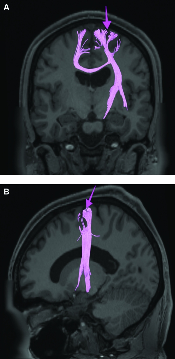
Structural connectivity of 6mp in the left hemisphere, shown on T1-weighted MR images. Coronal view A, showing projections into the contralateral hemisphere, and sagittal view B. Pink: white matter tracts of 6mp demonstrating connections with the pyramidal tracts and the contralateral hemisphere. Pink arrows designate the parcellation of interest.
What Is Known About Its Function?
Area 6mp was subdivided from adjacent parcellations due to differences in myelin thickness and functional activity.9 Specifically, area 6mp shows less activation compared to 6ma when an individual was given a visual instruction cue and when moving their feet. Compared to 6d, 6mp shows greater deactivation when listening to a story or solving a math problem. Lastly, compared to area 6a, area 6mp shows less activation in social interaction settings.9
Area SFL
Where Is It?
Area SFL is located on the posterior medial SFG straddling over the interhemispheric cleft.
What Are Its Borders?
Area SFL borders SCEF inferiorly. Its anterior inferior neighbor is area 8BM and its anterior superior neighbor is area 8BL. Areas 6ma and s6-8 are its lateral neighbors.
What Is Its Functional Connectivity?
Area SFL demonstrates functional connectivity to areas 8BL, 8AV, 9a, 9p, and 9m in dorsolateral frontal lobe, areas 8BM, d32, 44, 45, 47L, and 47s in the inferior frontal lobe, area 55b in the premotor areas, areas STSda, STSdp, STSva, STSvp, TE1a, and TGd in the temporal lobe, area PGi in the lateral parietal lobe, and areas 31pv and 31pv in the medial parietal lobe (Figure 28).
FIGURE 28.
Functional connectivity of SFL demonstrated on an inflated left hemisphere. A, Lateral and medial views. B, Rostral and caudal views. C, Dorsal and ventral views. Parcellations with the strongest functional connectivity are shown in yellow. Pink arrows designate the parcellation of interest.
What Are Its White Matter Connections?
Area SFL is structurally connected to pyramidal tracts, the FAT, and contralateral hemisphere. Connections to pyramidal tracts descend through the posterior limb of the internal capsule and cerebral peduncle to the brainstem. The FAT connects SFL with the inferior frontal gyrus, terminating at parcellations 44, IFSp, and MI. Contralateral connections course through the body of the corpus callosum to SCEF and 8BL. Local short association fibers connect with SCEF, 8BL, SFL, and 6ma (Figure 29).
FIGURE 29.
Structural connectivity of SFL in the left hemisphere, shown on T1-weighted MR images. Coronal views of A, caudal and B, rostral planes showing projections to the contralateral hemisphere. C, Sagittal view of SFL. Orange: white matter tracts of SFL demonstrating connections with the pyramidal tracts, the frontal aslant tract, and contralateral hemisphere. Pink arrows designate the parcellation of interest.
What Is Known About Its Function?
Area SFL was subdivided from adjacent parcellations due to differences in myelin thickness and functional activity.9 Area SFL is known to be hemispherically asymmetric. Specifically, the left hemisphere shows more activity when listening to stories and when a participant is matching objects based on a verbal cue. Compared to area 8BL, area SFL shows more activation when listening to a story, matching objects based on verbal cues and in social interaction settings. Compared to area s6-8, area SFL shows more activation in the left hemisphere when individuals listen to a story. In the right hemisphere, area SFL is activated in social interaction settings and is deactivated during object feature comparison tasks.9
PREMOTOR AREAS
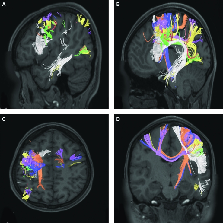
The premotor areas are largely subdivisions of Brodmann's area 6. They are predominantly located on the anterior precentral gyrus and in portions of the precentral sulcus. The frontal and PEFs are included here. The anatomic location of the parcellations comprised by the premotor area is shown in Figure 30. These parcellations include 6a, 6d, FEF, 55b, PEF, 6v, and 6r. This region has consistent white matter connections with the superior longitudinal fasciculus and contralateral hemisphere. Parcellations 6a and 6d also have consistent connections with the pyramidal tracts. The combined tractography of the parcellations is shown in Figure 31.
FIGURE 30.
Anatomical location of premotor area parcellations shown on the right hemisphere of a cadaver brain. A, Oblique view with dilated precentral sulcus to show extension of the parcellations into the sulcus. B, Dorsolateral view with dilated precentral sulcus. C, Superior view with dilated precentral sulcus.
FIGURE 31.
Combined structural connectivity of the premotor area parcellations in the left hemisphere, shown on T1-weighted MR images. Sagittal views of A, lateral and B, medial planes. C, Axial and D, coronal views showing projections to the contralateral hemisphere. Tracts correspond to areas 6a, 6d, FEF, 55b, PEF, 6v, and 6r.
Area 6a
Where Is It?
Area 6a (6 anterior) makes up the posterior superior most bank of the superior frontal sulcus and the adjacent portions of the superior frontal gyrus, principally forming this bank just as the sulcus forms the right angle with the precentral sulcus.
What Are Its Borders?
Area 6a borders areas 6d and 6mp posteriorly, and area 6ma medially. Its anterior border is made by s6-8, area 8AD, and i6-8. FEF is its inferior neighbor.
What Is Its Functional Connectivity?
Area 6a demonstrates functional connectivity to area 2 in the sensory strip, areas SCEF, PEF, FEF, 6ma, 6mp, 6d, and 6v in the premotor regions, areas a24prime, p32prime, 5mv, and 23c in the middle cingulate regions, areas IFSa, IFJa, i6-8, 46, p9-46v, and 9-46d in the lateral frontal lobe, areas OP4, PFcm, FOP4, and FOP2 in the superior insula opercular regions, areas PoI1 and PoI2 in the lower opercula and Heschl's gyrus regions, areas TE2p, PHA3, and PHT in the temporal lobe, areas AIP, MIP, VIP, LIPd, LIPv, PFop, PF, PFt, PGp IP2, IP1, IP0, IPS1, 7AL,7PL, and 7PC in the lateral parietal lobe, areas 7pm, 7am, DVT, and PCV in the medial parietal lobe, area V2 in the medial occipital lobe, and areas PH, TPOJ2, TPOJ3, and FST of the lateral occipital lobe (Figure 32).
FIGURE 32.
Functional connectivity of 6a demonstrated on an inflated left hemisphere. A, Lateral and medial views. B, Rostral and caudal views. C, Dorsal and ventral views. Parcellations with the strongest functional connectivity are shown in yellow. Pink arrows designate the parcellation of interest.
What Are Its White Matter Connections?
Area 6a is structurally connected to the pyramidal tracts and the parietal lobe. Connections to pyramidal tracts descend through the posterior limb of the internal capsule and cerebral peduncle to the brainstem. Parietal projections are portions of the SLF and connect with 3a, 3b, 7PC, and 7AL. Local short association fibers connect with FEF, i6-8, 55b, 8Av, 46, and 6r (Figure 33).
FIGURE 33.
Structural connectivity of 6a in the left hemisphere, shown on T1-weighted MR images. Sagittal views of A, lateral and C, medial planes. B, Coronal view showing projections to the contralateral hemisphere. Orange: white matter tracts of 6a demonstrating connections with the pyramidal tracts and the parietal lobe. Pink arrows designate the parcellation of interest.
What Is Known About Its Function?
Areas 6a and 6d are newly described subdivisions of the premotor cortex. While the precise function of these areas is unknown, the functions of the premotor cortex are well characterized in the literature. First, the premotor cortex is functionally divided into ventral and dorsal aspects. The dorsal premotor cortex is involved in associating informational cues with a particular body movement. These cues could be learned and arbitrary in nature or they can be based on other forms of somatosensation, such as visual or auditory sensation.2 The ventral premotor area is involved in hand movement manipulation of objects, eg when grasping or lifting. The ventral premotor area is also involved in more complex cortical functions such as when individuals learn actions or movements while observing others performing a task.2 Overall, the premotor cortex has significant function in the preparation of voluntary movements.14
Regarding area 6a more specifically, this region was distinguished from adjacent areas of the cortex based on differences in myelin thickness and functional activity.9 Compared to area s6-8, area 6a shows greater activation when solving math problems, in social interaction settings, and when performing object feature comparison tasks. Compared to area i6-8, area 6a shows greater activation in social interaction settings and relative deactivation in emotion identification and object feature comparison. Compared to area FEF, area 6a shows less activation in both gambling and object feature comparison.9
Area 6d
Where Is It?
Area 6d (6 dorsal) is located on the anterosuperior portion of the precentral gyrus, just inferior to its junction with the SFG. It makes up the posterior bank of the adjacent precentral sulcus.
What Are Its Borders?
Area 6d borders area 6mp superiorly and FEF inferiorly. Area 4 is its posterior border and area 6a forms its anterior border across the precentral sulcus.
What Is Its Functional Connectivity?
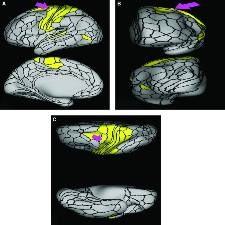
Area 6d demonstrates functional connectivity to areas 1, 2, 3a, and 3b in the sensory strip, area 4 in the motor strip, areas 6a, 6mp, and 6v in the premotor regions, areas 5L and 24dd in the middle cingulate regions, areas FOP2, OP4, OP1, A4, and PBelt in the insula opercula regions, areas 7PC, 7AL, and PFt in the parietal lobe, and area FST in the lateral occipital lobe (Figure 34).
FIGURE 34.
Functional connectivity of 6d demonstrated on an inflated left hemisphere. A, Lateral and medial views. B, Rostral and caudal views. C, Dorsal and ventral views. Parcellations with the strongest functional connectivity are shown in yellow. Pink arrows designate the parcellation of interest.
What Are Its White Matter Connections?
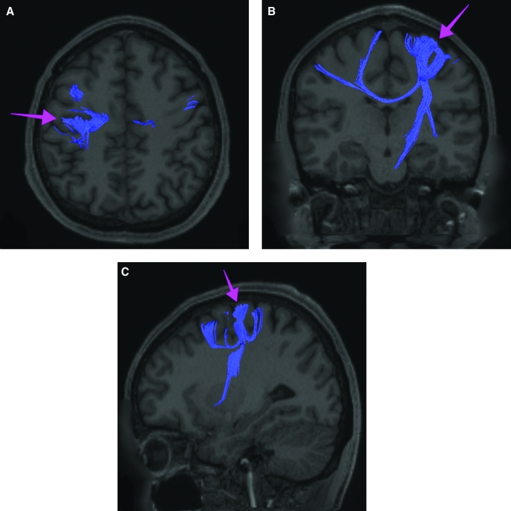
Area 6d is structurally connected to the pyramidal tracts and contralateral hemisphere. Connections to pyramidal tracts descend through the posterior limb of the internal capsule and cerebral peduncle to the brainstem. Contralateral connections course through the body of the corpus callosum to end at 6mp, FEF, and 55b. Local short association fibers connect with 4, 3a, 3b, 6a, FEF, i6-8, 8Av, 3a, 3b, 6ma, and 6d (Figure 35).
FIGURE 35.
Structural connectivity of 6d in the left hemisphere, shown on T1-weighted MR images. A, Axial and B, coronal views showing projections to the contralateral hemisphere. C, Sagittal view. Blue: white matter tracts of 6d demonstrating connections with the pyramidal tracts and the contralateral hemisphere. Pink arrows designate the parcellation of interest.
What Is Known About Its Function?
Area 6d was distinguished from adjacent areas of the cortex based on differences in myelin thickness and functional connectivity.9 Area 6d shows less activation compared to area FEF during gambling tasks, in social interaction settings, and during object feature comparison. Compared to area 6a, area 6d shows less activation in solving math problems and in social interaction settings.9
Area FEF
Where Is It?
Area FEF is located on the anterior half of the precental gyrus, approximately half way down its length along the convexity, just inferior to the junction point of the precental and superior frontal sulci. It also forms the adjacent floor of the precentral sulci and straddles slightly onto the posterior edge of the middle frontal gyrus.
What Are Its Borders?
Area FEF borders areas 6a and 6d superiorly and area 55b inferiorly. Area 4 is its posterior border and area i6-8 forms its anterior border on the middle frontal gyrus.
What Is Its Functional Connectivity?
Area FEF demonstrates functional connectivity to area 2 in the sensory strip, areas SCEF, PEF, 6r, and 6v in the premotor regions, areas a24prime, p32prime, 5mv, and 23c in the middle cingulate regions, areas IFSa, IFJa, 46, and 9-46d in the lateral frontal lobe, areas 43, OP4, PFcm, FOP1, FOP3, FOP4, and FOP5 in the superior insula opercular regions, areas STV, LBelt, PBelt, A4, MI, 52, RI, PoI1, and PoI2 in the lower opercula and Heschl's gyrus regions, areas TE2p and PHT in the temporal lobe, areas AIP, MIP, VIP, LIPd, LIPv, PFop, PF, PFt, PGp, IP0, IPS1, 7AL,7PL, and 7PC in the lateral parietal lobe, areas 7am, DVT, and PCV in the medial parietal lobe, areas V1, V2, V3, and V4 in the medial occipital lobe, areas V3a, V3b, V6, V6a, and V7 of the dorsal visual stream, areas V8 PIT, FFC, VVC, VMV1, VMV2, and VMV3 of the ventral visual stream, and areas V3cd, LO1, LO2, LO3, PH, TPOJ1, TPOJ2, TPOJ3, V4t, MST, and FST of the lateral occipital lobe (Figure 36).
FIGURE 36.
Functional connectivity of FEF demonstrated on an inflated left hemisphere. A, Lateral and medial views. B, Rostral and caudal views. C, Dorsal and ventral views. Parcellations with the strongest functional connectivity are shown in yellow. Pink arrows designate the parcellation of interest.
What Are Its White Matter Connections?
Area FEF is structurally connected to the contralateral hemisphere and superior longitudinal fasciculus. Contralateral connections course through the body of the corpus callosum to i6-8 and SFL. Connections with the superior longitudinal fasciculus connect FEF to the intraparietal sulcus and the inferior parietal lobe terminating at IP1, IP2, and PGs. Local short association fibers connect with 6d, 55b, i6-8, 8Av, 6a, and PEF (Figure 37).
FIGURE 37.
Structural connectivity of FEF in the left hemisphere, shown on T1-weighted MR images. A, Coronal view showing projections to the contralateral hemisphere. Sagittal views of B, lateral and C, medial planes. Purple: white matter tracts of FEF demonstrating connections with the contralateral hemisphere and superior longitudinal fasciculus. Pink arrows designate the parcellation of interest.
What Is Known About Its Function?
Area FEF is known to be involved in rapid eye movements between fixed points, also known as intentional saccadic movements. Area FEF has also been implicated in smooth eye movements that allow the eyes to follow a moving target, also known as smooth pursuit eye movements. Together, these movements help the FEF to create a salience map for visual attention.15-19
Area 55b
Where Is It?
Area 55b is located on the anterior half of the precental gyrus, approximately half way down its length along the convexity, just inferior to FEF. It also forms the adjacent floor of the precentral sulci and straddles slightly onto the posterior edge of the middle frontal gyrus.
What Are Its Borders?
Area 55b borders area FEF superiorly and PEF and area 6v inferiorly. Area 4 is its posterior border and areas 8AV and 8C form its anterior border across the precentral sulcus.
What Is Its Functional Connectivity?
Area 55b demonstrates functional connectivity to area 4 in the motor strip, areas SCEF and SFL in the premotor areas, areas IFSp, IFJa, 8AV, 44, 45, and 47L in the lateral frontal lobe, areas STSda and STSdp in the temporal lobe, areas PSL and STV in the posterior opercular cortices, and area TPOJ1 in the lateral occipital lobe (Figure 38).
FIGURE 38.
Functional connectivity of 55b demonstrated on an inflated left hemisphere. A, Lateral and medial views. B, Rostral and caudal views. C, Dorsal and ventral views. Parcellations with the strongest functional connectivity are shown in yellow. Pink arrows designate the parcellation of interest.
What Are Its White Matter Connections?
Area 55b is structurally connected to the contralateral hemisphere and the superior longitudinal fasciculus. Contralateral connections course through the body of the corpus callosum to 6ma, 6a, and 6mp. Connections with the superior longitudinal fasciculus connect 55b to parcellations PHT and PFm, and this tract terminates eventually in the temporal lobe at TGd. Local short association fibers connect with 8Av, 8C, IFJp, 3a, 3b, and PEF (Figure 39).
FIGURE 39.
Structural connectivity of 55b in the left hemisphere, shown on T1-weighted MR images. A, Axial view showing projections to the contralateral hemisphere. Sagittal views of B, medial and C, lateral planes. White: white matter tracts of 55b demonstrating connections with the contralateral hemisphere and the superior longitudinal fasciculus. Pink arrows designate the parcellation of interest.
What Is Known About Its Function?
Area 55b is a relatively uncharacterized region. In 1956, one of the only studies to characterize this region concluded that the area played a role in language processing.20
Area PEF
Where Is It?
Area PEF is a small area located in the floor of the precentral sulcus at the junction of the precentral and inferior frontal sulci. It spills slightly onto the adjacent precentral gyrus and unlike FEF and area 55b, it is mostly vertically oriented.
What Are Its Borders?
Area PEF borders area 55b superiorly and area 6r inferiorly. Area 6v is its posterior border and areas 8C and IFJp form its anterior border on the MFG and in the inferior frontal sulcus, respectively.
What Is Its Functional Connectivity?
Area PEF demonstrates functional connectivity to areas SCEF, FEF, 6ma, 6r, 6a, and 6v in the premotor regions, areas a24 prime, p32prime, and 23c in the middle cingulate regions, areas IFSa, IFJp, and 9-46d in the lateral frontal lobe areas 43, PFcm, FOP4, and FOP5 in the superior insula opercular regions, areas MI and PoI2 in the lower opercula and Heschl's gyrus regions, areas TE2p and PHT in the temporal lobe, areas AIP, MIP, LIPd, LIPv, PFop, PFt, PGp, IP0, 7PL, and 7PC in the lateral parietal lobe, area 7am in the medial parietal lobe, and areas PH and FST of the lateral occipital lobe (Figure 40).
FIGURE 40.
Functional connectivity of PEF demonstrated on an inflated left hemisphere. A, Lateral and medial views. B, Rostral and caudal views. C, Dorsal and ventral views. Parcellations with the strongest functional connectivity are shown in yellow. Pink arrows designate the parcellation of interest.
What Are Its White Matter Connections?
Area PEF is structurally connected to the superior longitudinal fasciculus. PEF does have connections to the contralateral hemisphere but this is inconsistent across individuals. Connections with the superior longitudinal fasciculus connect 55b to inferior parietal lobe parcellations PHT, TPOJ2, FST, and PFm. Local short association fibers connect with 6r, 8C, and IFJp (Figure 41).
FIGURE 41.
Structural connectivity of PEF in the left hemisphere, shown on T1-weighted MR images. Sagittal views of A, lateral and B, medial planes. Blue: white matter tracts of PEF demonstrating connections with the superior longitudinal fasciculus. Pink arrows designate the parcellation of interest.
What Is Known About Its Function?
Area PEF is known to be involved in reflexive eye movements, also called reflexive saccades.17,18,21
Area 6v
Where Is It?
Area 6v (6 ventral) makes up the anteroinferior one-third of the precentral gyrus. It only minimally forms the posterior bank of the precentral sulcus, which is primarily formed by area 6r.
What Are Its Borders?
Area 6v borders area 55b superiorly and area 43 inferiorly. Area 4 is its posterior border and areas 6r and PEF form its anterior border.
What Is Its Functional Connectivity?
Area 6v is connected to areas 1, 2, 3a, and 3b in the sensory strip, area 4 in the motor strip areas SCEF, FEF PEF, 6ma, 6mp, 6r, 6d, and 6v in the premotor regions, areas 24dd and p32prime in the middle cingulate regions, areas 43, OP4, OP2-3, OP1, PFcm, FOP1 FOP2, FOP3, and FOP4 in the superior insula opercular regions, areas A4, PBelt, RI, and PoI2 in the lower opercula and Heschl's gyrus regions, areas AIP, MIP, VIP, LIPv, PFop, PFt, 7AL, and 7PC, in the lateral parietal lobe, areas V3a, V3b, V6, V6a, and V7 of the dorsal visual stream, areas FFC of the ventral visual stream, and areas PH, TPOJ2, MST, and FST of the lateral occipital lobe (Figure 42).
FIGURE 42.
Functional connectivity of 6v demonstrated on an inflated left hemisphere. A, Lateral and medial views. B, Rostral and caudal views. C, Dorsal and ventral views. Parcellations with the strongest functional connectivity are shown in yellow. Pink arrows designate the parcellation of interest.
What Are Its White Matter Connections?
Area 6v is structurally connected to the contralateral hemisphere and the superior longitudinal fasciculus. Contralateral connections course through the body of the corpus callosum to FEF. Connections with the superior longitudinal fasciculus connect 6v to inferior parietal lobe parcellations PHT, FST, PH, and PF. Local short association fibers connect with 4, 6r, PEF, 43, 3a, and 3b (Figure 43).
FIGURE 43.
Structural connectivity of 6v in the left hemisphere, shown on T1-weighted MR images. A, Axial view showing projections to the contralateral hemisphere. Sagittal views of B, medial and C, lateral planes. Green: white matter tracts of V2 demonstrating connections with the contralateral hemisphere and the superior longitudinal fasciculus. Pink arrows designate the parcellation of interest.
What Is Known About Its Function?
Area 6v comprises what is classically known as the premotor cortex. It has many different functions. The ventral premotor area is known to be active in the control of hand movements that occur while manipulating objects (eg grasping and lifting objects), and the dorsal premotor area is involved in performing specific motor tasks based on visual cues.2
Area 6r
Where Is It?
Area 6r (6 rostral) is the inferior portion of the precentral sulcus, including its floor and both banks. The latter point implies that some of area 6r lies on both the anterior inferior portion of the precentral gyrus and the posterior portion of the pars opercularis of the inferior frontal gyrus. The inferior extent of the precentral sulcus forms a shovel shaped cup near the opercular edge and this cup is area 6r.
What Are Its Borders?
Area 6r borders area 44 anteriorly and area 43 posteriorly. Area 6v forms a posterosuperior border with it. PEF is its main superior neighbor, while IFJp and IFJa form an anterior superior border. FOP1 and FOP4 form its inferior border on its undersurface opercular surface.
What Is Its Functional Connectivity?
Area 6r demonstrates functional connectivity to areas SCEF, FEF PEF, 6ma, 6a, and 6v in the premotor regions, areas 23c, 5mv, a24prime, p24prime, and p32prime in the middle cingulate regions, areas 46, p9-46v, 9-46d, IFSa, IFJa, IFJp, and p47r in the lateral frontal lobe, areas 43, OP4, PFcm, FOP2 FOP3, FOP4, and FOP5 in the superior insula opercular regions, areas AVI, MI, PoI1, and PoI2 in the lower opercula and Heschl's gyrus regions, areas TE2p and PHT in the temporal lobe, areas AIP, MIP, LIPd, LIPv, PFop, PF, PFt, IP0, 7PL, 7AL, and 7PC, in the lateral parietal lobe, and areas PH and FST of the lateral occipital lobe (Figure 44).
FIGURE 44.
Functional connectivity of 6r demonstrated on an inflated left hemisphere. A, Lateral and medial views. B, Rostral and caudal views. C, Dorsal and ventral views. Parcellations with the strongest functional connectivity are shown in yellow. Pink arrows designate the parcellation of interest.
What Are Its White Matter Connections?
Area 6r is structurally connected to the superior longitudinal fasciculus and FAT. FAT connects 6r with the superior frontal gyrus at parcellation SFL. Connections with the superior longitudinal fasciculus connect 6r to posterior temporal parcellations TE1a and TE2a. Local short association fibers connect with 6v, 44, IFJa, IFJp, 8C, and IFSa (Figure 45).
FIGURE 45.
Structural connectivity of 6r in the left hemisphere, shown on T1-weighted MR images. A, Coronal view. Sagittal views of B, medial and C, lateral planes. White: white matter tracts of 6r demonstrating connections with the superior longitudinal fasciculus and the frontal aslant tract. Pink arrows designate the parcellation of interest.
What Is Known About Its Function?
Area 6r has not been extensively studied. The literature indicates that this area is functionally related to Broca's area in humans, which is a well-known cortical area essential to language processing.22,23
DISCUSSION
Brodmann described the sensorimotor region as essentially 6 areas.1 For some time, we have found this scheme to be unsatisfactory as a framework for describing how motor function occurs, as it reduces the system to essentially 6 parallel strips of cortex comprising the motor and sensory strips, the SMA, the premotor areas, the frontal eye fields, and the cingulate motor areas. The Human Connectome Project (HCP) data suggest that the motor cortex is far more complex than this, as it is unlikely that M1 can work without something driving it. Furthermore, these data suggest that the premotor brain has other functions. Most importantly, we think these new data give us a framework to begin to discuss motor mapping in a way beyond merely avoiding the primary motor cortex.
The SMA
The commonly published subdivisions of the SMA, which divide the SMA into the pre-SMA and SMA proper,24 do not provide much help toward understanding the organization or exact role of different parts of this region. The HCP division into 3 parts, 6ma, 6mp, and SFL, provides a better anatomic definition of the area and makes functional definition of its subparts possible.
Our group has specifically found that white matter fibers leave the SMA in 3 main bundles, which are spatially segregated.25 The medial paths connect to the homologous SMA region in the contralateral hemisphere. The middle paths join the descending fiber system which connect to the basal ganglia and the corticospinal tract. The lateral paths form a large part of the FAT which joins these areas to the inferior frontal gyrus and insula.
It is rare in the cortex that an area can be subdivided relatively neatly; however, all 3 SMA regions seem to have relatively describable connectivity patterns based on their functional and structural maps:
SFL is part of the language network and connects to language areas via the FAT.
6mp is part of the sensorimotor network and connects to this network, the basal ganglia, and the corticospinal tract.
6ma contributes to the FAT and the corticospinal tract. It demonstrates functional connectivity to several parts of the fronto-parietal network and the insula.
The SMA syndrome, which consists of mutism and hemiplegia,25 has a firm anatomic explanation in these findings.
Area 5
Area 5 is a modest area largely tucked into the superoposterior sensory regions. The literature suggests that this area plays a role in coordinating bilateral motor tasks.11 The connectivity patterns of area 5 subregions are entirely consistent with this idea, as the connections of 5m and 5l are mainly local with connections to their homolog in the contralateral hemisphere.
It is also notable that area 5mv, like area 23c on the other side of the cingulate sulcus, is one of the most highly functionally connected areas of the cortex, despite having only modest white matter pathways. The significance of this is unclear to us.
The Sensorimotor Network
The anatomy of the corticospinal tract and the ascending thalamocortical sensory fibers is well known, and our tractography did not uncover any new findings with respect to these fibers.26-28 What is worth noting is the degree of bilateral connectivity between primary motor areas via the corpus callosum, which we did not appreciate previously. It is also notable that these cortices are densely interconnected via U-fibers, and also highly connected to their neighboring premotor and parietal cortices. In addition, parts of area 3 subregions sit in the depths of the central sulcus. We note these observations to highlight the idea that transsulcal trajectories in these areas are not necessarily better simply because they enter less brain, as they could have the unintended consequence of disconnecting 2 areas which need to talk to each other for normal functioning, thereby causing more damage than a longer trajectory. This topic needs to be studied more intensely relative to the exact anatomy.
The most stunning finding we observed while studying the connectivity of these areas was the nature of the functional connectivity of the primary sensorimotor cortices. The primary sensory and motor areas all demonstrated evidence of functional connectivity to regions corresponding to the early auditory and visual areas with no real evidence of direct structural connections between these areas and the sensorimotor regions. The significance of this is unclear, but this was a notable finding in our data and given that it was seen in all 4 areas, it seems unlikely, at least to us, that this represents artifact or noise.
The Premotor Areas
Close examination of Brodmann's original maps demonstrates that a substantial portion of area 6 takes up the anterior half of the precentral gyrus, in addition to overlapping the adjacent precentral sulcus and neighboring gyri.1 Without parcellating and reducing this area to something more manageable, studying and explaining the connectional anatomy of area 6 would be difficult. Several observations follow from our analysis.
Areas 6a, 6d, 6r, and 6v
These areas are not contiguous as a significant area of cortex separates areas 6d from 6v along the superior-to-inferior axis of the precentral gyrus. Areas 6d and 6v are both part of the sensorimotor network based on their underlying functional connectivity, which is not surprising given that they are both located in the precentral gyrus. Meanwhile, areas 6a and 6r are not directly functionally in sync with the primary sensorimotor network. What is interesting is that while 6d largely contributes to the corticospinal tract, 6r and 6v primarily have long range connections via the SLF/arcuate system. Area 6a is a hybrid between these 2 extremes connecting to the parietal regions via the medial portions of the SLF and contributing to the corticospinal tract.
Dorsal and Ventral Premotor Areas: A 2-Stream System of Motor Planning
The dorsal and ventral premotor areas have been described previously.2 The dorsal premotor area is thought to be involved in the use of visual guidance for hand movement, and the ventral premotor area is thought to be involved in manipulating or grasping objects.2
Areas 6a and 6d seem the most likely candidates for the dorsal premotor areas, given their location and their substantial contributions to the corticospinal tract. In contrast, areas 6r and 6v seem more like ventral premotor areas. What may be initially confusing about this distinction is the fact that areas 6r and 6v are largely connected to semantic network areas (6r) and higher visual processing areas (6v). At first glance, this might make someone consider that these areas better fit the role of visual control of hand movements than an area like 6a which mostly connects to the anterior parietal lobe. Some explanation is needed. The posterior parietal cortex contains a variety of representations of 3-dimensional space which represents the binding of somatosensory, proprioceptive, visual, and auditory inputs.29 Injury to this area in some people causes the syndrome of optic ataxia, or difficulty guiding the hand using visual cues.30 Given the connections of area 6a in the medial portions of the SLF to parietal regions 7PC and 7AL, we hypothesize that area 6a is more likely to represent the dorsal premotor area with these connections serving as a site of injury leading to the syndrome of optic ataxia.
Conversely, apraxia represents the difficulty of using or manipulating tools or performing sequenced tasks. While it comes in numerous forms, the canonical form, ideomotor apraxia, has been localized to the same areas which govern language, usually co-occurring with forms of aphasia.31,32 Studies have localized lesions causing apraxia to the posterior temporal lobe, the inferior parietal lobule, and the posterior inferior frontal gyrus,31,33 which strongly suggests that the underlying apraxia network is contained within the SLF/arcuate areas of the parasylvian cortices. Given that 6r and 6d are part of this network, it seems plausible that they represent the ventral premotor area. The information carried could involve semantic information for tools and visual information about the mechanical properties of different structures and tools. In addition, it could provide information about the shapes and characteristics of objects provided by higher visual processing areas like FST. The fact that these 2 areas link between regions 44, 45, and the primary motor cortex deserves closer attention.
Finally, it should be noted that these systems, dorsal and ventral premotor, are largely separate from each other. While areas 6a and 6r share some local connections and some degree of functional connectivity, there is no evidence that 6d and 6v talk to each other in a meaningful way. In addition, the inputs these areas receive from the sensory system are quite different, with the dorsal system largely receiving input from the superior parietal lobule/medial SLF system, and the ventral system receiving input from the posterior temporal/inferior parietal lobule/arcuate system. This suggests the possibility of 2 distinct streams of sensorimotor transfer in the premotor cortex. As a result, at least one of the functions of the FAT seems to be to provide access for the ventral system to the SMA areas (presumably, the dorsal system can talk to the SMA areas directly as they are adjacent to one another).
FEF, PEF, and Area 55b
The anatomy of these 3 areas, which separate the dorsal and ventral premotor systems, was found to be among the most variable in the human neocortex by the HCP authors.9 Notably, area 55b can often be shifted relative to the hand motor portions of area 4, or alternately areas FEF and PEF can encroach on the location of 55b by invading this area from above and below to join in the middle and split 55b in half.9 The connectional anatomy of these small areas is similarly complex as they differ from each other significantly.
Area FEF seems to be part of the dorsal attention network.34 While it has functional connectivity to a wide number of brain regions, its principle long-range connections are to the inferior bank of the intraparietal sulcus and inferior parietal lobule. In some models, FEF plays a role as a salience map,19 which keep track of the previous areas of saccade when scanning a visual scene so that the most salient feature is not repeated as the target of saccades.
Area PEF, true to its name as the premotor eye field, seems to have a similar pattern of connection as the ventral premotor areas. Its most notable long range connections are to the lateral occipital regions and late stage visual processing areas such as FST and PHT, and multimodal areas such as TPOJ2 and PFm.
Area 55b is likely a language area with functional and structural connections to semantic areas such as PHT and to the superior temporal sulcus areas also involved in language processing.9 The fact that a new part of the language network was found wedged between 2 eye fields which occasionally interrupt, it demonstrates the power of the connectomic method for classifying anatomy.
Disclosures
Synaptive Medical assisted in the funding of all 18 chapters of this supplement. No other funding sources were utilized in the production or submission of this work.
Acknowledgments
Data were provided [in part] by the Human Connectome Project, WU-Minn Consortium (Principal Investigators: David Van Essen and Kamil Ugurbil; 1U54MH091657) funded by the 16 NIH Institutes and Centers that support the NIH Blueprint for Neuroscience Research; and by the McDonnell Center for Systems Neuroscience at Washington University. We would also like to thank Brad Fernald, Haley Harris, and Alicia McNeely of Synaptive Medical for their assistance in constructing the network figures for Chapter 18 and for coordinating the completion and submission of this supplement
REFERENCES
- 1. Zilles K, Amunts K. Centenary of Brodmann's map – conception and fate. Nat Rev Neurosci. 2010;11(2):139-145. [DOI] [PubMed] [Google Scholar]
- 2. Chouinard PA, Paus T. The primary motor and premotor areas of the human cerebral cortex. Neuroscientist. 2006;12(2):143-152. [DOI] [PubMed] [Google Scholar]
- 3. Coco M, Perciavalle V, Cavallari P, Perciavalle V. Effects of an exhaustive exercise on motor skill learning and on the excitability of primary motor cortex and supplementary motor area. Medicine (Baltimore). 2016;95(11):e2978. [DOI] [PMC free article] [PubMed] [Google Scholar]
- 4. Ploner M, Schmitz F, Freund HJ, Schnitzler A. Differential organization of touch and pain in human primary somatosensory cortex. J Neurophysiol. 2000;83(3):1770-1776. [DOI] [PubMed] [Google Scholar]
- 5. Hyvarinen J, Poranen A. Receptive field integration and submodality convergence in the hand area of the post-central gyrus of the alert monkey. J Physiol. 1978;283(1):539-556. [DOI] [PMC free article] [PubMed] [Google Scholar]
- 6. Vierck CJ, Whitsel BL, Favorov OV, Brown AW, Tommerdahl M. Role of primary somatosensory cortex in the coding of pain. Pain. 2013;154(3):334-344. [DOI] [PMC free article] [PubMed] [Google Scholar]
- 7. Huffman KJ, Krubitzer L. Area 3a: topographic organization and cortical connections in marmoset monkeys. Cereb Cortex. 2001;11(9):849-867. [DOI] [PubMed] [Google Scholar]
- 8. Martuzzi R, van der Zwaag W, Dieguez S, Serino A, Gruetter R, Blanke O. Distinct contributions of Brodmann areas 1 and 2 to body ownership. Soc Cogn Affect Neurosci. 2015;10(11):1449-1459. [DOI] [PMC free article] [PubMed] [Google Scholar]
- 9. Glasser MF, Coalson TS, Robinson ECet al. A multi-modal parcellation of human cerebral cortex. Nature. 2016;536(7615):171-178. [DOI] [PMC free article] [PubMed] [Google Scholar]
- 10. Scheperjans F, Palomero-Gallagher N, Grefkes C, Schleicher A, Zilles K. Transmitter receptors reveal segregation of cortical areas in the human superior parietal cortex: relations to visual and somatosensory regions. Neuroimage. 2005;28(2):362-379. [DOI] [PubMed] [Google Scholar]
- 11. Naito E, Scheperjans F, Eickhoff SBet al. Human superior parietal lobule is involved in somatic perception of bimanual interaction with an external object. J Neurophysiol. 2008;99(2):695-703. [DOI] [PubMed] [Google Scholar]
- 12. Kruger B, Bischoff M, Blecker Cet al. Parietal and premotor cortices: activation reflects imitation accuracy during observation, delayed imitation and concurrent imitation. Neuroimage. 2014;100:39-50. [DOI] [PubMed] [Google Scholar]
- 13. Ziegler W, Kilian B, Deger K. The role of the left mesial frontal cortex in fluent speech: evidence from a case of left supplementary motor area hemorrhage. Neuropsychologia. 1997;35(9):1197-1208. [DOI] [PubMed] [Google Scholar]
- 14. Li N, Chen TW, Guo ZV, Gerfen CR, Svoboda K. A motor cortex circuit for motor planning and movement. Nature. 2015;519(7541):51-56. [DOI] [PubMed] [Google Scholar]
- 15. Paus T. Location and function of the human frontal eye-field: a selective review. Neuropsychologia. 1996;34(6):475-483. [DOI] [PubMed] [Google Scholar]
- 16. Petit L, Clark VP, Ingeholm J, Haxby JV. Dissociation of saccade-related and pursuit-related activation in human frontal eye fields as revealed by fMRI. J Neurophysiol. 1997;77(6):3386-3390. [DOI] [PubMed] [Google Scholar]
- 17. Pierrot-Deseilligny C. Saccade and smooth-pursuit impairment after cerebral hemispheric lesions. Eur Neurol. 1994;34(3):121-134. [DOI] [PubMed] [Google Scholar]
- 18. Pierrot-Deseilligny C, Gaymard B, Muri R, Rivaud S. Cerebral ocular motor signs. J Neurol. 1997;244(2):65-70. [DOI] [PubMed] [Google Scholar]
- 19. Fecteau JH, Munoz DP. Salience, relevance, and firing: a priority map for target selection. Trends Cogn Sci. 2006;10(8):382-390. [DOI] [PubMed] [Google Scholar]
- 20. Glasser MF, Coalson TS, Robinson ECet al. A multi-modal parcellation of human cerebral cortex. Nature. 2016;536(7615):171-178. [DOI] [PMC free article] [PubMed] [Google Scholar]
- 21. Pierrot-Deseilligny C, Muri RM, Nyffeler T, Milea D. The role of the human dorsolateral prefrontal cortex in ocular motor behavior. Ann N Y Acad Sci. 2005;1039:239-251. [DOI] [PubMed] [Google Scholar]
- 22. Amunts K, Lenzen M, Friederici ADet al. Broca's Region: novel organizational principles and multiple receptor mapping. PLoS Biol. 2010;8(9):e1000489. [DOI] [PMC free article] [PubMed] [Google Scholar]
- 23. Amunts K, Zilles K. Architecture and organizational principles of Broca's region. Trends Cogn Sci. 2012;16(8):418-426. [DOI] [PubMed] [Google Scholar]
- 24. Kim JH, Lee JM, Jo HJet al. Defining functional SMA and pre-SMA subregions in human MFC using resting state fMRI: functional connectivity-based parcellation method. Neuroimage. 2010;49(3):2375-2386. [DOI] [PMC free article] [PubMed] [Google Scholar]
- 25. Baker CM, Burks JD, Briggs RGet al. The crossed frontal aslant tract: A possible pathway involved in the recovery of supplementary motor area syndrome. Brain Behav. 2018;8(3):e00926. [DOI] [PMC free article] [PubMed] [Google Scholar]
- 26. Mamata H, Mamata Y, Westin CFet al. High-resolution line scan diffusion tensor MR imaging of white matter fiber tract anatomy. AJNR Am J Neuroradiol. 2002;23(1):67-75. [PMC free article] [PubMed] [Google Scholar]
- 27. Hong JH, Son SM, Jang SH. Identification of spinothalamic tract and its related thalamocortical fibers in human brain. Neurosci Lett. 2010;468(2):102-105. [DOI] [PubMed] [Google Scholar]
- 28. Jang SH, Yeo SS. Thalamocortical connections between the mediodorsal nucleus of the thalamus and prefrontal cortex in the human brain: a diffusion tensor tractographic study. Yonsei Med J. 2014;55(3):709-714. [DOI] [PMC free article] [PubMed] [Google Scholar]
- 29. Andersen RA. Multimodal integration for the representation of space in the posterior parietal cortex. Philos Trans R Soc Lond B Bio Sci. 1997;352(1360):1421-1428. [DOI] [PMC free article] [PubMed] [Google Scholar]
- 30. Martin JA, Karnath HO, Himmelbach M. Revisiting the cortical system for peripheral reaching at the parieto-occipital junction. Cortex. 2015;64:363-379. [DOI] [PubMed] [Google Scholar]
- 31. Petreska B, Adriani M, Blanke O, Billard AG. Apraxia: a review. Prog Brain Res. 2007;164:61-83. [DOI] [PubMed] [Google Scholar]
- 32. Lehmkuhl G, Poeck K, Willmes K. Ideomotor apraxia and aphasia: An examination of types and manifestations of apraxic symptoms. Neuropsychologia. 1983;21(3):199-212. [DOI] [PubMed] [Google Scholar]
- 33. Watson CE, Buxbaum LJ. A distributed network critical for selecting among tool-directed actions. Cortex. 2015;65:65-82. [DOI] [PMC free article] [PubMed] [Google Scholar]
- 34. Ozaki TJ. Frontal-to-parietal top-down causal streams along the dorsal attention network exclusively mediate voluntary orienting of attention. PLoS One. 2011;6(5):e20079. [DOI] [PMC free article] [PubMed] [Google Scholar]


