ABSTRACT
In this supplement, we show a comprehensive anatomic atlas of the human cerebrum demonstrating all 180 distinct regions comprising the cerebral cortex. The location, functional connectivity, and structural connectivity of these regions are outlined, and where possible a discussion is included of the functional significance of these areas. In part 2, we specifically address regions relevant to the lateral frontal lobe.
Keywords: Anatomy, Cerebrum, Connectivity, DTI, Functional connectivity, Human, Parcellations
ABBREVIATIONS
- AVI
anterior ventral insula
- DLPFC
dorsolateral prefrontal cortex
- DMN
default mode network
- DVT
deep venous thrombosis
- FAT
frontal aslant tract
- FEF
frontal eye field
- IFG
inferior frontal gyrus
- IFOF
inferior fronto-occipital fasciculus
- IFS
inferior frontal sulcus
- MFG
middle frontal gyrus
- MR
magnetic resonance
- PEEF
premotor eye–ear field
- RSC
retrosplenial cortex
- SFG
superior frontal gyrus
- SFS
superior frontal sulcus
- SLF
superior longitudinal fasciculus
The human prefrontal cortex represents one of the great unknown frontiers in cerebral neurosurgery. Long deemed to be functionally “silent,”1 we would argue this region is not so silent as complex and redundant. The clinical problems resulting from its disruption are less obvious and hard to quantify. However, given that this is one of two regions significantly reorganized in human brains compared to lower nonhuman primates (the other being the inferior parietal lobule),2 and given this is the key region that develops from adolescence into adulthood,3 it is better thought of as incredibly complex rather than truly “silent.”
Brodmann divided the dorsolateral prefrontal lobe, meaning those areas on the lateral convex surface anterior to premotor area 6, into four major areas: 8, 9, 10, and 46.4 An additional 3 areas were located in the inferior frontal gyrus (IFG): 44, 45, and 47.4 Not surprisingly, a connectomic-based scheme subdivides the prefrontal lobe into far more subregions, 27 to be exact, suggesting a more complex picture than any previous scheme that has been proposed.4 As the Human Connectome Project authors noted themselves, describing this area as the “dorsolateral prefrontal cortex (DLPFC)” is so overly simplistic that the nomenclature becomes meaningless.5
The frontal lobe has certainly not received as much attention as the motor strip. However, given that damaging this lobe can radically alter the humanity of a patient in ways that we cannot presently predict or prevent, we welcome a nomenclature that allows for more serious study of this highly complex region.
Ethics Statement
Approval for this study was granted via our home institution's Institutional Review Board (IRB #3199). Use of cadaver brains was approved via our institution's Willed Body Program.
OVERALL OBSERVATIONS
More than any other part of the cerebrum (save for perhaps the cingulate gyrus), the naming scheme in the lateral prefrontal lobe utilized as much of the Brodmann classification scheme as possible. With this in mind, the following basic patterns are noted:
Frontal pole: area 10 subregions
Superior frontal gyrus (SFG): anterior: area 9 subregions, posterior: area 8 subregions
Middle frontal gyrus (MFG): anterior: areas 46 and 9-46 hybrid subregions, posterior: area 8 subregions
Inferior frontal sulcus: IFS subregions
IFG: anterior: area 47 subregions, posterior: areas 44 and 45
Note that the general pattern of many of these areas do not conform well to our views of gyri and sulci, but rather are often oriented in a series of superior to inferior columns that often straddle multiple gyri or sulci. Several areas also fold over the interhemispheric fissure or onto the orbitofrontal surface. In any case, we begin our discussion with the frontal polar regions (ie the divisions of area 10).
FRONTAL POLAR REGION
The frontal polar region primarily comprises the subdivisions of area 10, though some parts of area 10 are located in the inferomedial frontal lobe and are discussed with that part of the brain (see part 4). The four polar area 10 subregions are located on the more anterior and medial parts of the pole. The rostral parts of area 47 make up the anterior parts of the IFG and form the anterior boundary of the IFS.
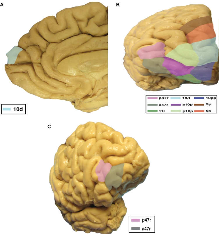
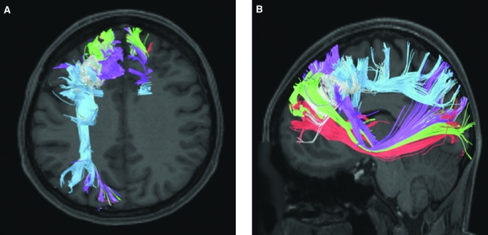
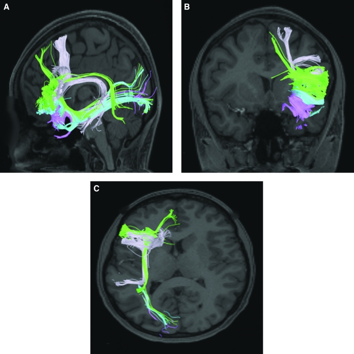
The parcellations that comprise the frontal polar region include 10pp, 10d, p10p, a10p, p47r, and a47r. The anatomic location of these parcellations is shown on a cadaver brain in Figure 1. This region has consistent white matter connections with the inferior fronto-occipital fasciculus (IFOF) and the contralateral hemisphere. The combined tractography of the parcellations is shown in Figure 2.
FIGURE 1.
Anatomic location of frontal polar parcellations shown on the right hemisphere of a cadaver brain. A, Medial view of the frontal lobe. B, Rostral view of the frontal pole. C, Oblique view of the frontal lobe. Corresponding labels are shown in the lower right corner of each figure.
FIGURE 2.
Combined structural connectivity of frontal polar parcellations. A, Sagittal, B, coronal, and C, axial views. Tracks include 10pp (dark blue), 10d (light blue), a10p (pink), p10p (yellow), a47r (gray), and p47r (light pink).
Area 10pp
Where is it?
Area 10pp is located at the anterior tip of the frontal pole. It is located at the fusiform junction of the anterior-most aspects of the superior and middle frontal gyri.
What are its Borders?
Area 10pp borders the orbitofrontal region on its inferoposterior boundary, and area 10d on its superior boundary. Area a10p is its lateral neighbor.
What is its Functional Connectivity?
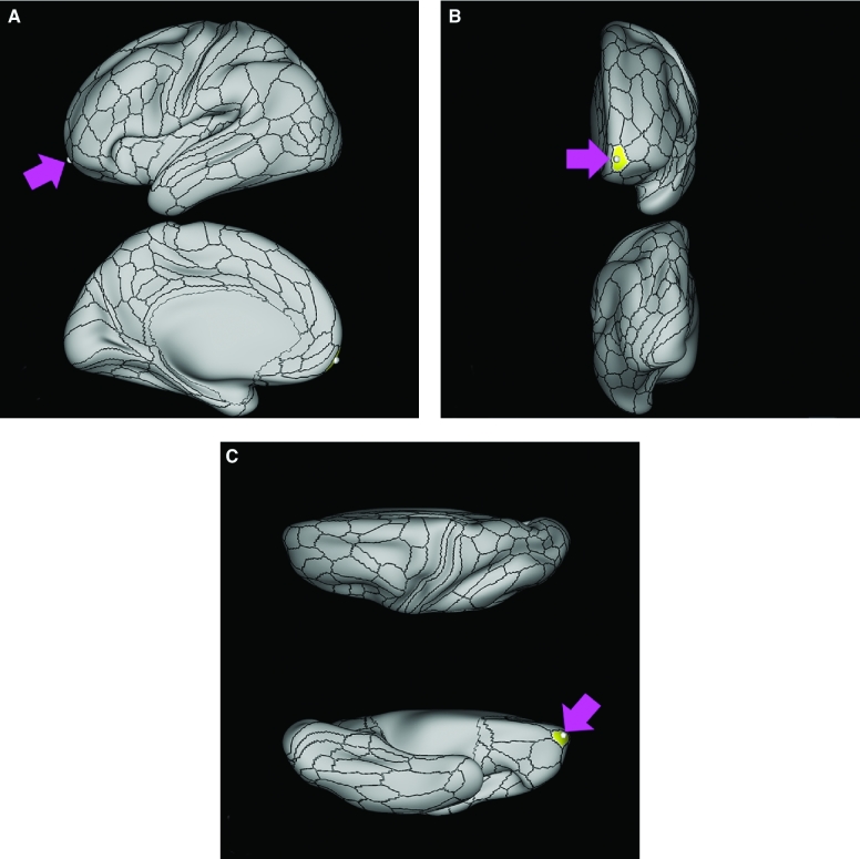
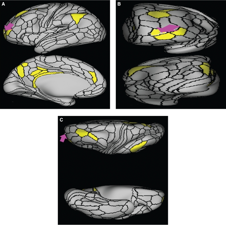
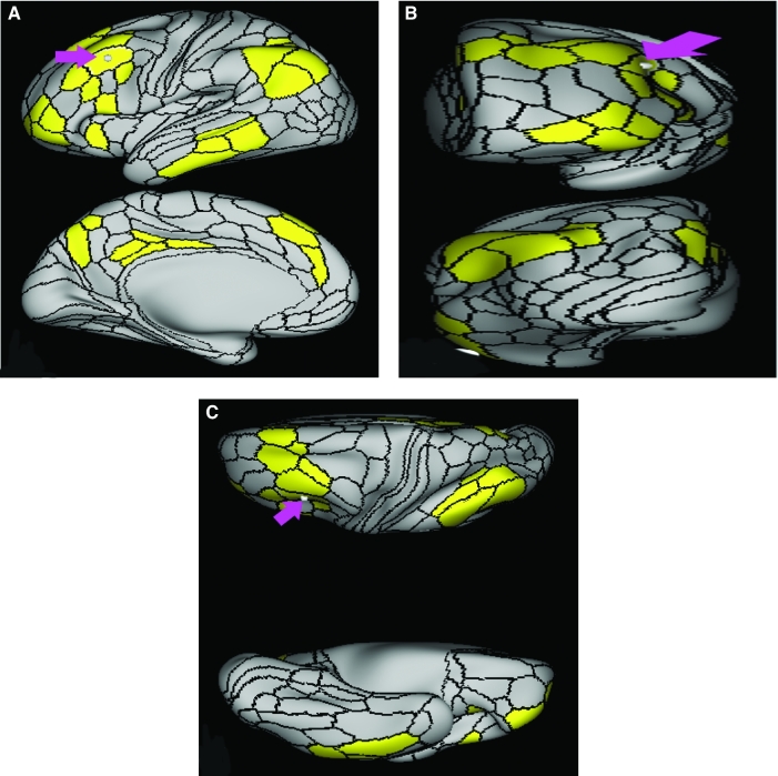
Area 10pp lacks strong evidence of functional connectivity to other areas, at least when using a z-score cutoff of 20. There is borderline evidence of functional connectivity with its neighbor a10p, as well as areas 8Av and 8C in the posterior dorsolateral frontal lobe, areas 31pd and 31pv in the posterior cingulate cortex, areas PFm, and PGi in the inferior parietal lobule, and area STSvp in the superior temporal sulcus (Figure 3).
FIGURE 3.
Functional connectivity of 10pp demonstrated on an inflated left hemisphere. A, Lateral and medial views. B, Rostral and caudal views. C, Dorsal and ventral views. Parcellations with the strongest functional connectivity are shown in yellow. Pink arrows designate the parcellation of interest.
What are its White Matter Connections?
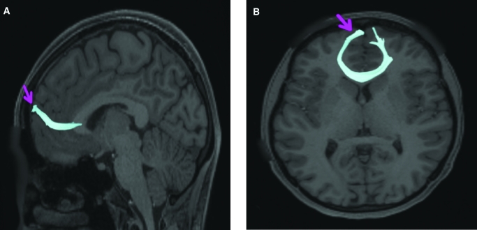
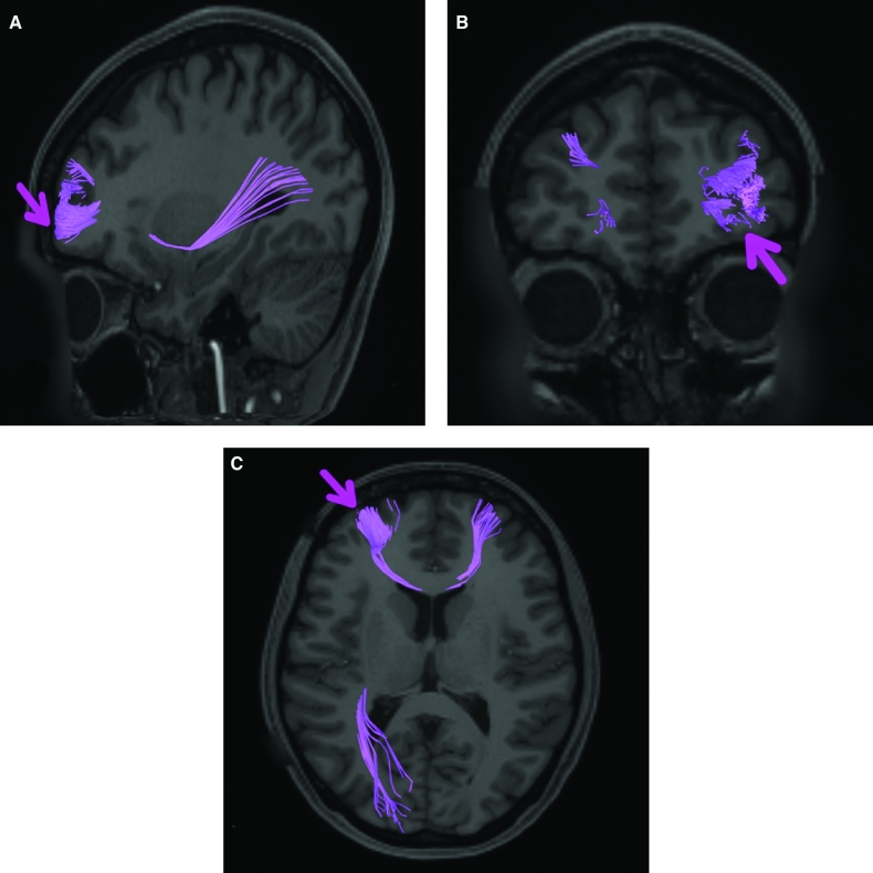
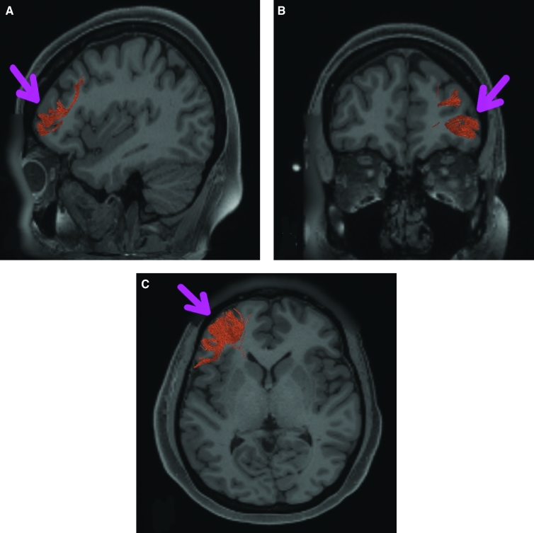
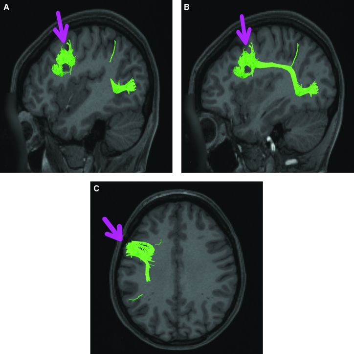
Area 10pp is structurally connected to the IFOF and contralateral hemisphere. IFOF connections travel from 10pp through the extreme/external capsule and continue posteriorly to end at occipital lobe parcellations V1 and V2. Contralateral connections course through the genu of the corpus callousum with the forceps minor to terminate at 10d and 10pp. Local short association bundles are connected with 10d (Figure 4).
FIGURE 4.
Structural connectivity of 10pp in the left hemisphere, shown on T1-weighted magnetic resonance (MR) images. A, Sagittal, B, coronal, and C, axial views. Dark blue: white matter tracts of 10pp demonstrating connections with IFOF, the contralateral hemisphere, and local parcellations.
What is Known About its Function?
Area 10pp is involved in episodic and working memory tasks. Brodmann area 10 more generally is activated in relation to increasing complexity of working memory tasks.7 This area also plays a role in abstract cognitive function.6
Area 10d
Where is it?
Area 10d (10 dorsal) is located in the anterior SFG. It wraps into the interhemispheric fissure, lying on the medial bank.
What are its Borders?
Area 10d borders area 10pp inferiorly, and area 9a posterior on the SFG. It also borders area 10r medially, and areas a10p and p10 laterally.
What is its Functional Connectivity?
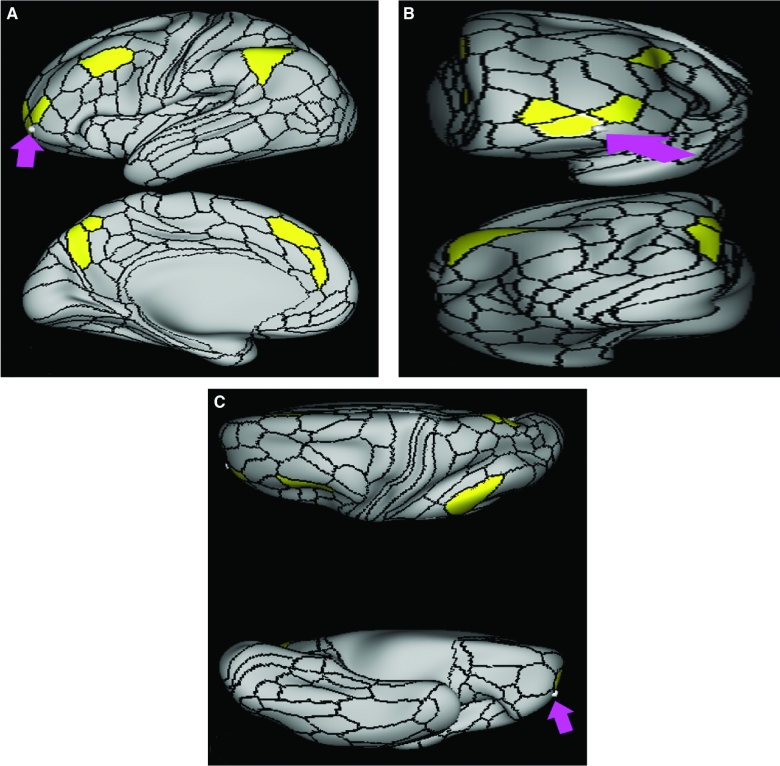
Area 10d demonstrates functional connectivity to areas 10r, 10v, 9M, 9P, 8BL, and 8AV in the anterior frontal lobe, areas s32, d32, a24 in the anterior cingulate cortex, areas POS1, 7M, 31a, 31pv, 31pd, and v23ab in the posterior cingulate area, area STSva and TE1a in the middle temporal gyrus region, areas PGs and PGi in the inferior parietal lobule, and the hippocampus. Many of these areas correspond to regions typically associated with the default mode network (DMN; Figure 5).
FIGURE 5.
Functional connectivity of 10d demonstrated on an inflated left hemisphere. A, Lateral and medial views. B, Rostral and caudal views. C, Dorsal and ventral views. Parcellations with the strongest functional connectivity are shown in yellow. Pink arrows designate the parcellation of interest.
What are its White Matter Connections?
Area 10d is structurally connected to the contralateral hemisphere. Connections to the contralateral hemisphere travel thorough the genu of the corpus callosum with the forceps minor to end at 9a and 10d. Local short association bundles are connected with 10pp, a10p, 9a, 10r, 9m, and p32 (Figure 6).
FIGURE 6.
Structural connectivity of 10d in the left hemisphere, shown on T1-weighted MR images. A, Sagittal and B, axial planes are shown. Light blue: white matter tracts of 10d demonstrating connections with local parcellations and the contralateral hemisphere.
What is Known About its Function?
Area 10d is involved in episodic and working memory tasks. Brodmann area 10 more generally is activated in relation to increasing complexity of working memory tasks.7 This area also plays a role in abstract cognitive function.6
Area a10p
Where is it?
Area a10p (anterior 10 polar) is located at the fusiform junction of the anterior most aspects of the superior and middle frontal gyri.
What are its Borders?
Area a10p borders area 11r on its inferior (orbitofrontal) boundary, and area p10p on its superior boundary. Areas a47r and p47r are its lateral neighbors, and area 10pp is its medial neighbor.
What is its Functional Connectivity?
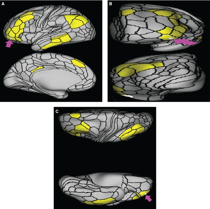
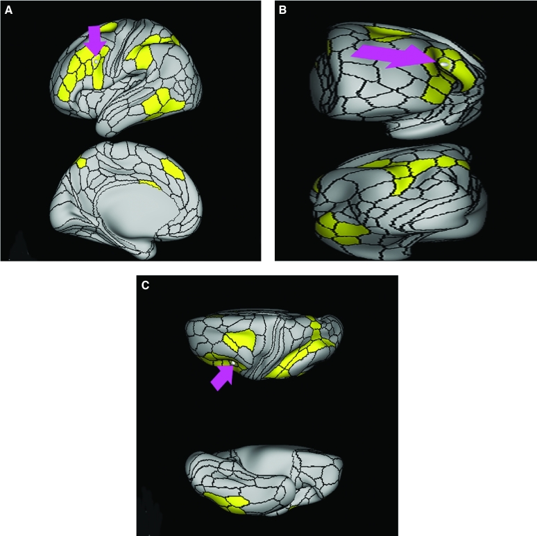
Area a10p demonstrates functional connectivity to areas p10p, a9-46v, 8BM, and 8C in the dorsolateral frontal lobe, d32 in the anterior cingulate region, area PFm in the lateral parietal lobe, and POS2 and 7pm in the medial parietal lobe (Figure 7).
FIGURE 7.
Functional connectivity of a10p demonstrated on an inflated left hemisphere. A, Lateral and medial views. B, Rostral and caudal views. C, Dorsal and ventral views. Parcellations with the strongest functional connectivity are shown in yellow. Pink arrows designate the parcellation of interest.
What are its White Matter Connections?
Area a10p is structurally connected to the IFOF and contralateral hemisphere. Contralateral connections travel through the genu of the corpus callosum with the forceps minor to end at 9a and p10p. IFOF connections travel from a10p through the extreme/external capsule and continue posteriorly to end at occipital lobe parcellations V1, V2, V3, V6, and V6a. Local short association bundles are connected to 10d, 10pp, p10p, a9-46v, and 9-46d (Figure 8).
FIGURE 8.
Structural connectivity of a10p in the left hemisphere, shown on T1-weighted MR images. A, Sagittal, B, coronal, and C, axial planes. Pink: white matter tracts of a10p demonstrating connections with IFOF, contralateral hemisphere, and local parcellations.
What is Known About its Function?
Area a10p is involved in episodic and working memory tasks. Brodmann area 10 more generally is activated in relation to increasing complexity of working memory tasks.7 This area also plays a role in abstract cognitive function.6
Area p10p
Where is it?
Area p10p (posterior 10 polar) is located on the lateral bank of the anterior aspect of the SFG, just posterior to a10p. It straddles the anterior most point of the superior frontal sulcus (SFS) that terminates here.
What are its Borders?
Area p10p borders area a10p on its anterior boundary, and area a9-46d and a small part of area 9a on its superior boundary. Area 9-46d is also its lateral neighbor. It is slightly triangular shaped and its superior apex is wedged between area 9a and area 9-46d.
What is its Functional Connectivity?
Area p10p demonstrates functional connectivity to a10p and a9-46v in the anterior frontal lobe, d32 in the anterior cingulate region, i6-8 in the posterior frontal lobe, areas 31a, 31pv, d23ab, POS2, retrosplenial cortex (RSC), and 7pm in the posterior cingulate region, and area PFm in the inferior parietal lobule (Figure 9).
FIGURE 9.
Functional connectivity of p10p demonstrated on an inflated left hemisphere. A, Lateral and medial views. B, Rostral and caudal views. C, Dorsal and ventral views. Parcellations with the strongest functional connectivity are shown in yellow. Pink arrows designate the parcellation of interest.
What are its White Matter Connections?
Area p10p is structurally connected to the IFOF and contralateral hemisphere. Contralateral connections travel through the genu of the corpus callosum with forceps minor to end at 9m. IFOF connections travel from p10p through the extreme/external capsule and continue posteriorly to end at occipital lobe parcellations V1 and V2. Local short association bundles are connected with 9-46d, 10d, a10p, a9-46v, and p9-46v (Figure 10).
FIGURE 10.
Structural connectivity of p10p in the left hemisphere, shown on T1-weighted MR images. A, Sagittal, B, coronal, and C, axial planes. Yellow: white matter tracts of p10p demonstrating connections with IFOF, contralateral hemisphere, and local parcellations.
What is Known About its Function?
Area p10p is involved in episodic and working memory tasks. Brodmann area 10 more generally is activated in relation to increasing complexity of working memory tasks.7 This area also plays a role in abstract cognitive function.6
Area a47r
Where is it?
Area a47r (anterior 47 rostral) is a j-shaped area located at the anterior inferior portion of the pars orbitalis of the IFG. It has a slight superior bend near its anterior extreme that blends into the anterior MFG.
What are its Borders?
Area a47r borders area p47r and area a9-46v; its superior end lies between their anterior surfaces. Its medial neighbor is area a10p. Its posterior edge is with 47l, and it borders 11r on its orbitofrontal side.
What is its Functional Connectivity?
Area a47r demonstrates functional connectivity to areas p47r, 8C, 8av 8BL, 8BM, i6-8, a9-46v, and p9-46v in the dorsolateral frontal lobe, areas IFSa, IFSp, 47l, and 45 in the IFG regions, areas IP1, IP2, PGi, PGs, and PFm in the inferior parietal lobule, areas TE1m, TE1p, TE2a, and STSv in the lateral temporal lobe, and area d23ab in the posterior cingulate region (Figure 11).
FIGURE 11.
Functional connectivity of a47r demonstrated on an inflated left hemisphere. A, Lateral and medial views. B, Rostral and caudal views. C, Dorsal and ventral views. Parcellations with the strongest functional connectivity are shown in yellow. Pink arrows designate the parcellation of interest.
What are its White Matter Connections?
Area a47r is structurally connected to the IFOF. IFOF connections travel from a47r through the extreme/external capsule and continue posteriorly to end at occipital lobe parcellations V1, V2, V3, V3a, V6, V6a, 7Am, and 7PL. Local short association bundles are connected with 47m and 11l (Figure 12).
FIGURE 12.
Structural connectivity of a47r in the left hemisphere, shown on T1-weighted MR images. A, Sagittal and B, axial planes area shown. Gray: white matter tracts of a47r demonstrating connections with IFOF and local parcellations.
What is Known About its Function?
Previous research has shown that the pars orbitalis is involved in controlled semantic retrieval.8
Area p47r
Where is it?
Area p47r (posterior 47 rostral) is located at the anterior end of the IFS in the anterosuperior most part of the IFG.
What are its Borders?
Area p47r borders area a47r anteriorly and IFSa posteriorly. Its entire medial border is with area a9-46v. It has a small lateral border with area 45.
What is its Functional Connectivity?
Area p47r demonstrates functional connectivity to areas a47r, a9-46v, p9-46v, 8BM, 8C, and i6-8 in the dorsolateral frontal lobe, areas IFSa, IFSp, IFJp, and 6r in the inferior frontal lobe, area anterior ventral insula (AVI) in the insula, areas TE1p and PHT in the temporal lobe, area PFm in the inferior parietal lobe, and areas IP2, IP1, and LIPd in the intraparietal area (Figure 13).
FIGURE 13.
Functional connectivity of p47r demonstrated on an inflated left hemisphere. A, Lateral and medial views. B, Rostral and caudal views. C, Dorsal and ventral views. Parcellations with the strongest functional connectivity are shown in yellow. Pink arrows designate the parcellation of interest.
What are its White Matter Connections?
Area p47r is structurally connected with surrounding parcellations. The white matter tracts from this parcellation are highly inconsistent. Some individuals seem to have connections with the IFOF. Local short association bundles are connected to a47r, p10p, 45, IFSa, s6-8, 9-47d, and 9a (Figure 14).
FIGURE 14.
Structural connectivity of p47r in the left hemisphere, shown on T1-weighted MR images. Sagittal planes in A, lateral and B, medial views, and C, coronal planes are shown. Light pink: white matter tracts of p47r demonstrating connections with local parcellations.
What is Known About its Function?
Brodmann area 47 is activated during the processing of linguistically complex words.9 In the recognition of spoken language, Brodmann area 47 increases in activity as the cohort of spoken words increases in size.10
SFG REGIONS
The anatomy of the SFG is simpler than its neighbors. The anterior half of this gyrus (assuming you are excluding the supplementary motor areas from this estimation) comprises the subdivisions of area 9: areas 9a, 9p, and 9m. Area 9m is principally located in the medial frontal lobe and, as such, is discussed in detail with that section. Areas 9a and 9p lie on the superior surface of the SFG.
The posterior half of the gyrus is comprised of the subdivisions of area 8: areas 8BL and 8AD. These two areas run in an anterior–posterior direction and parallel each other to create the medial and lateral parts of the upper surface of the SFG, respectively. This pattern between subdivisions of area 8 is consistent in other parts of the greater area 8 complex as well.
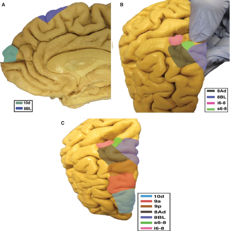
The parcellations that comprise the SFG include 9a, 9p, 8BL, and 8AD. The anatomic location of these parcellations is shown on a cadaver brain in Figure 15. This region has consistent white matter connections with the IFOF and contralateral hemisphere. There are also connections to the medial thalamus and frontal aslant tract (FAT), but these connections are unique to specific parcellations. The combined tractography of the parcellations is shown in Figure 16.
FIGURE 15.
Anatomic location of the frontal lobe parcellations of the SFG shown on the right hemisphere of a cadaver brain. A, Medial view of the frontal lobe. B, Dorsal view of the frontal lobe with widening of the precentral sulcus to show extension of 8Ad. C, Dorsal view of the frontal lobe. Corresponding labels are shown in the lower right corner of each figure.
FIGURE 16.
Combined structural connectivity of SFG parcellations. A, Axial and B, sagittal planes are shown. Tracks include 9a (red), 9p (green), 8BL (purple), and 8Ad (gray).
Area 9a
Where is it?
Area 9a (9 anterior) is located at the anterior portion of the superior surface of the SFG.
What are its Borders?
Area 9a borders area 10d anteriorly and area 9p posteriorly. Area 9m is its medial border. Its lateral border is area 9-46d.
What is its Functional Connectivity?
Area 9a demonstrates functional connectivity to areas 9p, 9m, 8BL, and 8AV in the dorsolateral frontal lobe, areas SFL and d32 in the medial frontal lobe, areas 44, 45, 47l, and 47s in the inferior frontal lobe, areas TGd, TE1a, STSva, and STSvp in the temporal lobe, areas PGi and PFm in the inferior parietal lobe, and areas 7m, 31pd, 31pv, and 23d in the medial parietal lobe (Figure 17).
FIGURE 17.
Functional connectivity of 9a demonstrated on an inflated left hemisphere. A, Lateral and medial views. B, Rostral and caudal views. C, Dorsal and ventral views. Parcellations with the strongest functional connectivity are shown in yellow. Pink arrows designate the parcellation of interest.
What are its White Matter Connections?
Area 9a is structurally connected to the IFOF and contralateral hemisphere. Some individuals also have medial thalamic connections but this is inconsistent. Contralateral connections travel through the genu of the corpus callosum with the forceps minor to end at 9a, 9p, p10p, 10d, and 9m. IFOF connections travel from 9a through the extreme/external capsule and continue posteriorly through the temporal lobe to end at occipital lobe parcellations V1, V2, and V3. Local short association bundles connect with 9p (Figure 18).
FIGURE 18.
Structural connectivity of 9a in the left hemisphere, shown on T1-weighted MR images. A, Sagittal, B, coronal, and C, axial planes. Red: white matter tracts of 9a demonstrating connections with IFOF, contralateral hemisphere, and local parcellations.
What is Known About its Function?
Brodmann area 9 is involved in the maintenance of behaviorally relevant working memory.11 Area 9 along with Brodmann areas 46 and 8 play a role in reestablishing executive control during automatic behaviors.12
Area 9p
Where is it?
Area 9p (9 posterior) is located at the anterior portion of the superior surface of the SFG.
What are its Borders?
Area 9p borders area 9a anteriorly and its superior triangular apex wedges in between areas 8BL and 8AD superiorly. Its medial border is area 9M. Its lateral border is area 9-46d.
What is its Functional Connectivity?
Area 9p demonstrates functional connectivity to areas 9a, 9m, 8BL, 8AD, and 8AV in the dorsolateral frontal lobe, areas SFL, 10r, a24, and d32 in the medial frontal lobe, areas 45, 47l, and 47s in the inferior frontal lobe, areas TGd, TE1a, STSva, and STSvp in the temporal lobe, areas PGi and PGs in the inferior parietal lobe, and areas 7m, 31pd, 31pv, 23d, d23ab, and v23ab in the medial parietal lobe (Figure 19).
FIGURE 19.
Functional connectivity of 9p demonstrated on an inflated left hemisphere. A, Lateral and medial views. B, Rostral and caudal views. C, Dorsal and ventral views. Parcellations with the strongest functional connectivity are shown in yellow. Pink arrows designate the parcellation of interest.
What are its White Matter Connections?
Area 9p is structurally connected to the IFOF, contralateral hemisphere and the medial thalamus. Contralateral connections course through the genu of the corpus callosum with the forceps minor to end at 8BL, 9a, 9p, and 9m. Connections with the thalamus travel through the anterior limb of the internal capsule. IFOF connections travel from 9p through the extreme/external capsule and continue posteriorly through the temporal lobe to end at occipital lobe parcellations V1, V2, V3, V3A, and V6. Local short association bundles are connected with 9a, 8BL, and 9-46d (Figure 20).
FIGURE 20.
Structural connectivity of 9p in the left hemisphere, shown on T1-weighted MR images. A, Sagittal, B, coronal, and C, axial planes. Orange: white matter tracts of 9p demonstrating connections with IFOF, contralateral hemisphere, and the medial thalamus.
What is Known About its Function?
Brodmann area 9 is involved in the maintenance of behaviorally relevant working memory.11 Area 9 along with Brodmann areas 46 and 8 plays a role in reestablishing executive control during automatic behaviors.12
Area 8BL
Where is it?
Area 8BL (8B lateral) is located at the posterior half of the superior surface of the SFG. It is the lateral subdivision of area 8B from a previously described parcellation of area 8 into 8A, 8B, and 8C.
What are its Borders?
Area 8BL has a medial border with area 8BM. Its lateral border is area 8ad. It also has an anterior apex that is wedged between the posterior edges of areas 9p and 9m. Its posterior boundary includes area s6-8 and area SFL.
What is its Functional Connectivity?
Area 8BL demonstrates functional connectivity to areas 9a, 9m, 8BM, 8C, and 8AV in the dorsolateral frontal lobe, areas SFL, 10d, 10v, and d32 in the medial frontal lobe, areas a47r, 44, 45, 47l, and 47s in the inferior frontal lobe, areas TGd, TE1a, TE1m, TE2a, STSdp, STSva, and STSvp in the temporal lobe, areas PGi and PGs in the inferior parietal lobe, and areas 7m, 31pd, 31pv, 23d, d23ab, and v23ab in the medial parietal lobe (Figure 21).
FIGURE 21.
Functional connectivity of 8BL demonstrated on an inflated left hemisphere. A, Lateral and medial views. B, Rostral and caudal views. C, Dorsal and ventral views. Parcellations with the strongest functional connectivity are shown in yellow. Pink arrows designate the parcellation of interest.
What are its White Matter Connections?
Area 8BL is structurally connected to the IFOF, contralateral hemisphere, medial thalamus and FAT. Contralateral connections course through the genu of the corpus callosum with the forceps minor to end at 8BM and 9m. Connections to the medial thalamus travel through the anterior limb of the internal capsule. IFOF connections travel from 8BL pass through the extreme/external capsule and continue posteriorly to end at occipital lobe parcellations V2, V3, 7PL, MIP, V6, and V6A. From 8BL, the FAT projects to the IFG to terminate at 44. From this tract there are also connections with p9-46v. Local short association bundles connect with SFL, 8BM, 9a, and 9p (Figure 22).
FIGURE 22.
Structural connectivity of 8BL in the left hemisphere, shown on T1-weighted MR images. A, Sagittal, B, coronal, and C, axial planes. Dark blue: white matter tracts of 8BL demonstrating connections with IFOF, contralateral hemisphere, medial thalamus, and FAT.
What is Known About its Function?
Area 8B in macaques represents the premotor eye–ear field (PEEF). The PEEF has two divisions—the core and the belt—that are involved in the coordination of eye and ear movements in relation to space and the topographical representation of that space.13 Areas 8 and 6 also play a role in the maintenance of spatial information.14 The posterior part of the SFS anterior to the frontal eye field (FEF) is specialized for handling spatial working memory.15
Area 8AD
Where is it?
Area 8AD (8A dorsal) is located at the posterior lateral part of the SFG on the banks of the SFS. It fills the depths of the posterior SFS and makes up some of the posterior MFG bank of this sulcus. There is a classic right angle between the SFS and precentral sulci that has long been described as a landmark for locating the precentral gyrus. Area 8AD makes up the banks and sulcal depths of the SFS as it is about to join that right angle.
What are its Borders?
Area 8AD’s anterior border is a wedge that contacts areas 9P, 9-46d, and 46, from medial to lateral. Its medial border is area 8BL. Its lateral border is area 8AV, which it contacts on the medial bank of the MFG. Its posterior boundaries are with areas s6-8 and i-6-8, which form the mouth of the SFS junction with the precentral sulcus.
What is its Functional Connectivity?
Area 8AD demonstrates functional connectivity to areas 9p, s6-8, i6-8, 10d, p10p, 8C, and 8AV in the dorsolateral frontal lobe, areas 10r, a24, p32, s32, and d32 in the medial frontal lobe, area 47m in the orbitofrontal region, areas STSva, PHA1, PHA2, PreS, EC, TE1p, TE1m, TE1a, and the hippocampus in the temporal lobe, areas PFm, PGi, and PGs in the inferior parietal lobe, and areas 7m, 7pm, 31pd, 31pv, 31a, RSC, PCV, 23d, d23ab, and v23ab in the medial parietal lobe (Figure 23).
FIGURE 23.
Functional connectivity of 8Ad demonstrated on an inflated left hemisphere. A, Lateral and medial views. B, Rostral and caudal views. C, Dorsal and ventral views. Parcellations with the strongest functional connectivity are shown in yellow. Pink arrows designate the parcellation of interest.
What are its White Matter Connections?
Area 8AD is structurally connected to local parcellations. White matter tracts from this parcellation are highly inconsistent and in some individuals this parcellation connects with the FAT and contralateral hemisphere. Local short association bundles are abundant and connect with 9a, 9p, s6-8, 8Av, 6a, and p10p (Figure 24). White matter connections of 8AD in the right hemisphere have more consistent connections with the contralateral hemisphere.
FIGURE 24.
Structural connectivity of 8Ad in the left hemisphere, shown on T1-weighted MR images. A, Sagittal, B, coronal, and C, axial planes. Gray: white matter tracts of 8Ad demonstrating connections with local parcellations.
What is Known About its Function?
Area 8AD plays a role in integrating information related to peripheral vision and audition in the context of spatial cognition.16 The SFS is also involved in the spatial processing of sounds.17 The posterior part of the SFS anterior to the FEF is specialized for handling spatial working memory.15
MFG AND SFS REGIONS
There are only a few subregions occupying the MFG and SFS; however, this region is still quite complex. The reasons for this complexity are numerous: the subregions here have hybrid names, some of these areas have complex borders, and several of these areas are not well confined to easily recognized anatomy. As such, these areas can be difficult to learn at first glance.
The present scheme can best be described as dividing this region into area 9 proper (areas 9a, 9p, and 9m on the SFG) and the 9-46 hybrid regions. These include area 9-46d, area 46, and two ventral hybrid regions, areas a9-46v and p9-46v. The latter subregions are described in this section. The posterior half comprises subdivisions of area 8: areas 8AV and 8C. These two areas run in an anterior–posterior direction and parallel to each other to create the medial and lateral parts of the upper surface of the MFG, respectively. Finally, the SFS terminates at a right angle with the precentral sulcus. There are two small 6-8 hybrid regions that sit at the mouth of this sulcal confluence, areas s6-8 and i6-8.
In summary, the parcellations that comprise the MFG and SFS include 9-46d, 46, a9-46v, p9-46v, 8Av, 8C, i6-8, and s6-8. The anatomic location of these parcellations is shown on a cadaver brain in Figure 25. This region has consistent white matter connections with the contralateral hemisphere and the arcuate/superior longitudinal fasciculus (SLF). There are also connections with the FAT but this is specific to s6-8. The combined tractography of the parcellations is shown in Figure 26.
FIGURE 25.
Anatomic location of the frontal lobe parcellations of the MFG and SFS shown on the right hemisphere of a cadaver brain. A, Lateral view of the frontal lobe. B, Dorsal view of the frontal lobe with dilation of the IFS. C, Dorsal view of the frontal lobe with dilation of the SFS. D, Dorsal view of the frontal lobe. Corresponding labels are shown in the lower right corner of each figure.
FIGURE 26.
Combined structural connectivity of MFG and SFS parcellations. A, Sagittal, B, dorsal-oblique view, C, coronal, and D, axial planes are shown. Tracks include 9-46d (green), 46 (gray), a9-46v (brown), p9-46v (purple), 8Av (blue), 8c (pink), s6-8 (orange), and i6-8 (brown).
Area 9-46d
Where is it?
Area 9-46d (9-46 dorsal) occupies a location difficult to describe. It runs across the SFS at a slightly oblique, anterior-to-posterior angle. The approximate centroid of the area lies in the depths of the anterior SFS. A significant portion of the anterior-to-posterior length of this region lies on the lateral bank of the SFG; however, anteriorly a lateral wedge rises out of the sulcus onto the MFG.
What are its Borders?
Area 9-46d has a number of areas on its medial border. From anterior to posterior these include area p10p, area 9a, area 9p, and a small posterior interface with area 8AD. Its lateral border is a slight wedge between area a9-46v and area 46. Its anterior most point forms a wedge between area p10p and area a9-46v.
What is its Functional Connectivity?
Area 9-46d demonstrates functional connectivity to areas 46, a9-46v, p9-46v, and FEF in the dorsolateral frontal lobe, areas SCEF, a32prime, p32prime, p24, and a24prime in the medial frontal lobe, areas 6a, 6r, and PEF in the premotor regions, areas FOP1, FOP3, FOP4, FOP5, PSL, PFop, PFcm, AVI, 43 PoI1, and MI in the insula-opercular region, area PHT in the temporal lobe, areas PF, 7AL, 7PL, and LIPd in the parietal lobe, and areas POS2, PCV, 7am, 7pm, 23c, and deep venous thrombosis (DVT) in the medial parietal lobe. It is also connected to much of the occipital lobe, showing connectivity to V1, V2, V3, V4, and V6 (Figure 27).
FIGURE 27.
Functional connectivity of 9-46d demonstrated on an inflated left hemisphere. A, Lateral and medial views. B, Rostral and caudal views. C, Dorsal and ventral views. Parcellations with the strongest functional connectivity are shown in yellow. Pink arrows designate the parcellation of interest.
What are its White Matter Connections?
Area 9-46d is structurally connected to local parcellations and the contralateral hemisphere. Some individuals have connections with the IFOF, though this is inconsistent. Contralateral connections travel through the corpus callosum to end at 9m and 9p. There are abundant local short association bundles that connect with 8BL, 9p, 9-47d, 46, a9-46v, 8Ad, p47r, and a10p (Figure 28).
FIGURE 28.
Structural connectivity of 9-46d in the left hemisphere, shown on T1-weighted MR images. A, Sagittal, B, coronal, and C, axial planes. Green: white matter tracts of 9-46d demonstrating connections with the contralateral hemisphere and local parcellations.
What is Known About its Function?
Area 9-46d, like area 46, plays a role in goal-directed higher-order cognitive processes.18 The mid-DLPFC, which includes areas 9-46 and 46, is also involved in the conscious, active control of planned behavior.19
Area 46
Where is it?
Area 46 parallels area 9-46d along its slightly oblique course. It begins in the depths of the SFS posteriorly and its anterior extent spills onto the MFG.
What are its Borders?
Area 46 borders all three 9-46 hybrid regions, as well as several others, giving it a central place in the ventral prefrontal cortex. Its medial border is mainly area 9-46d and its lateral border is mainly area p9-46v, both of which roughly parallel it along its oblique course. It has an anterolateral border with IFSa, as IFSa rises onto the lateral bank of MFG to meet it. Its anterior limit is mainly with area a9-46v. Its posterior limit forms a wedge with areas 8AV and 8AD.
What is its Functional Connectivity?
Area 46 demonstrates functional connectivity to areas a9-46v, p9-46v, and IFSa in the dorsolateral frontal lobe, areas SCEF, a32prime, p24prime, and a24prime in the medial frontal lobe, areas 6ma, 6a, and 6r in the premotor regions, area 11L in the orbitofrontal region areas FOP1, FOP3, FOP4, FOP5, 52 PFop, PFcm, 43 PoI2, PoI1, and MI in the insula-opercular region, area PHT in the temporal lobe, areas PF, PFt, PGp, AIP, MIP 7AL, 7PL, and LIPd IP2 and IP0 in the parietal lobe, and areas 23c, PCV, POS2, PCV, 7am, 7pm, 23c, and DVT in the medial parietal lobe. It is also connected to much of the occipital lobe, showing connectivity to V1, V2, V3, V4, V3a, and V6 (Figure 29).
FIGURE 29.
Functional connectivity of 46 demonstrated on an inflated left hemisphere. A, Lateral and medial views. B, Rostral and caudal views. C, Dorsal and ventral views. Parcellations with the strongest functional connectivity are shown in yellow. Pink arrows designate the parcellation of interest.
What are its White Matter Connections?
Area 46 is structurally connected to local parcellations and the contralateral hemisphere. Contralateral connections travel through the corpus callosum to end at 9-46d and p9-47v. There are abundant local short association bundles connecting with 9-46d, a9-46v, p9-46v, IFSp, and IFSa (Figure 30).
FIGURE 30.
Structural connectivity of 46 in the left hemisphere, shown on T1-weighted MR images. A, Sagittal, B, coronal, and C, axial planes. Gray: white matter tracts of 46 demonstrating connections with the contralateral hemisphere, and local parcellations.
What is Known About its Function?
Area 46 plays a role in goal-directed higher-order cognitive processes.18 The mid-DLPFC, which includes areas 9-46 and 46, is also involved in the conscious, active control of planned behavior.19
Area a9-46v
Where is it?
Area a9-46v (anterior 9-46 ventral) is located at the anterior portior of the MFG.
What are its Borders?
Area a9-46v borders area 46 posteriorly, and area 9-46d medially. Its lateral border is mainly with area p47r and a slight contribution with IFSa. Its anterior limit forms a wedge with areas p10p and a47r.
What is its Functional Connectivity?
Area a9-46v demonstrates functional connectivity to area 46, 9-46d, p9-46v, a10p, p10p, 6ma, i6-8, a47r, p47r, 8C, and IFSa in the dorsolateral frontal lobe, areas 8BM, and a32prime in the medial frontal lobe, area 11L in the orbitofrontal region, area TE1p in the temporal lobe, area AVI in the insula, areas LIPd, PF, IP1, and IP2 in the parietal lobe, and areas 7pm, 31a, and RSC in the medial parietal lobe (Figure 31).
FIGURE 31.
Functional connectivity of a9-46v demonstrated on an inflated left hemisphere. A, Lateral and medial views. B, Rostral and caudal views. C, Dorsal and ventral views. Parcellations with the strongest functional connectivity are shown in yellow. Pink arrows designate the parcellation of interest.
What are its White Matter Connections?
Area a9-46v is structurally connected to local parcellations. White matter tracts from this parcellation are highly variable. Short association bundles connect with 8C, 9-46d, 46, IFSa, and p47r (Figure 32).
FIGURE 32.
Structural connectivity of a9-46v in the left hemisphere, shown on T1-weighted MR images. A, Sagittal, B, coronal, and C, axial planes. Brown: white matter tracts of a9-46v demonstrating connections with local parcellations.
What is Known About its Function?
Area 9-46d, like Area 46, plays a role in goal-directed higher-order cognitive processes.18 The mid- DLPFC, which includes areas 9-46 and 46, is also involved in the conscious, active control of planned behavior.19
Area p9-46v
Where is it?
Area p9-46v (posterior 9-46 ventral) is a small triangular shaped region located in the MFG.
What are its Borders?
Area p9-46v borders area 8C posteriorly. Its medial border is area 46. Its lateral border is made of IFSp and IFJa.
What is its Functional Connectivity?
Area p9-46v demonstrates functional connectivity to area 46, 9-46d, a9-46v a10p, p10p, 6ma, i6-8, a47r, p47r, 8C, IFJp, IFJa, IFSp, and IFSa in the dorsolateral frontal lobe, areas 8BM, and 33prime in the medial frontal lobe, areas 6a and 6r in the premotor area, area 11L in the orbitofrontal region, areas TE1p, TE2p, PH, and PHT in the temporal lobe, area AVI in the insula, areas LIPd, AIP, MIP, PFm, 7PL, IP0, IP1, and IP2 in the parietal lobe, area 7pm in the medial parietal lobe, and area V1 in the occipital lobe (Figure 33).
FIGURE 33.
Functional connectivity of p9-46v demonstrated on an inflated left hemisphere. A, Lateral and medial views. B, Rostral and caudal views. C, Dorsal and ventral views. Parcellations with the strongest functional connectivity are shown in yellow. Pink arrows designate the parcellation of interest.
What are its White Matter Connections?
Area p9-46v is structurally connected to the arcuate/SLF. Connections with the arcuate/SLF project posteriorly and wrap around the sylvian fissure to the inferior temporal gyrus to end at TE2a. Local short association bundles connect with 46, a9-46v, IFJa, IFSa, IFSp, 8C, and 9-46d (Figure 34).
FIGURE 34.
Structural connectivity of p9-46v in the left hemisphere, shown on T1-weighted MR images. A, Sagittal, B, coronal, and C, axial planes. Purple: white matter tracts of p9-46v demonstrating connections with SLF and local parcellations.
What is Known About its Function?
Area 9-46d, like area 46, plays a role in goal-directed higher-order cognitive processes.18 The mid-DLPFC, which includes areas 9-46 and 46, is also involved in the conscious, active control of planned behavior.19
Area 8AV
Where is it?
Area 8AV (8A ventral) is located at the posterior part of the MFG. It is an anterior-to-posterior band that is medial to area 8C.
What are its Borders?
Area 8AV borders area 46 anteriorly, and area 55b and the FEF posteriorly, as well as i6-8. Its medial border is area 8AD. Its lateral border is area 8C. There is a small anterior border with area 46.
What is its Functional Connectivity?
Area 8AV demonstrates functional connectivity to areas 9a, 9p, 9m, 8BL, 8AD, and 8C, i6-8 and s6-8 in the dorsolateral frontal lobe, areas 8BM, SFL, 10d, and d32 in the medial frontal lobe, areas 44 45, A47r 47l, and 47s in the inferior frontal lobe, area 55b in the premotor region areas TGd, TE1a, TE1m, TE1p, TE2a, STSva, and STSvp in the temporal lobe, areas PFm, IP1, PGi, and PGs in the inferior parietal lobe, and areas 7m, 31pd, 31pv, 31a, 23d, d23ab, and v23ab in the medial parietal lobe (Figure 35).
FIGURE 35.
Functional connectivity of 8Av demonstrated on an inflated left hemisphere. A, Lateral and medial views. B, Rostral and caudal views. C, Dorsal and ventral views. Parcellations with the strongest functional connectivity are shown in yellow. Pink arrows designate the parcellation of interest.
What are its White Matter Connections?
Area 8AV is structurally connected to the arcuate/SLF and the contralateral hemisphere. Connections to the contralateral hemisphere travel through the body of the corpus callosum to connect to SFL. Connections with the arcuate/SLF project posteriorly and wrap around the sylvian fissure to the parietal lobule to end at 6a, 7PC, MIP, PFm, and 2. Local short association bundles are connected with 8C, 8Ad, i6-8, and 46 (Figure 36).
FIGURE 36.
Structural connectivity of 8Av in the left hemisphere, shown on T1-weighted MR images. A, Sagittal, B, coronal, and C, axial planes. Blue: white matter tracts of 8Av demonstrating connections with SLF, the contralateral hemisphere, and local parcellations.
What is Known About its Function?
Within the context of spatial working memory, area 8AV is involved in the interpretation of complex visual information and attention.16 Areas 8 and rostral 6, as part of the posterior dorsolateral frontal areas, are also involved in the maintenance of spatial information.14
Area 8C
Where is it?
Area 8C is located at the posterior part of the MFG. It is an anterior-to-posterior band that is lateral to area 8AV.
What are its Borders?
Area 8C has area 8AV as its main medial border. Its lateral border is with 3 IFS areas: IFSp, IFJa, and IFJp. Area 55b and PEF (precentral eyefield) are its posterior borders (and discussed in a different section). Area p9-46v and to a lesser extent area 46 are its anterior neighbors.
What is its Functional Connectivity?
Area 8C demonstrates functional connectivity to areas s6-8, i6-8, a9-46v, p9-46v, a10p, 8BL, 8AD, and 8AV in the dorsolateral frontal lobe, areas 8BM and d32 in the medial frontal lobe, areas IFSp, IFJp, a47r, p47r, and 44 in the inferior frontal lobe, area AVI in the insula, areas TE1m, TE1p, TE2a, STSva, and STSvp in the temporal lobe, areas IP1, IP2, LIPd, PFm, PGi, and PGs in the inferior parietal lobe, and areas 7pm, 31pv, 31a, POS2, 23d, and d23ab in the medial parietal lobe (Figure 37).
FIGURE 37.
Functional connectivity of 8C demonstrated on an inflated left hemisphere. A, Lateral and medial views. B, Rostral and caudal views. C, Dorsal and ventral views. Parcellations with the strongest functional connectivity are shown in yellow. Pink arrows designate the parcellation of interest.
What are its White Matter Connections?
Area 8C is structurally connected to the arcuate/SLF and the contralateral hemisphere. Contralateral connections travel through the corpus callosum to end at 8C. Connections with the arcuate/SLF project posteriorly and wrap around the sylvian fissure to the posterior temporal lobe to end at PH and PHT (Figure 38).
FIGURE 38.
Structural connectivity of 8C in the left hemisphere, shown on T1-weighted MR images. A, Sagittal and B, axial planes. Pink: white matter tracts of 8C demonstrating connections with SLF, the contralateral hemisphere, and local parcellations.
What is Known About its Function?
Within the context of spatial working memory, area 8C is involved in the interpretation of complex visual information and attention.16 Areas 8 and rostral 6, as part of the posterior dorsolateral frontal areas, are also involved in the maintenance of spatial information.14
Area s6-8
Where is it?
Area s6-8 (superior 6-8) is located at the posterior most SFG, on its lateral bank near where the SFS joins with the precentral sulcus.
What are its Borders?
Area s6-8 borders areas 8AD and 8BL anteriorly. Its medial border is SFL (discussed in later sections). Its posterior border is made of areas 6ma and 6a.
What is its Functional Connectivity?
Area s6-8 demonstrates functional connectivity to areas i6-8, 8C, 8AD, and 8AV in the dorsolateral frontal lobe, areas 8BM and d32 in the medial frontal lobe, areas TE1m andTE1p in the temporal lobe, areas PFm and PGs in the inferior parietal lobe, and areas 7pm, 31a, and d23ab in the medial parietal lobe (Figure 39).
FIGURE 39.
Functional connectivity of s6-8 demonstrated on an inflated left hemisphere. A, Lateral and medial views. B, Rostral and caudal views. C, Dorsal and ventral views. Parcellations with the strongest functional connectivity are shown in yellow. Pink arrows designate the parcellation of interest.
What are its White Matter Connections?
Area s6-8 is structurally connected to the FAT. Connections from the FAT project to the IFG to terminate at 44, FOP4, and 6r. Local short association bundles connect to 8Av (Figure 40).
FIGURE 40.
Structural connectivity of s6-8 in the left hemisphere, shown on T1-weighted MR images. A, Sagittal, B, coronal, and C, axial planes. Orange: white matter tracts of s6-8 demonstrating connections with FAT and local parcellations.
What is Known About its Function?
Areas s6-8 and i6-8 represent transitional areas of cortex between Brodmann areas 6 and 8, previously described by Economo and Koskinas.20 Areas 8 and rostral 6, as part of the posterior dorsolateral frontal areas, are involved in the maintenance of spatial information.14 Brodmann area 6 has also been subdivided into areas including the premotor and supplementary motor areas that influence motor control. The premotor area, which encompasses the lateral part of area 6, is further divided into ventral and dorsal premotor portions, named PMv and PMd, respectively.21
PMv has a significant contribution to the control of object manipulation with the hands, specifically when it comes to the force applied with precision grip. In addition, PMv contains mirror neurons that play a role in understanding actions performed by others.21
PMd is involved in selecting motor responses based on both arbitrary and spatial cues. For example, the PMd integrates sensory information to guide motor tasks such as reaching for an object. In addition to sensory connections, the PMd also communicates with the prefrontal cortex. Within this subdivision, the caudal portion of the PMd connects to the primary motor area, and the rostral portion analyzes cues to select motor responses.21
Area i6-8
Where is it?
Area i6-8 (inferior 6-8) is located at the posterior medial MFG near where the SFS joins with the precentral sulcus.
What are Its Borders?
Area i6-8 borders area 6a medially, and area 55b and FEF laterally. Area 8AV is its lateral border.
What is its Functional Connectivity?
Area i6-8 demonstrates functional connectivity to areas s6-8, a9-46v, p9-46v, p10p, 8C, 8AD, and 8AV in the dorsolateral frontal lobe, areas 8BM and d32 in the medial frontal lobe, areas IFSp, IFJp, a47r, p47r, and 44 in the inferior frontal lobe, area 6a in the premotor region, areas TE1m, TE1p, TE2a, PHA2, and PreS the temporal lobe, areas IP0, IP1, IP2, LIPd, PFm, and PGs in the inferior parietal lobe, and areas 7pm, 7m, 31a, POS2, POS1, and d23ab in the medial parietal lobe (Figure 41).
FIGURE 41.
Functional connectivity of i6-8 demonstrated on an inflated left hemisphere. A, Lateral and medial views. B, Rostral and caudal views. C, Dorsal and ventral views. Parcellations with the strongest functional connectivity are shown in yellow. Pink arrows designate the parcellation of interest.
What are its White Matter Connections?
Area i6-8 is structurally connected to surrounding parcellations. White matter tracts from this parcellation are variable. Local short association bundles connect with 8Av, 8Ad, 9-46d, 6a, and FEF (Figure 42).
FIGURE 42.
Structural connectivity of i6-8 in the left hemisphere, shown on T1-weighted MR images. A, Sagittal, B, coronal, and C, axial planes. Brown: white matter tracts of i6-8 demonstrating connections with local parcellations.
What is Known About its Function?
Areas s6-8 and i6-8 represent transitional areas of cortex between Brodmann areas 6 and 8, previously described by Economo and Koskinas.20 Areas 8 and rostral 6, as part of the posterior dorsolateral frontal areas, are involved in the maintenance of spatial information.14 Brodmann area 6 has also been subdivided into areas including the premotor and supplementary motor areas that influence motor control. The premotor area, which encompasses the lateral part of area 6, is further divided into ventral and dorsal portions, named PMv and PMd, respectively.21
IFS REGIONS
The IFS contains 4 distinct regions aligned in an anterior to posterior fashion. The parcellations that comprise the IFS include IFSa, IFSp, IFJa, and IFJp. The anatomic location of these parcellations is shown on a cadaver brain in Figure 43. This region has consistent white matter connections with the arcuate/SLF. The combined tractography of the parcellations is shown in Figure 44.
FIGURE 43.

Anatomic location of the frontal lobe parcellations of the IFS shown on the right hemisphere of a cadaver brain. A, Lateral view of the frontal lobe with dilation of the pars opercularis. B, Lateral view of the frontal lobe with widening of the lateral sulcus. Corresponding labels are shown in the lower right corner of each figure.
FIGURE 44.

Combined structural connectivity of IFS parcellations. A, Sagittal and B, axial planes are shown. Tracks include IFSa (orange), IFSp (dark blue), IFJa (yellow), and IFJp (green).
Area IFSa
Where is it?
Area IFSa is located at the anterior portion of the IFS. It comprises part of the inferior bank of the MFG in its upper portions. It is superior to the pars orbitalis portion of the IFG.
What are its Borders?
Area IFSa borders area p47r anterior and IFSp posteriorly. Its inferior border is area 45 and its superior border is made up of areas a9-46v, 46, and p9-46v.
What is its Functional Connectivity?
Area IFSa demonstrates functional connectivity to areas a47r, p47r, IFSp, IFJa, IFJp, a9-46v, p9-46v, and 46 in the dorsolateral frontal lobe, areas 8BM and 33prime in the medial frontal lobe, areas 6ma, 6a, 6r, PEF, and FEF in the premotor areas FOP4, FOP5, PFop, MI, and PoI2 in the insula opercular area, area 11L in the orbitofrontal region, areas PH, PHT, TE1p, TE2p PeEc, and PHA3 in the temporal lobe, areas 7PL, IP0, IP1, IP2, PF, PGp, PFt, AIP, MIP, and LIPd in the inferior parietal lobe, and areas 7am and 23c in the medial parietal lobe (Figure 45).
FIGURE 45.
Functional connectivity of IFSa demonstrated on an inflated left hemisphere. A, Lateral and medial views. B, Rostral and caudal views. C, Dorsal and ventral views. Parcellations with the strongest functional connectivity are shown in yellow. Pink arrows designate the parcellation of interest.
What are its White Matter Connections?
Area IFSa is structurally connected with the arcuate/SLF and surrounding parcellations. Connections with the arcuate/SLF project posteriorly and wrap around the Sylvian fissure to the inferior temporal gyrus to end at TE2a, there are also connections from the arcuate/SLF that end at 4. Local short association bundles connect with p47r, 46, p9-46v, 45, IFSa, 8C, 44, IFSp, and p47r, a9-46v (Figure 46).
FIGURE 46.
Structural connectivity of IFSa in the left hemisphere, shown on T1-weighted MR images. Sagittal planes in A, medial and B, lateral views, and C, axial planes are shown. Orange: white matter tracts of IFSa demonstrating connections with SLF and local parcellations.
What is Known About its Function?
Areas in the midventrolateral prefrontal cortex interact with posterior areas of the brain to retrieve specific auditory memories.22 The IFS also plays a specific role in creating procedural representations in working memory from verbal instructions.23
Area IFSp
Where is it?
Area IFSp is located at the anterior portion of the IFS. It comprises part of the inferior bank of the MFG in its upper portions. It is roughly superior to the pars triangularis portion of the IFG.
What are its Borders?
Area IFSp borders area IFSa anteriorly and IFJa posteriorly. Its inferior border is a wedge interrupting the upper borders of areas 45 and 44 and its superior border is made up of areas p9-46v and 8C.
What is its Functional Connectivity?
Area IFSp demonstrates functional connectivity to areas a47r, p47r, IFSa, IFJa, IFJp, p9-46v, 47l, 44, 45, i6-8, and 8C in the dorsolateral frontal lobe, area 8BM in the medial frontal lobe, area 55b in the premotor areas, area 47m in the orbitofrontal region, areas PH, TE1p, STSdp, and STSvp in the temporal lobe, areas IP0, IP1, TPOJ1, and LIPd in the inferior parietal lobe, and no areas in the medial parietal lobe (Figure 47).
FIGURE 47.
Functional connectivity of IFSp demonstrated on an inflated left hemisphere. A, Lateral and medial views. B, Rostral and caudal views. C, Dorsal and ventral views. Parcellations with the strongest functional connectivity are shown in yellow. Pink arrows designate the parcellation of interest.
What are its White Matter Connections?
Area IFSp is structurally connected with the arcuate/SLF and surrounding parcellations. Connections with the arcuate/SLF project posteriorly and wrap around the Sylvian fissure to the middle and inferior temporal gyrus to end at TE1a, TE1m, and TE2a. Local short association bundles connect to 46, IFJa, IFSa, IFSp, TE2a, TE1m, TE1a, 9-46d, p9-46v, 8C, and 8Av (Figure 48).
FIGURE 48.
Structural connectivity of IFSp in the left hemisphere, shown on T1-weighted MR images. A, Sagittal and B, coronal planes are shown. Dark blue: white matter tracts of IFSp demonstrating connections with SLF and local parcellations.
What is Known About its Function?
Areas in the midventrolateral prefrontal cortex interact with posterior areas of the brain to retrieve specific auditory memories.22 The IFS also plays a specific role in creating procedural representations in working memory from verbal instructions.23
Area IFJa
Where is it?
Area IFJA is located in the posterior portion of the IFS. It comprises part of the inferior bank of the MFG in its upper portions. It is roughly superior to the pars opercularis portion of the IFG.
What are its Borders?
Area IFJa borders area IFSp anteriorly and IFJp posteriorly. Its inferior border is area 44 and its superior border is area 8C.
What is its Functional Connectivity?
Area IFJa demonstrates functional connectivity to areas 44, IFSa, IFSp, IFJp, and p9-46v in the dorsolateral frontal lobe, area SCEF in the medial frontal lobe, areas FEF, 55b PEF, and 6r in the premotor areas, area FOP5 and PSL in the insular opercular regions, areas PH, PHT, and TE2p in the temporal lobe, areas MIP, TPOJ1, and LIPd in the inferior parietal lobe, and no areas in the medial parietal lobe (Figure 49).
FIGURE 49.
Functional connectivity of IFJa demonstrated on an inflated left hemisphere. A, Lateral and medial views. B, Rostral and caudal views. C, Dorsal and ventral views. Parcellations with the strongest functional connectivity are shown in yellow. Pink arrows designate the parcellation of interest.
What are its White Matter Connections?
Area IFJa is structurally connected with the arcuate/SLF and surrounding parcellations. Connections with the arcuate/SLF project posteriorly and wrap around the Sylvian fissure to the middle and inferior temporal gyrus to end at TE1a, TE1m, and TE2a. There are also fibers that project superiorly form IFJa to end at SFL. These fibers are likely portions of the FAT that has the majority of its inferior terminations at 44, a neighbor of IFJa. Local short association bundles connect to 8c, IFJa, IFSp, 44, and 8Av (Figure 50).
FIGURE 50.
Structural connectivity of IFJa in the left hemisphere, shown on T1-weighted MR images. Sagittal planes in A, lateral and B, medial views, and C, coronal planes are shown. Yellow: white matter tracts of IFJa demonstrating connections with SLF and local parcellations.
What is Known About its Function?
Areas in the midventrolateral PFC interact with posterior areas of the brain to retrieve specific auditory memories.22 The IFJ, in particular, serves as an important crossroads between bottom-up and top-down processing in the lateral prefrontal cortex.24
Area IFJp
Where is it?
Area IFJp is at the posterior most part of the IFS. It is roughly superior to pars opercularis of the IFG.
What are its Borders?
Area IFJp borders IFJa anteriorly and the PEF posteriorly. Area 8C is its medial border and its inferior border is wedged between then upper borders of areas 6R and 6V.
What is its Functional Connectivity?
Area IFJp demonstrates functional connectivity to areas p47r, IFSa, IFJa, IFJp, p9-46v, i6-8, and 8C in the dorsolateral frontal lobe, areas 8BM and 33prime in the medial frontal lobe, areas 6r, 6a, and PEF in the premotor areas, areas PH, PHT, TE1p, and TE2p the temporal lobe, areas 7PL, PFt, PF, IP0, IP1, IP2, AIP, MIP, and LIPd in the inferior parietal lobe, and area 7PM in the medial parietal lobe (Figure 51).
FIGURE 51.
Functional connectivity of IFJp demonstrated on an inflated left hemisphere. A, Lateral and medial views. B, Rostral and caudal views. C, Dorsal and ventral views. Parcellations with the strongest functional connectivity are shown in yellow. Pink arrows designate the parcellation of interest.
What are its White Matter Connections?
Area IFJp is structurally connected with the arcuate/SLF and surrounding parcellations. Connections with the arcuate/SLF project posteriorly and wrap around the Sylvian fissure to the posterior temporal gyrus to end at PHT and FST. There are also connections from the arcuate/SLF to PFm. Local short association bundles connect with 8C, IFJa, IFJp, IFSp, 44, 6r, and PEF (Figure 52).
FIGURE 52.
Structural connectivity of IFJp in the left hemisphere, shown on T1-weighted MR images. Sagittal planes in A, lateral and B, medial views, and C, axial planes are shown. Green: white matter tracts of IFJp demonstrating connections with SLF and local parcellations.
What is Known About its Function?
Areas in the midventrolateral PFC interact with posterior areas of the brain to retrieve specific auditory memories.22 The IFJ, in particular, serves as an important crossroads between bottom-up and top-down processing in the lateral prefrontal cortex.24
IFG REGIONS
The regions of the IFG have names familiar to most neurosurgeons as they include several areas that have not changed significantly overtime and are often associated with expressive language function. These areas include 44, 45, and the posterior parts of area 47.
The parcellations that comprise the IFG include 44, 45, 47L, and 47S. The anatomic location of these parcellations is shown on a cadaver brain in Figure 53. This region has consistent white matter connections with the arcuate/SLF, IFOF, and uncinate fasciculus. The combined tractography of the parcellations is shown in Figure 54.
FIGURE 53.
Anatomic location of the frontal lobe parcellations of the IFG shown on the right hemisphere of a cadaver brain. A, Basal view of the frontal lobe. B, Lateral view of the frontal lobe with dilation of the pars opercularis. C, Lateral view of the frontal lobe with widening of the lateral sulcus. Corresponding labels are shown in the lower right corner of each figure.
FIGURE 54.
Combined structural connectivity of MFG and SFS parcellations. A, Sagittal, B, coronal, and C, axial planes are shown. Tracks include 44 (gray), 45 (green), 47l (light blue), and 47s (pink).
Area 44
Where is it?
Area 44 is at the posterior most part of the IFG. It is the anterior bank of pars opercularis of the IFG.
What are its Borders?
Area 44 borders area 45 anteriorly and area 6r posteriorly. Area 8C is its medial border and its inferior border is wedged between then upper borders of areas 6R and 6V. Its superior edge borders IFSp and IFJa. Its opercular surface is FOP4.
What is its Functional Connectivity?
Area 44 demonstrates functional connectivity to areas SFL, IFSp, IFJa, 45, 47s, 47L, 9a, 9m, 8AV, 8BL, and 8C in the dorsolateral frontal lobe, area 8BM in the medial frontal lobe, area 55b in the premotor areas, areas FOP5, AVI, and PSL in the insula-opercular region, areas TGd, STSdp, and STSvp in the temporal lobe, areas PFm and PGi in the inferior parietal lobe, and no areas in the medial parietal lobe (Figure 55).
FIGURE 55.
Functional connectivity of 44 demonstrated on an inflated left hemisphere. A, Lateral and medial views. B, Rostral and caudal views. C, Dorsal and ventral views. Parcellations with the strongest functional connectivity are shown in yellow. Pink arrows designate the parcellation of interest.
What are its White Matter Connections?
Area 44 is structurally connected to the arcuate/SLF and the FAT. Connections with the arcuate/SLF project posteriorly and wrap around the Sylvian fissure to the middle temporal gyrus to end at TE1a and TE1m. There are also projections from the arcuate/SLF before it terminates to parcellations A5 and STSdp. The majority of the inferior connections of the FAT end at 44, the tract is connected superiorly to SFG parcellations SFL, 6ma and s6-8. Local short association bundles are connected with 45 and 8C (Figure 56). White matter tracts from 44 in the right hemisphere have less consistent connections with the arcuate/SLF.
FIGURE 56.
Structural connectivity of 44 in the left hemisphere, shown on T1-weighted MR images. Sagittal planes in A, lateral and B, medial views, and C, coronal planes are shown. Gray: white matter tracts of 44 demonstrating connections with FAT and SLF.
What is Known About its Function?
Area 44 translates abstract and intentional information in the prefrontal cortex to more detailed representations to help guide the production of verbal and manual actions.25 Area 44, in addition to its known association with Broca's area, is sometimes represented as part of “Broca's complex,” including Brodmann areas 45, 46, 47, and the mesial supplementary motor area of 6, which contribute to a frontal-subcortical circuit.26 The right pars opercularis has also been implicated in cognitive inhibition in the overall context of working memory.27
Area 45
Where is it?
Area 45 is the lateral surface of pars triangularis of the IFG.
What are its Borders?
Area 45 borders area 47L anteriorly and area 44 posteriorly. Its superior edge borders area p47r, IFSa, and IFSp. Its opercular surface is conveniently named FOP5.
What is its Functional Connectivity?
Area 45 demonstrates functional connectivity to areas SFL, IFSp, 44, a47r, 47s, 47L, 9a, 9p, 9m, 8AV, and 8BL in the dorsolateral frontal lobe, area 8BM in the medial frontal lobe, area 55b in the premotor areas, areas FOP5, and PSL in the insula-opercular region, areas TGd, TGv, TE1a, STSva, STSdp, and STSvp in the temporal lobe, area PGi in the inferior parietal lobe, and area 31pd in the medial parietal lobe (Figure 57).
FIGURE 57.
Functional connectivity of 45 demonstrated on an inflated left hemisphere. A, Lateral and medial views. B, Rostral and caudal views. C, Dorsal and ventral views. Parcellations with the strongest functional connectivity are shown in yellow. Pink arrows designate the parcellation of interest.
What are its White Matter Connections?
Area 45 is structurally connected to the arcuate/SLF and IFOF. However, arcuate/SLF connections are not consistent across individuals. Connections with the arcuate/SLF project posteriorly and wrap around the Sylvian fissure to the middle temporal gyrus to end at TE1p. There are also projections from the arcuate/SLF before it terminates to parcellations A4 and PBelt. IFOF connections travel from 45 through the extreme/external capsule and continue posteriorly through the temporal lobe to end at occipital lobe parcellations V1, V2, V3, and V4. Local short association bundles connect with 44 and FOP4 (Figure 58).
FIGURE 58.
Structural connectivity of 45 in the left hemisphere, shown on T1-weighted MR images. A, Sagittal and B, coronal planes are shown. Green: white matter tracts of 45 demonstrating connections with IFOF and SLF.
What is Known About its Function?
Area 45, in addition to its known association with Broca's area, is sometimes represented as part of “Broca's complex,” including Brodmann areas 45, 46, 47, and the mesial supplementary motor area of 6, which contribute to a frontal-subcortical circuit.26
Area 47L
Where is it?
Area 47L is a part of the pars orbitalis of the IFG. It lies on the inferolateral border near the posterior boundary of this region.
What are its Borders?
Area 47L is at the inferior termination of area 45, which comprises its superoposterior boundary. It has some boundary with FOP5 on its small posterior opercular surface. Its inferior boundary is with 47s and 47m. Anteriorly, it contacts areas a47r and p47r as they curve superiorly in the polar regions of the IFG.
What is its Functional Connectivity?
Area 47L demonstrates functional connectivity to areas SFL, IFSp, 45, 44, a47r, 47s, 9a, 9p, 9m, 8AV, and 8BL in the dorsolateral frontal lobe, areas 8BM and 10v in the medial frontal lobe, area 55b in the premotor areas, areas TGd, TE1a, STSda, STSdp, and STSva in the temporal lobe, area PGi in the inferior parietal lobe, and areas 31pd and 31pv in the medial parietal lobe (Figure 59).
FIGURE 59.
Functional connectivity of 47l demonstrated on an inflated left hemisphere. A, Lateral and medial views. B, Rostral and caudal views. C, Dorsal and ventral views. Parcellations with the strongest functional connectivity are shown in yellow. Pink arrows designate the parcellation of interest.
What are its White Matter Connections?
Area 47L is structurally connected to the IFOF and uncinate fasciculus. IFOF connections travel from 47L through the extreme/external capsule and continue posteriorly through the temporal lobe to end at occipital lobe parcellations V1, V2, V3, and V4. Uncinate fibers course inferiorly through the limen insulae to the termporal pole to end at TGd and STGa. Local short association bundles connect with 45 and FOP5 (Figure 60). White matter tracts from 47L in the right hemisphere do not have consistent connections with the uncinate fasciculus.
FIGURE 60.
Structural connectivity of 47l in the left hemisphere, shown on T1-weighted MR images. Sagittal planes in A, lateral and B, medial views, and C, axial planes are shown. Light blue: white matter tracts of 47l demonstrating connections with IFOF and the uncinate fasciculus.
What is Known About its Function?
Area 47 is involved in several language processes, including language production and semantic processing.28 Area 47, in addition to its known association with Broca's area, is sometimes represented as part of “Broca's complex,” including Brodmann areas 45, 46, 47, and the mesial supplementary motor area of 6, which contribute to a frontal-subcortical circuit.26
Area 47s
Where is it?
Area 47s is located on the posterior bank of the pars orbitalis of the IFG as it folds over onto the orbitofrontal surface.
What are its Borders?
Area 47s borders insular regions AVI and Pir (piriform cortex) posteriorly. Area 13r is its medial border, and its anterior and lateral borders are with 47L and 47m.
What is its Functional Connectivity?
Area 47s demonstrates functional connectivity to areas SFL, 45, 44, 47L, 9a, 9p, 9m, 8AV, and 8BL in the dorsolateral frontal lobe, area d32 in the medial frontal lobe, area AAIC in the insula, areas TGd, TE1a, STSdp, STSvp, and STSva in the temporal lobe, area PGi in the inferior parietal lobe, and areas 7M, 31pd, and 31pv in the medial parietal lobe (Figure 61).
FIGURE 61.
Functional connectivity of 47s demonstrated on an inflated left hemisphere. A, Lateral and medial views. B, Rostral and caudal views. C, Dorsal and ventral views. Parcellations with the strongest functional connectivity are shown in yellow. Pink arrows designate the parcellation of interest.
What are its White Matter Connections?
Area 47s is structurally connected to the IFOF and uncinate fasciculus. IFOF connections travel from 47s through the extreme/external capsule and continue posteriorly to end at occipital lobe parcellations V1, V2, and V3. Uncinate fibers course inferiorly through the limen insulae to the termporal pole to end at TGd. There are also anterior uncinate projections that end at frontal polar parcellations 10v and 10pp (Figure 62).
FIGURE 62.
Structural connectivity of 47s in the left hemisphere, shown on T1-weighted MR images. Sagittal planes in A, lateral and B, medial views, and C, axial planes are shown. Pink: white matter tracts of 47s demonstrating connections with IFOF and the uncinate fasciculus.
What is Known About its Function?
Area 47 is involved in several language processes, including language production and semantic processing.28 Area 47, in addition to its known association with Broca's area, is sometimes represented as part of “Broca's complex,” including Brodmann areas 45, 46, 47, and the mesial supplementary motor area of 6, which contribute to a frontal-subcortical circuit.26
DISCUSSION
The areas in this section are heavily studied in cognitive neuroscience under the name of the DLPFC, which classically has been described as playing a role in working memory, executive function, and weighing the merits of different possible actions.15,29 The DLPFC with its anatomic complexity and mysterious relationship between pathoanatomy and executive dysfunction was the initial motivating factor for the senior author to begin exploring connectomics, as it seems unlikely that executive functions can be reduced to a simple circuit whose preservation permits conservation of this vital ability. Rather, the DLPFC is likely characterized by multiple parallel circuits, redundancy, and high interindividual variability, which to us, implies it can only be appropriated evaluated by computers and complex data analyses.
It is difficult to reduce this anatomy to generalities, however, in the interest of simplifying these connections to learnable patterns in some meaningful way, we suggest envisioning a rotated “T” created by planes parallel to the FAT and area 8C. Creating these lines involves drawing a line over the lateral surface from area 8BL superiorly to area 44 inferiorly, and drawing another anterior-to-posterior line perpendicular to this line through area 8C towards the motor strip. After drawing this “T,” connections can be summarized as follows:
Anterior to the “T”: mainly IFOF connections, including to the orbitofrontal cortex.
Superior to the “T”: SLF connections to the parietal lobe.
Inferior to the “T”: arcuate connections to the temporal-opercular regions.
A brief discussion of these regions follows.
The IFOF Regions (Anterior to the “T”)
The IFOF is a fascinating tract. In short, it mainly links subregions of areas 9, 10, 11, 45, and 47 to the early visual and dorsal stream areas, as well as a few select parts of the intraparietal sulcus, and superior parietal lobule. Why the higher abstract thought areas need direct interaction with early visual areas and the “where” pathway is an interesting question. This is especially true given that the literature we found that links area 8 subregions, which for the most part are not IFOF areas (areas 8BM and 8BL are the exception), with spatial working memory,15 suggesting that connections to the dorsal stream may be more useful to these areas. Whether these IFOF connections are involved in top down regulation of visual processing or provide visual information to these frontal areas for processing prior to transmitting this information on to neighboring area 8 subregions is worth investigating.
Similarly, while some studies suggest that area 47 is involved in semantic processing,26,28 which would suggest that visual information would be useful, it is unclear how the IFOF would be transmitting this information based on the connections identified in our analysis that are not to anything in the “what” pathway, any late-stage visual stream areas, or any of the semantic areas of the temporal lobe.
Other than IFOF, these regions are mainly locally connected, with many areas primarily connected to immediate neighbors or their contralateral homolog. They are characterized by sparse connectivity and low degrees of myelination compared to areas like the motor cortex.5 This is interesting given the traditional role attributed to many of these regions, ie high-order processing and abstract thought, which our intuition felt should provide these areas with faster and more extensive connections. Instead, the opposite is true.
The Frontoparietal Regions (Superior to the “T”) and the Extended DMN
Subregions of area 8 (Areas 8AV, 8AD, 8BM, and 8BL) generally play a role in spatial executive function and spatial working memory.13-15 Thus, it is not surprising that they have long range connections with intraparietal sulcus areas (8AV) and visual areas via IFOF (8BL and 8BM). Whether these areas are involved in processing this information or supervising other areas that perform such processing is part of the mystery at the center of what the prefrontal cortex is doing.
Of equal interest is the observation that the connectivity patterns of many of these areas strongly resemble the DMN, coactivating areas in the posterior cingulate, the anterior cingulate, medial frontal lobe, and the posterior parietal areas associated with the DMN (PFm, PGs, and PGi).30 However, traditional anatomic representations of the DMN usually do not include areas on the dorsolateral surface, so this suggests that these areas represent an extension of the network. This extended DMN, has direct connections with PFm, which seems to be part of the lateral parietal DMN, via the SLF. As described in other areas, the pathway of communication between the medial and lateral parts of the DMN is unclear; however, the extended DMN model provides one possible explanation for how they talk to each other.
Finally, it is interesting that the MFG areas just anterior to the area 8 subregions demonstrate functional connectivity with area PF, which seems anticorrelated with the DMN, suggesting that some of the 9-46 hybrid areas contribute to the central executive network.
The Arcuate Regions (Inferior to the “T”)
The arcuate fasciculus is classically a speech pathway on the left and probably part of the spatiotemporal system on the right.31,32 Thus, it is not surprising that our studies found that it links areas 44 and 45 to superior temporal sulcus areas, areas in the middle temporal gyrus, and areas in the temporal pole, all of which have been linked to language production in various studies.33 What is notable is that areas 44 and 45 are also extensively linked to the primary and early auditory areas in and around Heschl's gyrus. The multiple, parallel connections between auditory and speech planning areas suggest that these systems exert multilevel feedforward and feedback processing at several steps. This also highlights the complexity of the speech network, and the problems with 2 or 3 part models of language production that we have used for over a century.33-35
Similarly, the four areas identified in the IFS have similar functions and patterns of connectivity, suggesting they play some role in language processing and production. For example, the IFS areas are thought to be involved in retrieving auditory memories and in working memory for verbal commands.22,23 The IFJ areas serve as a crossroad between top-down and bottom-up processing.24 Their proximity to areas 44 and 45, and the fact that they have similar connection patterns suggests that they are involved in helping these areas in processing of this information. Area 44 is thought to play a role in transforming intent into a more detailed and complex representation,25 and the complexity of these areas suggests that there is more to this process than merely deciding that muscles to move in order to talk. In other words, there are likely several areas that are part of this process to transform auditory information and intent into a motor plan. Thus, while the arcuate regions are part of language, it seems that there are several more cortical sites in this network than previously thought.
Disclosures
Synaptive Medical assisted in the funding of all 18 chapters of this supplement. No other funding sources were utilized in the production or submission of this work.
Acknowledgments
Data were provided [in part] by the Human Connectome Project, WU-Minn Consortium (Principal Investigators: David Van Essen and Kamil Ugurbil; 1U54MH091657) funded by the 16 NIH Institutes and Centers that support the NIH Blueprint for Neuroscience Research; and by the McDonnell Center for Systems Neuroscience at Washington University. We would also like to thank Brad Fernald, Haley Harris, and Alicia McNeely of Synaptive Medical for their assistance in constructing the network figures for Chapter 18 and for coordinating the completion and submission of this supplement.
REFERENCES
- 1. Tandon PN. Not so "silent": the human prefrontal cortex. Neurol India. 2013;61(6):578-580. [DOI] [PubMed] [Google Scholar]
- 2. Orban GA, Claeys K, Nelissen Ket al. Mapping the parietal cortex of human and non-human primates. Neuropsychologia. 2006;44(13):2647-2667. [DOI] [PubMed] [Google Scholar]
- 3. Griffin A. Adolescent neurological development and implications for health and well-being. Healthcare. 2017;5(4):62. [DOI] [PMC free article] [PubMed] [Google Scholar]
- 4. Zilles K, Amunts K. Centenary of Brodmann's map–conception and fate. Nat Rev Neurosci. 2010;11(2):139-145. [DOI] [PubMed] [Google Scholar]
- 5. Glasser MF, Coalson TS, Robinson ECet al. A multi-modal parcellation of human cerebral cortex. Nature. 2016;536(7615):171-178. [DOI] [PMC free article] [PubMed] [Google Scholar]
- 6. Bludau S, Eickhoff SB, Mohlberg Het al. Cytoarchitecture, probability maps and functions of the human frontal pole. Neuroimage. 2014;93(pt 2):260-275. [DOI] [PMC free article] [PubMed] [Google Scholar]
- 7. Chahine G, Diekhof EK, Tinnermann A, Gruber O. On the role of the anterior prefrontal cortex in cognitive 'branching': an fMRI study. Neuropsychologia. 2015;77:421-429. [DOI] [PubMed] [Google Scholar]
- 8. Coderre EL, Smith JF, van Heuven WJ, Horwitz B. The functional overlap of executive control and language processing in bilinguals. Biling (Camb Engl). 2016;19(3):471-488. [DOI] [PMC free article] [PubMed] [Google Scholar]
- 9. Fonteneau E, Bozic M, Marslen-Wilson WD. Brain network connectivity during language comprehension: interacting linguistic and perceptual subsystems. Cereb Cortex. 2015;25(10):3962-3976. [DOI] [PMC free article] [PubMed] [Google Scholar]
- 10. Zhuang J, Tyler LK, Randall B, Stamatakis EA, Marslen-Wilson WD. Optimally efficient neural systems for processing spoken language. Cereb Cortex. 2014;24(4):908-918. [DOI] [PMC free article] [PubMed] [Google Scholar]
- 11. Roux F, Wibral M, Mohr HM, Singer W, Uhlhaas PJ. Gamma-band activity in human prefrontal cortex codes for the number of relevant items maintained in working memory. J Neurosci. 2012;32(36):12411-12420. [DOI] [PMC free article] [PubMed] [Google Scholar]
- 12. Kubler A, Dixon V, Garavan H. Automaticity and reestablishment of executive control-an fMRI study. J Cogn Neurosci. 2006;18(8):1331-1342. [DOI] [PubMed] [Google Scholar]
- 13. Lanzilotto M, Perciavalle V, Lucchetti C. Evidence for a functional subdivision of premotor ear-eye field (area 8B). Front Behav Neurosci. 2014;8:454. [DOI] [PMC free article] [PubMed] [Google Scholar]
- 14. Petrides M, Pandya DN. Dorsolateral prefrontal cortex: comparative cytoarchitectonic analysis in the human and the macaque brain and corticocortical connection patterns. Eur J Neurosci. 1999;11(3):1011-1036. [DOI] [PubMed] [Google Scholar]
- 15. Courtney SM, Petit L, Haxby JV, Ungerleider LG. The role of prefrontal cortex in working memory: examining the contents of consciousness. Philos Trans R Soc Lond B Biol Sci. 1998;353(1377):1819-1828. [DOI] [PMC free article] [PubMed] [Google Scholar]
- 16. Reser DH, Burman KJ, Yu HHet al. Contrasting patterns of cortical input to architectural subdivisions of the area 8 complex: a retrograde tracing study in marmoset monkeys. Cereb Cortex. 2013;23(8):1901-1922. [DOI] [PMC free article] [PubMed] [Google Scholar]
- 17. Arnott SR, Binns MA, Grady CL, Alain C. Assessing the auditory dual-pathway model in humans. Neuroimage. 2004;22(1):401-408. [DOI] [PubMed] [Google Scholar]
- 18. Nitschke K, Kostering L, Finkel L, Weiller C, Kaller CP. A Meta-analysis on the neural basis of planning: Activation likelihood estimation of functional brain imaging results in the Tower of London task. Hum Brain Mapp. 2017;38(1):396-413. [DOI] [PMC free article] [PubMed] [Google Scholar]
- 19. Petrides M. Lateral prefrontal cortex: architectonic and functional organization. Philos Trans R Soc Lond B Biol Sci. 2005;360(1456):781-795. [DOI] [PMC free article] [PubMed] [Google Scholar]
- 20. von Economo CF, Koskinas GN. Die Cytoarchitektonik der Hirnrinde des Erwachsenen Menschen. J Anat; 1927;61(Pt 2):264-266. [Google Scholar]
- 21. Chouinard PA, Paus T. The primary motor and premotor areas of the human cerebral cortex. Neuroscientist. 2006;12(2):143-152. [DOI] [PubMed] [Google Scholar]
- 22. Kostopoulos P, Petrides M. Selective memory retrieval of auditory what and auditory where involves the ventrolateral prefrontal cortex. Proc Natl Acad Sci U S A. 2016;113(7):1919-1924. [DOI] [PMC free article] [PubMed] [Google Scholar]
- 23. Demanet J, Liefooghe B, Hartstra E, Wenke D, De Houwer J, Brass M. There is more into 'doing' than 'knowing': The function of the right inferior frontal sulcus is specific for implementing versus memorising verbal instructions. Neuroimage. 2016;141:350-356. [DOI] [PubMed] [Google Scholar]
- 24. Muhle-Karbe PS, Derrfuss J, Lynn MTet al. Co-Activation-Based parcellation of the lateral prefrontal cortex delineates the inferior frontal junction area. Cereb Cortex. 2016;26(5):2225-2241. [DOI] [PMC free article] [PubMed] [Google Scholar]
- 25. van Schie HT, Toni I, Bekkering H. Comparable mechanisms for action and language: neural systems behind intentions, goals, and means. Cortex. 2006;42(4):495-498. [DOI] [PubMed] [Google Scholar]
- 26. Ardila A, Bernal B, Rosselli M. How localized are language brain areas? A review of brodmann areas involvement in oral language. Arch Clin Neuropsychol. 2016;31(1):112-122. [DOI] [PubMed] [Google Scholar]
- 27. Nissim NR, O'Shea AM, Bryant V, Porges EC, Cohen R, Woods AJ. Frontal structural neural correlates of working memory performance in older adults. Front Aging Neurosci. 2016;8:328. [DOI] [PMC free article] [PubMed] [Google Scholar]
- 28. Ardila A, Bernal B, Rosselli M. Should Broca's area include Brodmann area 47? Psicothema. 2017;29(1):73-77. [DOI] [PubMed] [Google Scholar]
- 29. Funahashi S. Prefrontal Contribution to Decision-Making under Free-Choice Conditions. Front Neurosci. 2017;11:431. [DOI] [PMC free article] [PubMed] [Google Scholar]
- 30. Mak LE, Minuzzi L, MacQueen G, Hall G, Kennedy SH, Milev R. The default mode network in healthy Individuals: A systematic review and Meta-Analysis. Brain Connectivity. 2017;7(1):25-33. [DOI] [PubMed] [Google Scholar]
- 31. Bernal B, Ardila A. The role of the arcuate fasciculus in conduction aphasia. Brain. 2009;132(9):2309-2316. [DOI] [PubMed] [Google Scholar]
- 32. Karnath HO, Rorden C. The anatomy of spatial neglect. Neuropsychologia. 2012;50(6):1010-1017. [DOI] [PMC free article] [PubMed] [Google Scholar]
- 33. Chang EF, Raygor KP, Berger MS. Contemporary model of language organization: an overview for neurosurgeons. J Neurosurg. 2015;122(2):250-261. [DOI] [PubMed] [Google Scholar]
- 34. Tremblay P, Dick AS. Broca and Wernicke are dead, or moving past the classic model of language neurobiology. Brain Lang. 2016;162:60-71. [DOI] [PubMed] [Google Scholar]
- 35. Hickok G, Poeppel D. Dorsal and ventral streams: a framework for understanding aspects of the functional anatomy of language. Cognition. 2004;92(1-2):67-99. [DOI] [PubMed] [Google Scholar]


