ABSTRACT
In this supplement, we build on work previously published under the Human Connectome Project. Specifically, we show a comprehensive anatomic atlas of the human cerebrum demonstrating all 180 distinct regions comprising the cerebral cortex. The location, functional connectivity, and structural connectivity of these regions are outlined, and where possible a discussion is included of the functional significance of these areas. In part 4, we specifically address regions relevant to the medial frontal lobe, anterior cingulate gyrus, and orbitofrontal cortex.
Keywords: Anatomy, Cerebrum, Connectivity, DTI, Functional connectivity, Human, Parcellations
ABBREVIATIONS
- ACC
anterior cingulate cortex
- DMN
default mode network
- OFC
orbitofrontal cortex
- PCC
posterior cingulate cortex
- pOFC
posterior OFC
- RSC
retrosplenial cortex
- SCEF
supplementary and cingulate eye field
- SFG
superior frontal gyrus
This section groups the medial frontal and anterior cingulate regions with the orbitofrontal cortices not only due to their anatomic proximity, but also due to the common association of many of these areas with limbic and emotional function.1-5 As brain mapping efforts repeatedly link areas like the anterior cingulate and orbitofrontal cortex (OFC), and as surgeons increase their interest in intervening in these areas, a common nomenclature will become essential for sharing results and studying this area in a scientific fashion.
One word of caution: 2 of the orbitofrontal regions, OFC and posterior OFC (pOFC), were noted by the Human Connectome Project authors to be only marginally well parcellated due to technical difficulties with imaging this region near the midline skull base.6 These regions largely lie within the gyrus rectus. We think it is important to note these limitations, though we do provide some discussion of the connectivity of these regions for the sake of completeness.
ANTERIOR CINGULATE REGIONS
It is well established that the cingulate gyrus is far from a unitary structure. Since Brodmann divided it into 6 areas (areas 23 24, 25, 31, 32, and 33),7 others have continued to subdivide the gyrus based on various characteristics.8 The present classification scheme divides the cingulate gyrus into 21 distinct regions. The anterior portion contains 13 of these: 6 regions as part of area 24, 5 regions as part of area 32, and 1 region each for areas 25 and 33. The regions comprised by the posterior cingulate cortex (PCC) are discussed in Chapter 8 of this series with the medial parietal lobe.
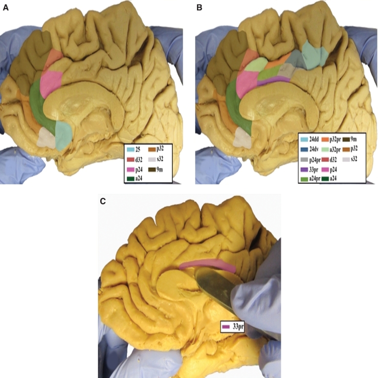
Though confusing at first glance, the schema for these areas is actually quite simple to learn. There are essentially 3 parallel, C-shaped rows of areas which follow the shape of the anterior cingulate from posterior to anterior, bending inferiorly to reach the subcallosal region. Immediately adjacent to the corpus callosum, in the depths of the callosal sulcus, is area 33prime, which represents the inner row. The middle row comprises area 24 subregions which extend superiorly in their posterior aspect into the paracentral lobule. The outer row comprises area 32 subregions which make up the superior border of the anterior cingulate gyrus and portions of the cingulate sulcus. Both area 24 subregions and area 32 subregions have prime and standard areas, with the prime areas being located posterior to the standard pairs. Thus, the area 24 row, from posterior to anterior, has the 24d regions, areas 24dd and 24dv, followed by the prime regions, area posterior 24prime and area anterior 24prime, followed by the standard pairs, p24 and a24. The area 32 row has, from posterior to anterior, p32prime, a32prime, followed by 3 standard regions, d32, p32, and s32. These parcellations all lead to subcallosal area 25. The anatomic location of these parcellations is shown on a cadaver brain in Figure 1. The combined tractography of the parcellations is shown in Figure 2.
FIGURE 1.
Anatomical location of medial frontal lobe parcellations shown on the right hemisphere of a cadaver brain. A, Medial view of the frontal lobe highlighting anterior cingulate parcellations. B, Medial view of the frontal lobe highlighting middle cingulate parcellations. C, Medial view of frontal lobe with widening of the sulcus of corpus callosum to show area 33pr. Corresponding labels are shown in the lower right corner of each figure.
FIGURE 2.
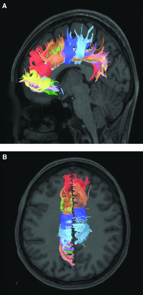
Combined structural connectivity of anterior cingulate parcellations. A, Sagittal and B, axial planes are shown. Tracks include 33pr (pink), 24dd (light blue), 24dv (dark blue), p24pr (gray), a24pr (light green), p24 (pink), a24 (dark green), p32pr (orange), a32pr (green), d32 (red), p32 (yellow), s32 (gray), and 25 (light blue).
Inner Row
Area 33prime
Where is it?
Area 33prime is located in the depths of the anterior callosal sulcus.
What are its borders?
Area 33prime borders the retrosplenial cortex (RSC) posteriorly and 3 area 24 subdivisions superiorly, areas a24pr, p24pr, and p24.
What is its functional connectivity?
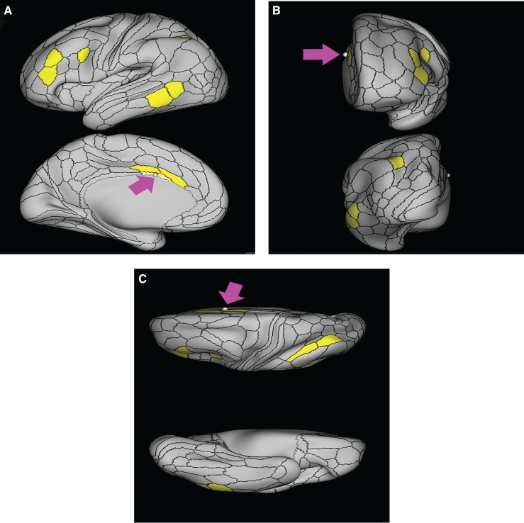
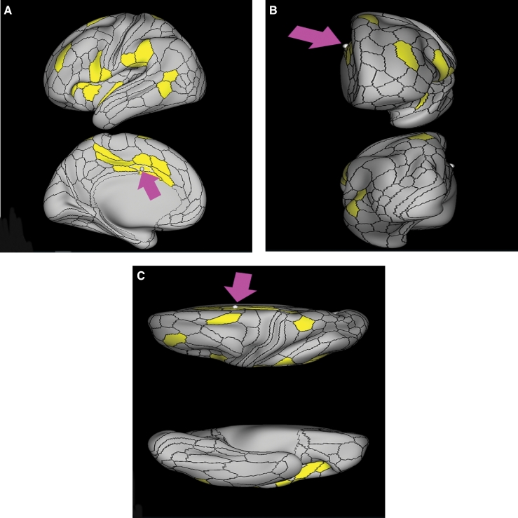
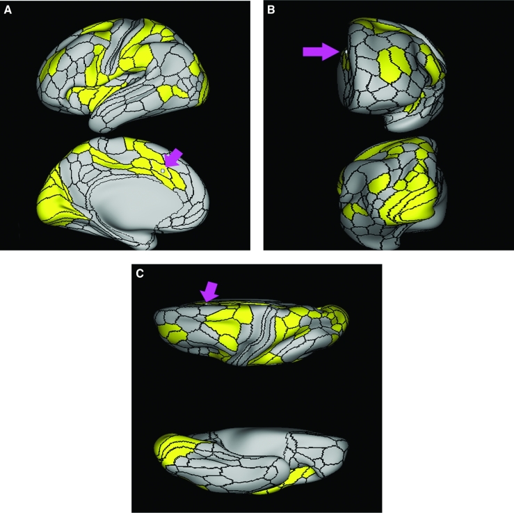
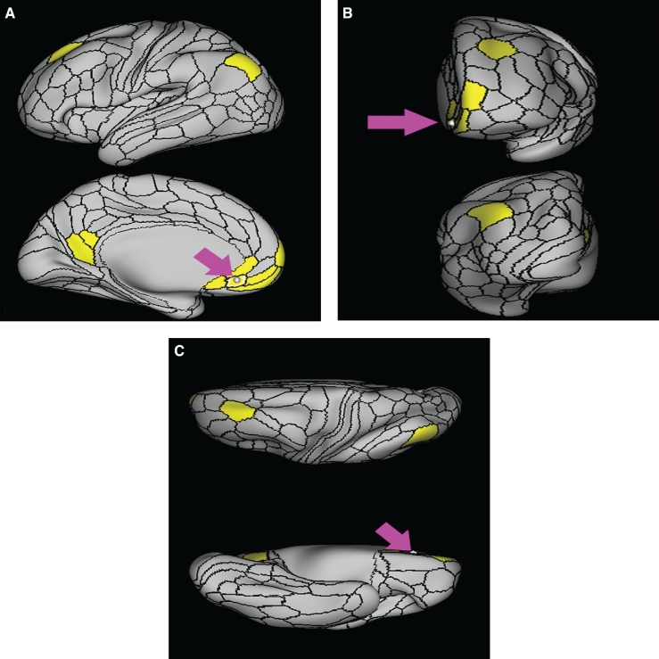
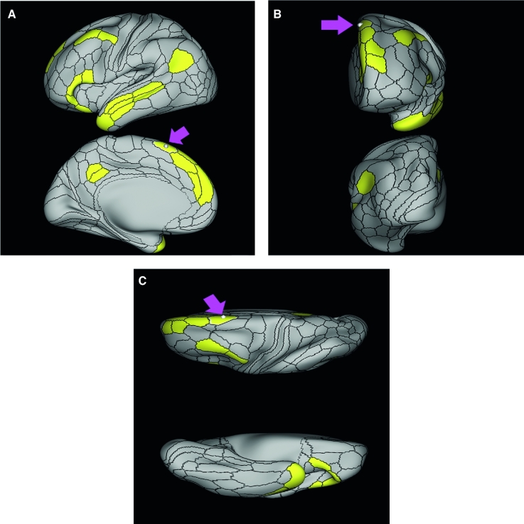
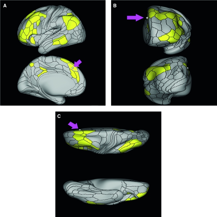
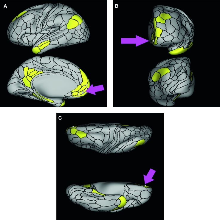
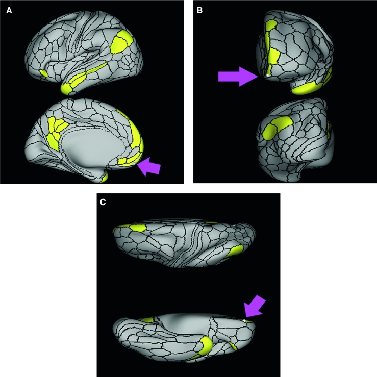
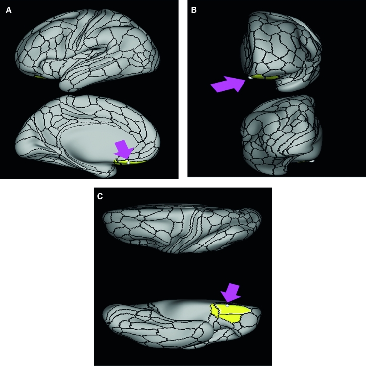
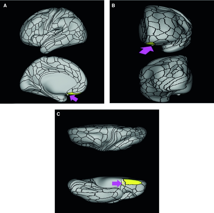
Area 33prime is connected to areas IFSa, IFJa, and p9-46v in the lateral frontal lobe, areas a24prime and p24prime in the medial frontal lobe, areas TE1p and PHT in the temporal lobe, and areas IP1 and IP2 in the parietal lobe (Figure 3).
FIGURE 3.
Functional connectivity of 33pr demonstrated on an inflated left hemisphere. A, Lateral and medial views. B, Rostral and caudal views. C, Dorsal and ventral views. Parcellations with the strongest functional connectivity are shown in yellow. Pink arrows designate the parcellation of interest.
What are its white matter connections?
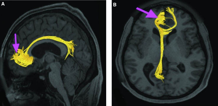
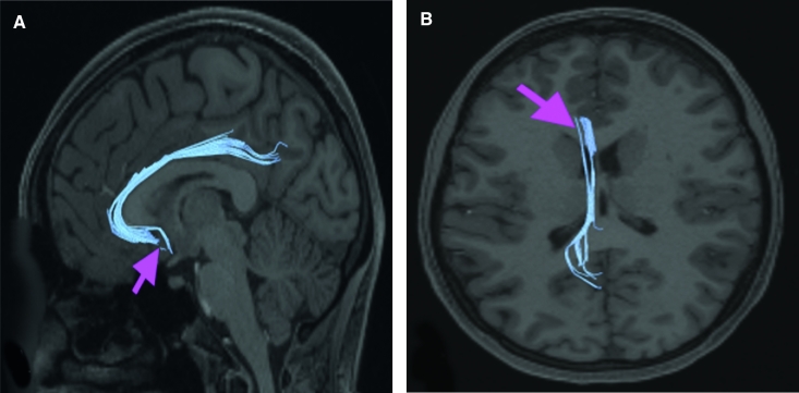
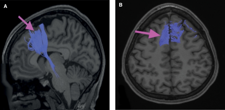
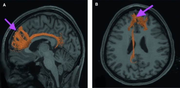
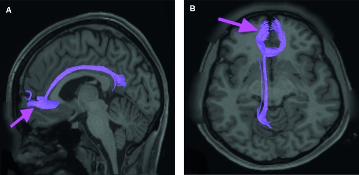
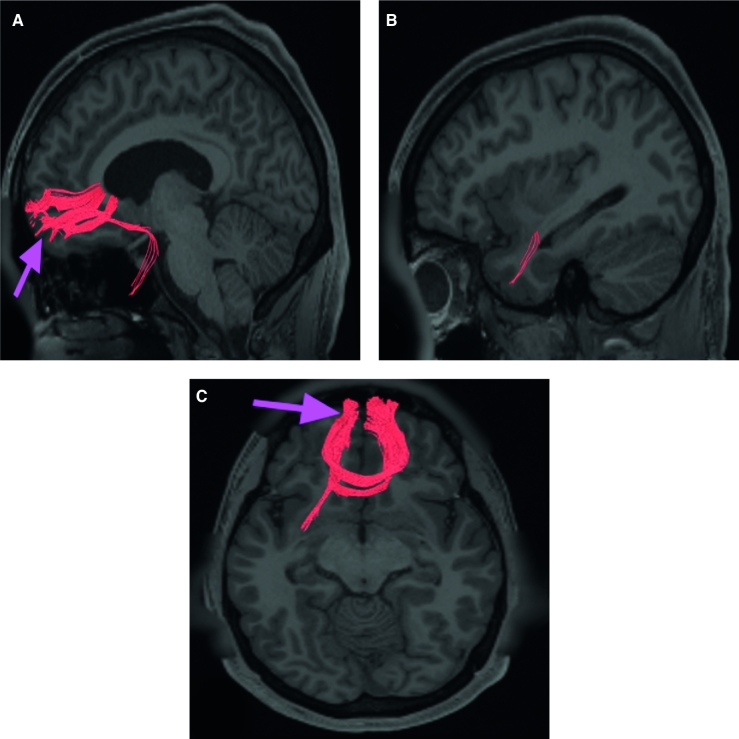
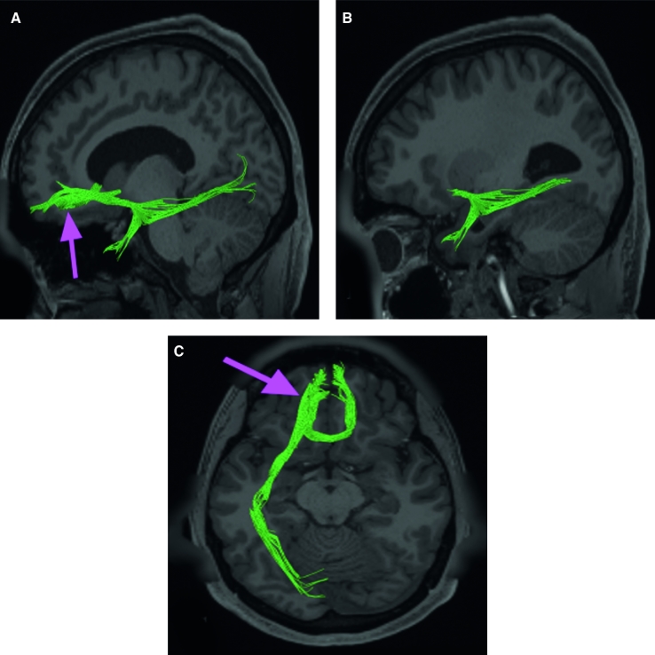
Area 33 prime is structurally connected to the cingulum. Fibers project anteriorly above the corpus callosum to end at a24, p32, and 10r, anterior fibers also curve around the rostrum of the corpus callosum to end at 25. Posterior cingulum fibers terminate at the precuneus to end at areas 7m and v23ab. Posterior fibers also curve around the splenium of the corpus callosum to end at the RSC (Figure 4).
FIGURE 4.
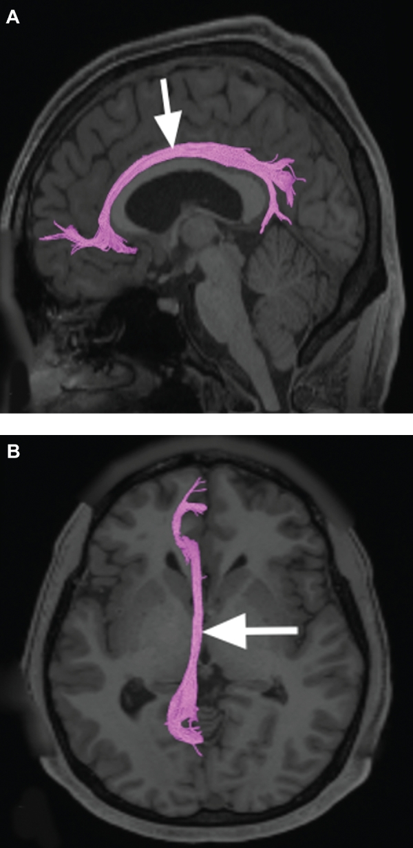
Structural connectivity of 33pr in the left hemisphere, shown on T1-weighted MR images. A, Sagittal and B, axial planes showing. Pink: white matter tracts of 33pr demonstrating connections with the cingulum.
What is known about its function?
Area 33prime is located on the ventral surface of the anterior cingulate cortex (ACC) and plays a major role in coordinating autonomic, visceromotor, and endocrine activity that accompany emotion.1
Middle Row
Area 24dd
Where is it?
Area 24dd (24 dorsal-dorsal) is located in the anterior inferior paracentral lobule straddling into the upper bank of the cingulate sulcus.
What are its borders?
Area 24dd has as its superior boundaries, from anterior to posterior, supplementary and cingulate eye field (SCEF), area 6mp, area 4, and area 5m. It continues into the marginal ramus of the cingulate sulcus, bordering area 5mv posteriorly and inferiorly. Its inferior border includes area 24dv and area 23c.
What is its functional connectivity?
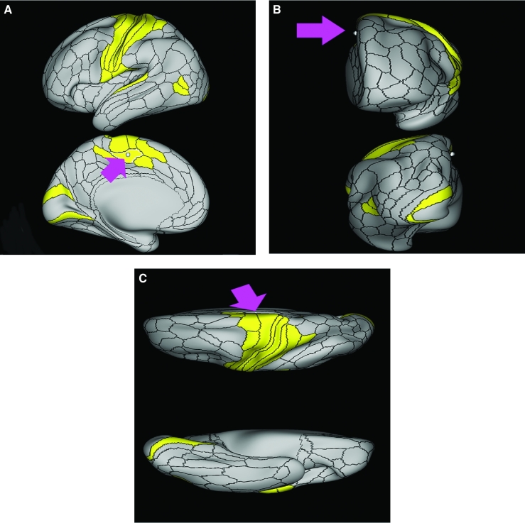
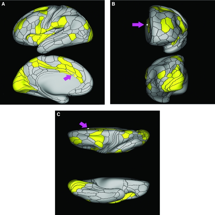
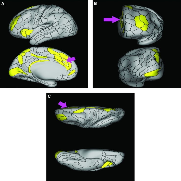
Area 24dd demonstrates functional connectivity to areas 1, 2, 3a, and 3b in the sensory strip, area 4 in the motor strip, areas 6mp, 6v, and 6d in the premotor regions, areas 5mv, SCEF, and 24dv in the middle cingulate regions, areas OP4, OP1, and 43 in the superior opercular areas, areas PBelt and A4 in the lower opercula and Heschl's gyrus regions, area 7PC in the parietal lobe, area V2 in the primary visual areas, and areas FST and MST in the lateral occipital lobe (Figure 5).
FIGURE 5.
Functional connectivity of 24dd demonstrated on an inflated left hemisphere. A, Lateral and medial views. B, Rostral and caudal views. C, Dorsal and ventral views. Parcellations with the strongest functional connectivity are shown in yellow. Pink arrows designate the parcellation of interest.
What are its white matter connections?
Area 24dd is structurally connected to the contralateral hemisphere. Fibers from 24dd project through the body of the corpus callosum to end at 24dd, 4, 5mv, 23c, SCEF, 6mp, and 24dv. Local short association bundles connect with 4, 5mv, 23c, SCEF, and 6mp (Figure 6).
FIGURE 6.
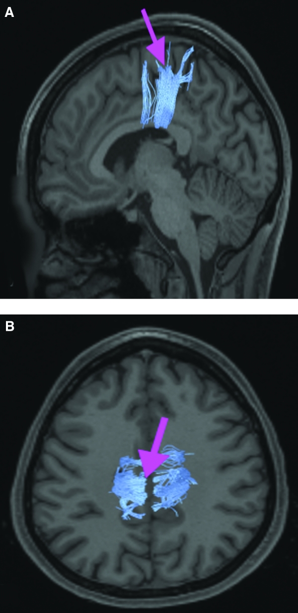
Structural connectivity of 24dd in the left hemisphere, shown on T1-weighted MR images. A, Sagittal and B, axial planes showing. Light blue: white matter tracts of 24dd demonstrating connections with the contralateral hemisphrere and local parcellations.
What is known about its function?
Area 24dd has been implicated in complex motor planning and regulation of muscles in the lower limb and lower trunk through coordination with the supplemental motor area and connections to the spinal cord.9
Area 24dv
Where is it?
Area 24dv (24 dorsal-ventral) is located in the anterior inferior paracentral lobule and straddles into the upper bank of the cingulate sulcus.
What are its borders?
Area 24dv has 24dd as its superior border, p32pr and a small portion of SCEF as its anterior border, p24pr as its inferior border, and 24dd as its posterior border. Note that this area causes the 24 areas and the 32 areas to be slightly out of sync in their anterior to posterior alignment.
What is its functional connectivity?
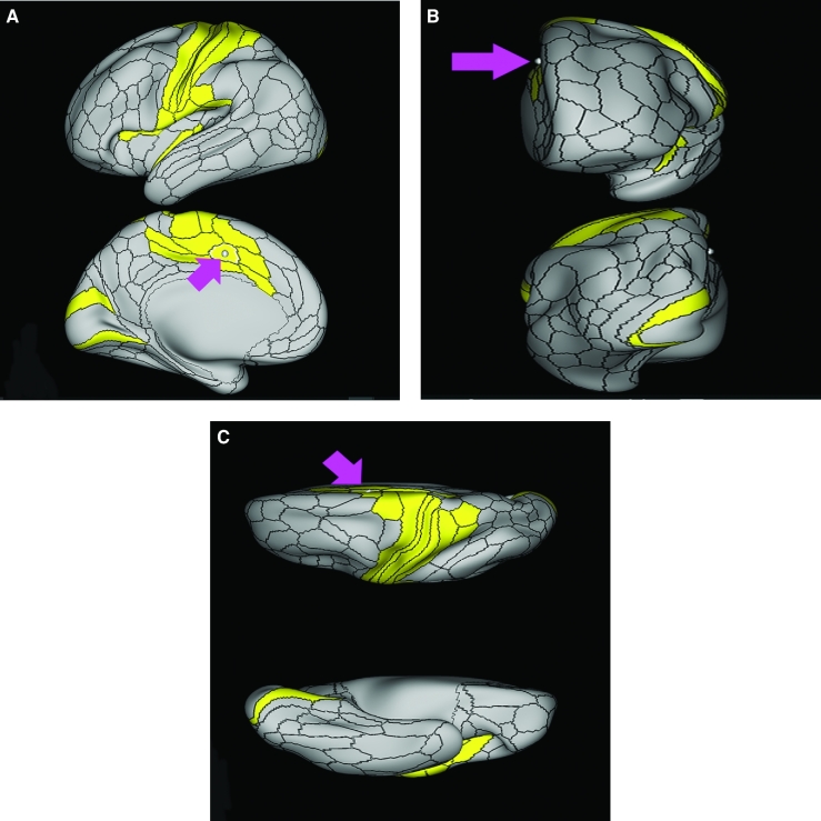
Area 24dv demonstrates functional connectivity to areas 1, 2, 3a, and 3b in the sensory strip, area 4 in the motor strip, area 6mp in the premotor regions, areas 5mv, SCEF, a24prime, p24prime, p32prime, 23c, and 24dd in the middle cingulate regions, areas FOP1, FOP3, FOP4, PFcm, OP4, OP1, and 43 in the superior opercular areas, areas PoI1 and 52 in the lower opercula and Heschl's gyrus regions, areas 7AL, 7PC, and PFop in the parietal lobe, and area V2 in the primary visual areas (Figure 7).
FIGURE 7.
Functional connectivity of 24dv demonstrated on an inflated left hemisphere. A, Lateral and medial views. B, Rostral and caudal views. C, Dorsal and ventral views. Parcellations with the strongest functional connectivity are shown in yellow. Pink arrows designate the parcellation of interest.
What are its white matter connections?
Area 24dv is structurally connected to the contralateral hemisphere, marginal branch of the cingulate sulcus and precuneus. Contralateral connections course through the body of the corpus callosum to 24dv, SCEF, and p32pr. Fibers from 24dv project posterior to the marginal branch of the cingulate sulcus and precuneus areas to 23c and 31a. Local short association fibers connect with SCEF, p32pr, and 24dv (Figure 8).
FIGURE 8.
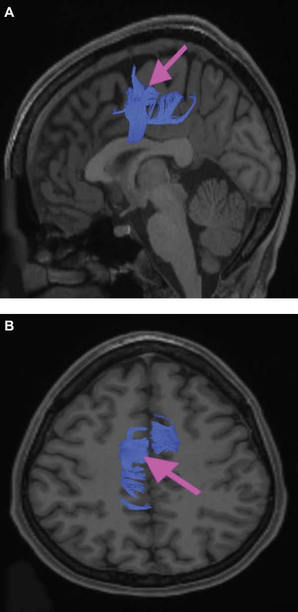
Structural connectivity of 24dv in the left hemisphere, shown on T1-weighted MR images. A, Sagittal and B, axial planes showing. Dark blue: white matter tracts of 24dv demonstrating connections with the contralateral hemisphere, marginal branch of the cingulate sulcus, and precuneus.
What is known about its function?
Area 24dv has been implicated in complex motor planning and regulation of muscles in the upper limb and upper trunk muscles through coordination with the supplemental motor area and connections to the spinal cord.9
Area p24pr
Where is it?
Area p24pr (posterior 24 prime) is located in the middle cingulate gyrus.
What are its borders?
Area p24pr borders area 24dv superiorly, a24pr anteriorly, 33prime inferiorly, and 23d posteriorly.
What is its functional connectivity?
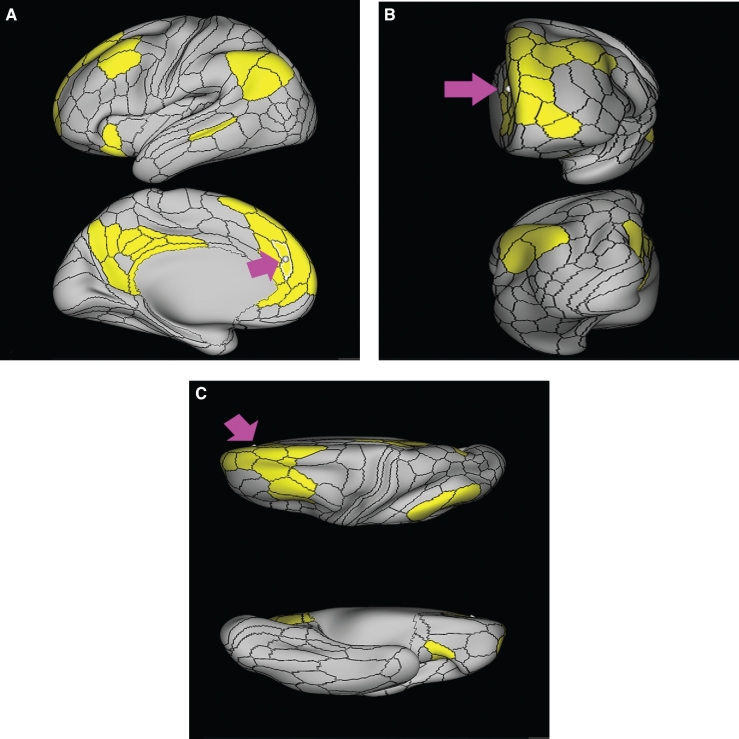
Area p24pr demonstrates functional connectivity to areas 33prime, 24dd, 24dv, 5mv, 23d, a24prime, and p32prime in the cingulate areas, areas 6ma and 6r in the premotor areas, area 46 in the lateral frontal lobe, areas FOP4, FOP5, PFcm, MI, 43, and PoI1 in the insula opercular areas, areas PF, PFop, and 7AL in the parietal lobe, and area TPOJ2 in the lateral occipital lobe (Figure 9).
FIGURE 9.
Functional connectivity of p24pr demonstrated on an inflated left hemisphere. A, Lateral and medial views. B, Rostral and caudal views. C, Dorsal and ventral views. Parcellations with the strongest functional connectivity are shown in yellow. Pink arrows designate the parcellation of interest.
What are its white matter connections?
Area p24pr is structurally connected to the marginal branch of the cingulate sulcus and precuneus. Fibers from p24pr project posterior to the marginal branch of the cingulate sulcus and precuneus to end at parcellations 23c, 31a, 31pd, and 7m (Figure 10).
FIGURE 10.
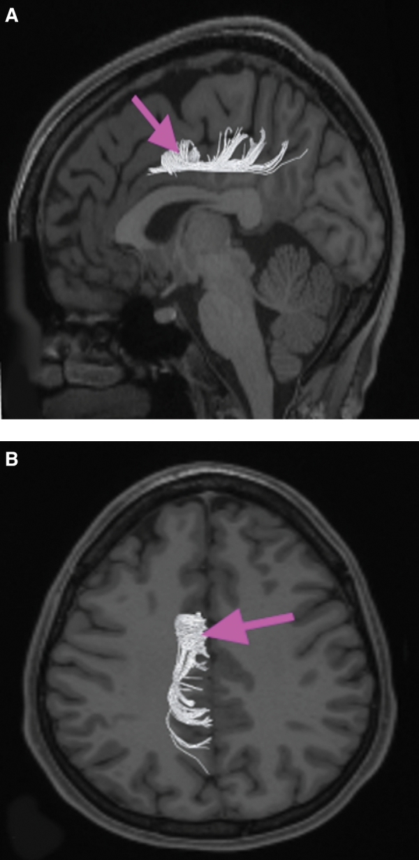
Structural connectivity of p24pr in the left hemisphere, shown on T1-weighted MR images. A, Sagittal and B, axial planes showing. Gray: white matter tracts of p24pr demonstrating connections with the marginal branch of the cingulate sulcus and precuneus.
What is known about its function?
Area p24pr has been implicated as part of the “cognitive division” of the ACC and is involved in stimulus and response selection during cognitively demanding tasks that may require movement.2,10
Area a24pr
Where is it?
Area a24pr (anterior 24prime) is located in the middle cingulate gyrus. It is primarily located in the superior half of the gyrus, and straddles into the inferior bank of the cingulate sulcus.
What are its borders?
Area a24pr borders area 33prime inferiorly, p24pr posteriorly, p24 anteriorly, and it shares its portion of the cingulate with p32pr superiorly.
What is its functional connectivity?
Area a24pr is connected to areas 33prime, 5mv, 23d, 24dv, p24, p24prime, a32prime, and p32prime in the cingulate areas, areas SCEF, FEF, PEF, 6a, and 6r in the premotor areas, area 9–46d, and 46 in the lateral frontal lobe, areas FOP1, FOP3, FOP4, FOP5, OP4, PFcm, MI, 43, 52, PoI2, and PoI1 in the insula opercular areas, area PHT in the temporal lobe, areas PGp, PF, PFop, and 7AL in the lateral parietal lobe, areas DVT, 7am, and PCV in the medial parietal lobe, areas V1, V2, V3, and V4 in the medial occipital lobe, and areas V6, V6a, V3a, V3b, and V7 in the dorsal visual stream areas (Figure 11).
FIGURE 11.
Functional connectivity of a24pr demonstrated on an inflated left hemisphere. A, Lateral and medial views. B, Rostral and caudal views. C, Dorsal and ventral views. Parcellations with the strongest functional connectivity are shown in yellow. Pink arrows designate the parcellation of interest.
What are its white matter connections?
Area a24pr is structurally connected to the cingulum. Some individuals have contralateral connections through the body of the corpus callosum but this tract is inconsistent. Fibers project anteriorly above the corpus callosum to end at p32, anterior cingulum fibers also curve around the rostrum of the corpus callosum to end at 25. Posterior cingulum fibers end at the precuneus to end at areas POS1 and 31pv, posterior fibers also curve around the splenium of the corpus callosum to end at RSC (Figure 12).
FIGURE 12.
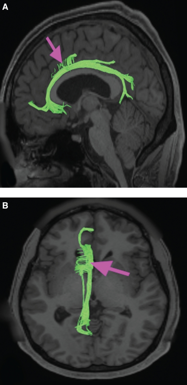
Structural connectivity of a24pr in the left hemisphere, shown on T1-weighted MR images. A, Sagittal and B, axial planes showing. Light green: white matter tracts of a24pr demonstrating connections with the cingulum.
What is known about its function?
Area a24pr shows little response to affective processes, but is implicated in cognitive response selection, and has been implicated in word and sentence selection during language-based tasks.2
Area p24
Where is it?
Area p24 (posterior 24) is located in the anterior cingulate gyrus. It covers the entire gyrus and is just anterosuperior to the genu of the corpus callosum.
What are its borders?
Area p24 borders areas d32 and a32pr superiorly and area a24 anteroinferiorly. It has a posterior boundary with a24pr and 33pr. Its inferior border is the callosal sulcus.
What is its functional connectivity?
Area p24 demonstrates functional connectivity to areas a24, d32, 23c, a24prime, and a32prime in the medial frontal lobe, area 9–46d in the lateral frontal lobe, areas RSC and POS2 in the medial parietal lobe, and area V1 in the medial occipital lobe (Figure 13).
FIGURE 13.
Functional connectivity of p24 demonstrated on an inflated left hemisphere. A, Lateral and medial views. B, Rostral and caudal views. C, Dorsal and ventral views. Parcellations with the strongest functional connectivity are shown in yellow. Pink arrows designate the parcellation of interest.
What are its white matter connections?
Area p24 is structurally connected to the cingulum. Cingulum fibers project anteriorly to end at frontal lobe parcellations p32 and 10r, anterior fibers also curve around the rostrum of the corpus callosum to end at 25. Posterior cingulum fibers end at the precuneus, near the splenium of the corpus callosum to areas POS1, v23ab and RSC (Figure 14). Local short association bundles connect with a24, d32, and a32pr.
FIGURE 14.
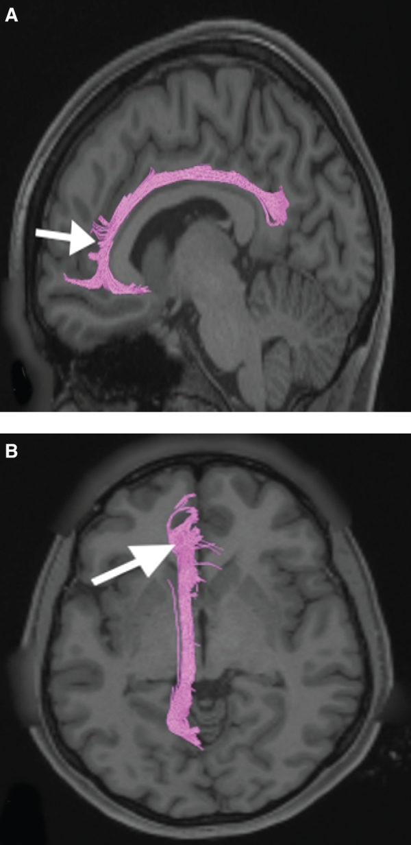
Structural connectivity of p24 in the left hemisphere, shown on T1-weighted MR images. A, Sagittal and B, axial planes showing. Pink: white matter tracts of p24 demonstrating connections with the cingulum.
What is known about its function?
Area p24 is functionally distinct from its anterior counterpart in that it plays a more prominent role in selective attention, coordination of conscious eye movements with complicated finger movement sequences, and stimulus/response selection.4,8
Area a24
Where is it?
Area a24 (anterior 24) is located in the anterior cingulate gyrus just anterior to the genu of the corpus callosum.
What are its borders?
Area a24 borders area p24 superiorly and area 25 posteriorly. It abuts the genu of the corpus callosum. Its inferior border is s32 and its anterior border is made up of p32 and 9m.
What is its functional connectivity?
Area a24 demonstrates functional connectivity to areas p24, d32, p32, s32, 10d, 10r, 9p, and 9m in the medial frontal lobe, area 8ad in the lateral frontal lobe, area TE1a in the temporal lobe, area PGs in the lateral parietal lobe, and areas RSC, 31pv, 31pd, 23d, d23ab, v23ab, 7m, and POS1 in the posterior cingulate areas (Figure 15).
FIGURE 15.
Functional connectivity of a24 demonstrated on an inflated left hemisphere. A, Lateral and medial views. B, Rostral and caudal views. C, Dorsal and ventral views. Parcellations with the strongest functional connectivity are shown in yellow. Pink arrows designate the parcellation of interest.
What are its white matter connections?
Area a24 is structurally connected to the cingulum. Cingulum fibers project anteriorly to end near the rostrum of the corpus callosum at 25. Posterior cingulum fibers end at the precuneus, near the splenium of the corpus callosum to areas 31pv and 23d (Figure 16). Local short association bundles connect with p32 and 9m.
FIGURE 16.
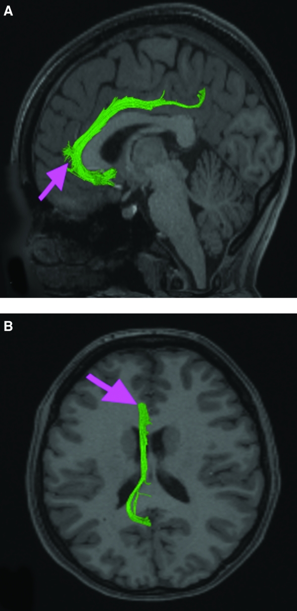
Structural connectivity of a24 in the left hemisphere, shown on T1-weighted MR images. A, Sagittal and B, axial planes showing. Dark green: white matter tracts of a24 demonstrating connections with the cingulum.
What is known about its function?
Area a24 has been implicated as part of the “affect division” of the ACC, and has been linked to analysis of internal and external states to play a role in emotional expression and motivation.2,3
Outer Row
Area p32pr
Where is it?
Area p32pr (posterior 32 prime) is located in the posterior inferior portion of the superior frontal gyrus (SFG). It wraps into the superior bank of the cingulate sulcus.
What are its borders?
Area p32pr borders SCEF superiorly, 24dv posteriorly, a24pr inferiorly, and a32pr anteriorly.
What is its functional connectivity?
Area p32pr demonstrates functional connectivity to area 2 in the sensory strip, areas 5mv, 23d, 24dv, p24, p24prime, and a32prime, and a32prime in the cingulate areas, areas SCEF, PEF, 6a, 6v, 6ma, and 6mp in the premotor areas, area 9–46d, 46 in the lateral frontal lobe, areas FOP1, FOP3, FOP4, FOP5, OP4, PFcm, MI, 43, 52, PoI2, and PoI1 in the insula opercular areas, area PHT in the temporal lobe, areas PGp, PFt, PF, PFop, LIPd, 7PC, 7PL, and 7AL in the lateral parietal lobe, areas DVT and 7am in the medial parietal lobe, areas V1, V2, V3, and V4 in the medial occipital lobe, areas V6, V6a, V3a, V3b, and V7 in the dorsal visual stream areas, and area FST in the lateral occipital lobe (Figure 17).
FIGURE 17.
Functional connectivity of p32pr demonstrated on an inflated left hemisphere. A, Lateral and medial views. B, Rostral and caudal views. C, Dorsal and ventral views. Parcellations with the strongest functional connectivity are shown in yellow. Pink arrows designate the parcellation of interest.
What are its white matter connections?
Area p32pr is structurally connected to the cingulum and contralateral hemisphere. Cingulum fibers project posteriorly to from p32pr to end at precuneus areas 31a, 31pd, 31pv, PCV, and v23ab. Contralateral connections course through the body of the corpus callosum to end at p32pr, SCEF, and a32pr. Local short association fibers connect with p32pr, SCEF, and a32pr (Figure 18).
FIGURE 18.
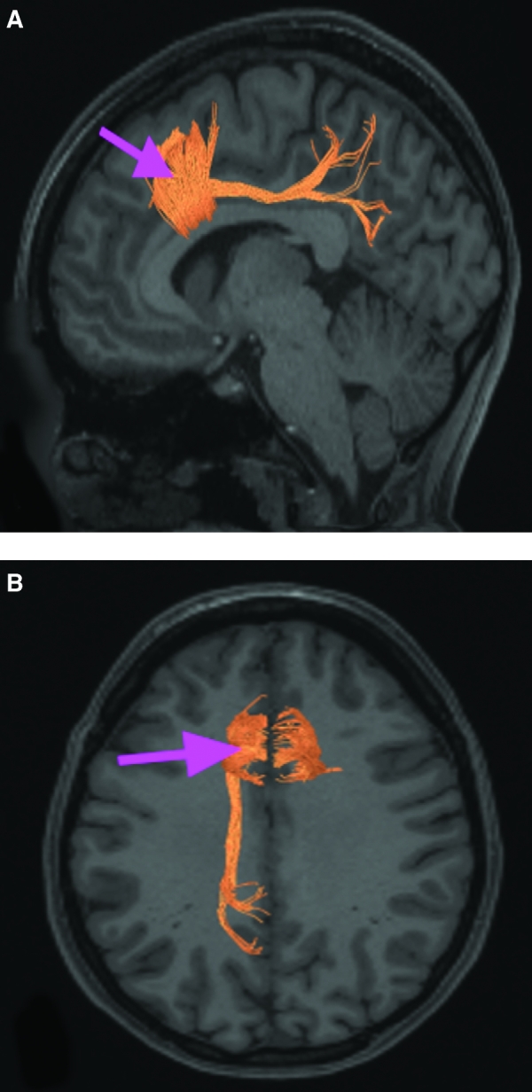
Structural connectivity of p32pr in the left hemisphere, shown on T1-weighted MR images. A, Sagittal and B, axial planes showing. Orange: white matter tracts of p32pr demonstrating connections with the contralateral hemisphere and cingulum.
What is known about its function?
Area p32pr has been implicated as part of the “cognitive division” of the ACC and is involved in stimulus and response selection in tasks that require attention for linguistic and sensory information.2,10
Area a32pr
Where is it?
Area a32pr (anterior 32 prime) is located in the posterior inferior portion of the SFG. It wraps into the superior bank of the cingulate sulcus.
What are its borders?
Area a32pr borders area 8BM superiorly, p32pr posteriorly, a24pr and p24 inferiorly, and d32 anteriorly.
What is its functional connectivity?
Area a32pr demonstrates functional connectivity to areas 8BM, SCEF, p24, d32, 23c, p24prime, and p32prime in the medial frontal lobe, areas a9-46v, 9–46d, and 46 in the lateral frontal lobe, areas FOP4, FOP5, AVI, and MI in the insula opercular areas, areas 7pm, RSC, and POS2 in the medial parietal lobe, and area V1 in the medial occipital lobe (Figure 19).
FIGURE 19.
Functional connectivity of a32pr demonstrated on an inflated left hemisphere. A, Lateral and medial views. B, Rostral and caudal views. C, Dorsal and ventral views. Parcellations with the strongest functional connectivity are shown in yellow. Pink arrows designate the parcellation of interest.
What are its white matter connections?
Area a32pr is structurally connected to the cingulum and contralateral hemisphere. Contralateral connections course through the body of the corpus callosum to end at a32pr, p32pr, 8BM, and 9m. Cingulum fibers project posteriorly from a32pr to end at precuneus areas 7m, 31a, 31pd, 31pv, RSC, and v23ab. Local short association fibers connect with p24, 8BM, SCEF, and d32 (Figure 20)
FIGURE 20.
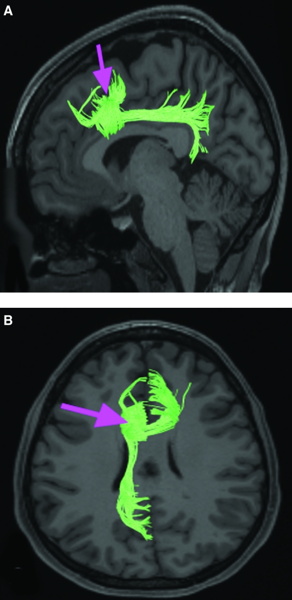
Structural connectivity of a32pr in the left hemisphere, shown on T1-weighted MR images. A, Sagittal and B, axial planes showing. Green: white matter tracts of a32pr demonstrating connections with the contralateral hemisphere and cingulum.
What is known about its function?
Research on this particular region suggests that a32pr helps to guide behavior by evaluating motivation, anticipating outcomes, recognizing reward values, and encoding errors to influence attention allocation and motor preparation.11,12
Area d32
Where is it?
Area d32 (dorsal 32) is a vertically oriented area in the inferior SFG. It is somewhat different in orientation to the other cingulate areas.
What are its borders?
Area d32 borders area 8BM superiorly, and area 9m anteriorly. Its posterior border is a32pr and its inferior border is p24.
What is its functional connectivity?
Area d32 demonstrates functional connectivity to areas p32, a24, p24, a32prime, p32, 9m, 10d, 8BM, and SFL in the medial frontal lobe, areas 8AV, 8AD, 8BL, 8C, 9a, 9p, a10p, p10p, i6-8, s6-8, and 47s in the lateral frontal lobe, area AVI in the insula, area STSvp in the temporal lobe, areas PFm, PGs, and PGi in the lateral parietal lobe, and areas RSC, 31a, 31pv, 31pd, d23ab, 7m, POS2, and POS1 in the posterior cingulate areas (Figure 21).
FIGURE 21.
Functional connectivity of d32 demonstrated on an inflated left hemisphere. A, Lateral and medial views. B, Rostral and caudal views. C, Dorsal and ventral views. Parcellations with the strongest functional connectivity are shown in yellow. Pink arrows designate the parcellation of interest.
What are its white matter connections?
Area d32 is structurally connected to the contralateral hemisphere, the cingulum, and abundant local parcellations. Connections to the contralateral hemisphere course through the corpus callosum to areas d32, 8BM, and 9m. Cingulum fibers project posteriorly to precuneus areas 31pv, POS1, v23ab, and RSC. Local short association bundles connect with 8BM, 9m a32pr, and 10d (Figure 22).
FIGURE 22.
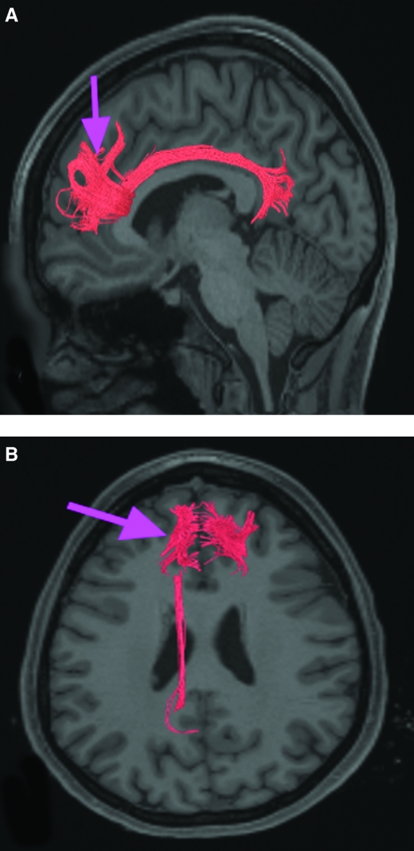
Structural connectivity of d32 in the left hemisphere, shown on T1-weighted MR images. A, Sagittal and B, axial planes showing. Red: white matter tracts of d32 demonstrating connections with the contralateral hemisphere, local parcellations, and cingulum.
What is known about its function?
Area d32 has been implicated in the evaluation of reinforcement and punishment in assigning value to social information.4
Area p32
Where is it?
Area p32 (posterior 32) is the central portion of the medial SFG. It borders the anterior bend of the callosal sulcus.
What are its borders?
Area p32 borders area 9m superiorly, and 10d anteriorly. Its inferior borders include areas 10r and s32. Its posterior border includes a24 and a small portion of s32.
What is its functional connectivity?
Area p32 demonstrates functional connectivity to areas d32, a24, p24, a32prime, and 10r in the medial frontal lobe, area 8AD in the lateral frontal lobe, and areas RSC, 31a, 31pv, POS2, and POS1 in the posterior cingulate areas (Figure 23).
FIGURE 23.
Functional connectivity of p32 demonstrated on an inflated left hemisphere. A, Lateral and medial views. B, Rostral and caudal views. C, Dorsal and ventral views. Parcellations with the strongest functional connectivity are shown in yellow. Pink arrows designate the parcellation of interest.
What are its white matter connections?
Area p32 is structurally connected to the contralateral hemisphere and cingulum. Connections to the contralateral hemisphere course through the genu of the corpus callosum to areas p32, 9m, 10d, and 10v.
Cingulum fibers project posteriorly to precuneus areas POS1, 31pv, RSC, and v23ab. Local short association bundles connect with 9m, 10d, 10r, and p24 (Figure 24).
FIGURE 24.
Structural connectivity of p32 in the left hemisphere, shown on T1-weighted MR images. A, Sagittal and B, axial planes showing. Yellow: white matter tracts of p32 demonstrating connections with the contralateral hemisphere and cingulum.
What is known about its function?
Area p32 has been shown to play a role in the emotional and cognitive integration of information during social interaction tasks, as well as play some role in error monitoring.4,5
Area s32
Where is it?
Area s32 (subcallosal 32) lies in the subcallosal gyri.
What are its borders?
Area s32 has a24, p32, and 10r as its anterior and superior borders. Area 10v and the OFC region are its inferior boundaries. Its posterior boundary is area 25.
What is its functional connectivity?
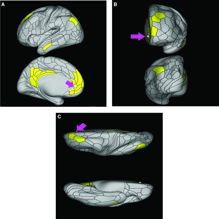
Area s32 demonstrates functional connectivity to areas a24, 25, 10d, and 10r in the medial frontal lobe, area 8AD in the lateral frontal lobe, area PGs in the lateral parietal lobe, and areas v23ab and POS1 in the posterior cingulate areas (Figure 25).
FIGURE 25.
Functional connectivity of s32 demonstrated on an inflated left hemisphere. A, Lateral and medial views. B, Rostral and caudal views. C, Dorsal and ventral views. Parcellations with the strongest functional connectivity are shown in yellow. Pink arrows designate the parcellation of interest.
What are its white matter connections?
Area s32 is structurally connected to local parcellations. S32 has posterior connections to 25 and anterior connections to p32 (Figure 26).
FIGURE 26.
Structural connectivity of s32 in the left hemisphere, shown on T1-weighted MR images. A, Sagittal and B, axial planes showing. Gray: white matter tracts of s32 demonstrating connections with the local parcellations.
What is known about its function?
Area s32 is heavily interconnected to other areas of the limbic system and thus plays a higher order role in emotional affect as well as reward expectation.4,5
Area 25
Where is it?
Area 25 is located in the most posterior portion of the subcallosal area.
What are its borders?
Area 25 borders area a24 and s32 anteriorly. Its inferior and posterior borders include the OFC and pOFC areas.
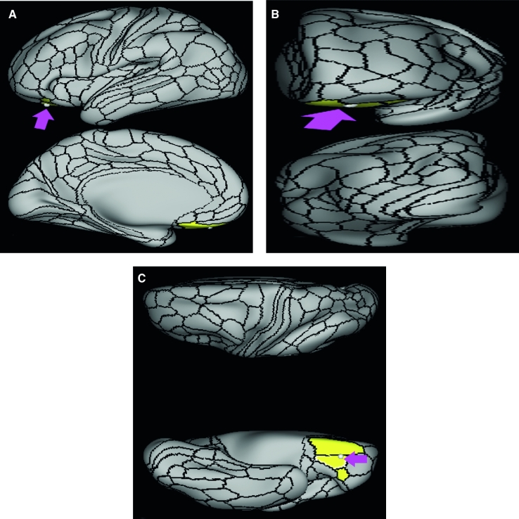
What is its functional connectivity?
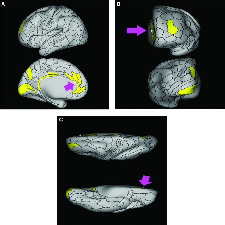
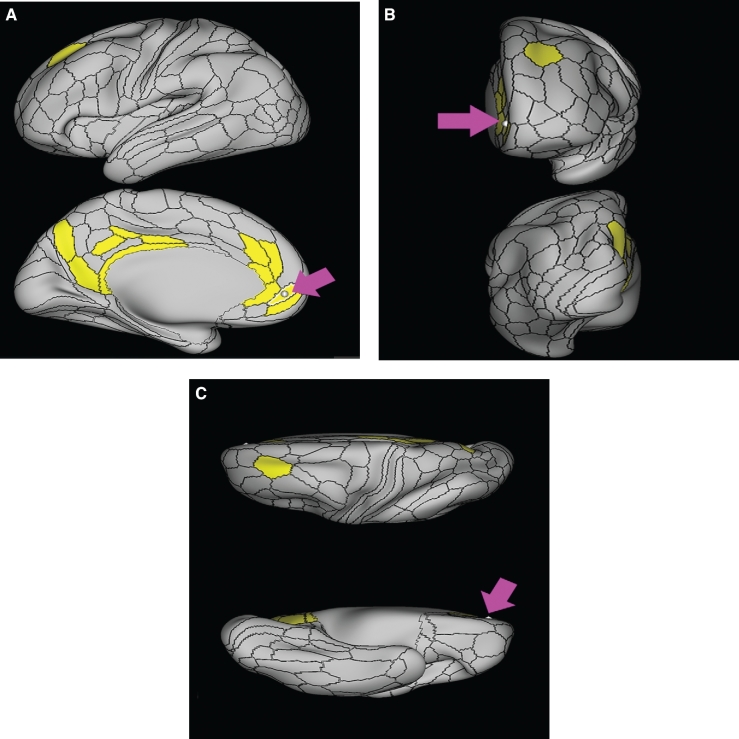
Area 25 demonstrates functional connectivity to area s32 (Figure 27).
FIGURE 27.
Functional connectivity of area 25 demonstrated on an inflated left hemisphere. A, Lateral and medial views. B, Rostral and caudal views. C, Dorsal and ventral views. Parcellations with the strongest functional connectivity are shown in yellow. Pink arrows designate the parcellation of interest.
What are its white matter connections?
Area 25 is connected to the cingulum. White matter tracts from this parcellation are variable. Some individual tracts connect to anterior parcellations; however, this is inconsistent across individuals. Fibers from 25 project posterior above the corpus callosum to areas v23ab and 31pv (Figure 28).
FIGURE 28.
Structural connectivity of area 25 in the left hemisphere, shown on T1-weighted MR images. A, Sagittal and B, axial planes showing. Light blue: white matter tracts of area 25 demonstrating connections with the cingulum.
What is known about its function?
Area 25 has been implicated as part of the “affect division” of the ACC and has been linked to conditioned emotional learning, emotional expression, assessment of motivational content, assignment of emotional valence to internal and external stimuli, and maternal-infant interactions.2,3
MEDIAL SFG REGIONS
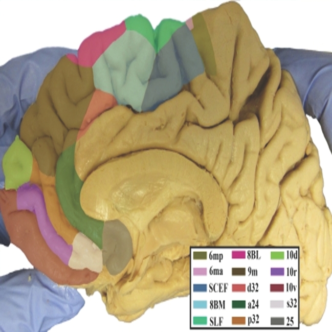
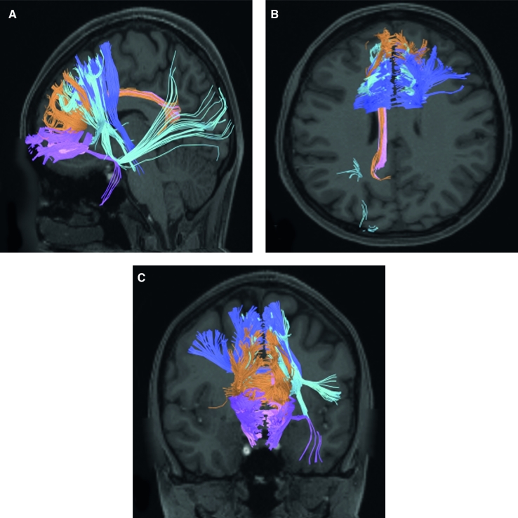
After accounting for the areas comprising the lateral frontal lobe and anterior cingulate gyrus, the remaining areas of the medial frontal bank of the SFG are quite simple. They are large areas that are mainly simple divisions of areas 8, 9, and 10. The anatomic location of these parcellations is shown on a cadaver brain in Figure 29. The combined tractography of the parcellations is shown in Figure 30.
FIGURE 29.
Anatomical location of medial frontal lobe parcellations shown on the right hemisphere of a cadaver brain. Medial view of the frontal lobe. Corresponding labels are shown in the lower right corner of each figure.
FIGURE 30.
Combined structural connectivity of medial superior frontal gyrus parcellations. A, Sagittal, B, axial, and C, coronal planes are shown. Tracks include SCEF (dark blue), 8Bm (blue), 9m (brown), 10r (purple), and 10v (red).
Inner Row
Area SCEF
Where is it?
Area SCEF (supplementary and cingulate eye field) is located in the posterior medial SFG.
What are its borders?
Area SCEF borders area 8BM anteriorly, areas 6ma and SFL superiorly, areas 6mp and 24dd posteriorly, and areas 24dv and p32pr inferiorly.
What is its functional connectivity?
Area SCEF demonstrates functional connectivity to areas 1, 2, 3a, and 3b in the sensory strip, area 4 in the motor strip, areas PEF, FEF, 55b, 6ma, 6mp, 6a, 6r, and 6v in the premotor regions, areas a24prime, p32prime, a32prime, 5mv, and 23c in the middle cingulate regions, areas IFJa, 46, and 9–46d in the lateral frontal lobe areas OP4, OP1, PFcm, 43, FOP1, FOP2, FOP3 FOP4, and FOP5 in the superior insula opercular regions, areas PSL, 52, A4, MI, PoI1, and PoI2 in the lower opercula and Heschl's gyrus regions, area PHT in the temporal lobe, areas AIP, MIP, VIP, LIPd, LIPv, PFop, PF, PFt, IP0, IPS1, 7AL, 7PL, and 7PC in the lateral parietal lobe, areas 7am and DVT in the medial parietal lobe, areas V1, V2, V3, and V4 in the medial occipital lobe, areas V3a, V3b, V6, V6a, and V7 in the dorsal visual stream, area FFC of the ventral visual stream, and areas PH, TPOJ2, LO3, MST, and FST of the lateral occipital lobe (Figure 31).
FIGURE 31.
Functional connectivity of SCEF demonstrated on an inflated left hemisphere. A, Lateral and medial views. B, Rostral and caudal views. C, Dorsal and ventral views. Parcellations with the strongest functional connectivity are shown in yellow. Pink arrows designate the parcellation of interest.
What are its white matter connections?
Area SCEF is structurally connected to the contralateral hemisphere and thalamus. Contralateral connections course through the body of the corpus callosum to end at SCEF, 8BL, SFL, and 8BM. Thalamic projections travel through the ventral thalamus to the brainstem (Figure 32). Local short association bundles connect with SF, 8BM, SFL, and 8BL.
FIGURE 32.
Structural connectivity of SCEF in the left hemisphere, shown on T1-weighted MR images. A, Medial sagittal view and B, axial view. Dark blue: white matter tracts of SCEF demonstrating connections with the contralateral hemisphere and thalamus.
What is known about its function?
Area SCEF is a higher order oculomotor center implicated in appraising all possible oculomotor behaviors to direct primary oculomotor centers in goal-directed behavior.13
Area 8BM
Where is it?
Area 8BM (8B medial) is located in the posterior medial SFG.
What are its borders?
Area 8BM borders area 9m anteriorly and SCEF posteriorly. It borders the following area 24 subdivisions superiorly: a24pr, p24pr, and p24. Its inferior border contains areas d32 and a32pr, and its superior boundary includes 8BL and SFL.
What is its functional connectivity?
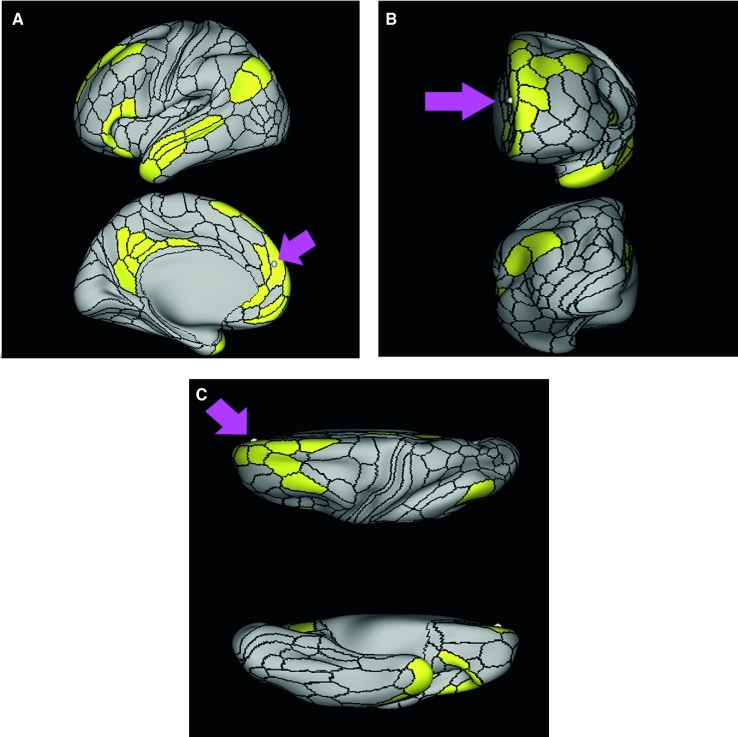
Area 8BM demonstrates functional connectivity to areas i6-8, s6-8, a10p, a9-46v, p9-46v, 8C, 8BL, 8AD, and 8AV in the dorsolateral frontal lobe, areas SFL, a32prime, and d32 in the medial frontal lobe, areas IFSa, IFSp, IFJp, 44, 45, a47r, and p47r in the inferior frontal lobe, area 55b in the premotor areas, area AVI in the insula, areas TE1m, TE1p, and STSvp in the temporal lobe, areas LIPv, IP1, IP2, PFm, PGi, and PGs in the lateral parietal lobe, and areas 7pm, 31a, and d23ab in the medial parietal lobe (Figure 33).
FIGURE 33.
Functional connectivity of 8Bm demonstrated on an inflated left hemisphere. A, Lateral and medial views. B, Rostral and caudal views. C, Dorsal and ventral views. Parcellations with the strongest functional connectivity are shown in yellow. Pink arrows designate the parcellation of interest.
What are its white matter connections?
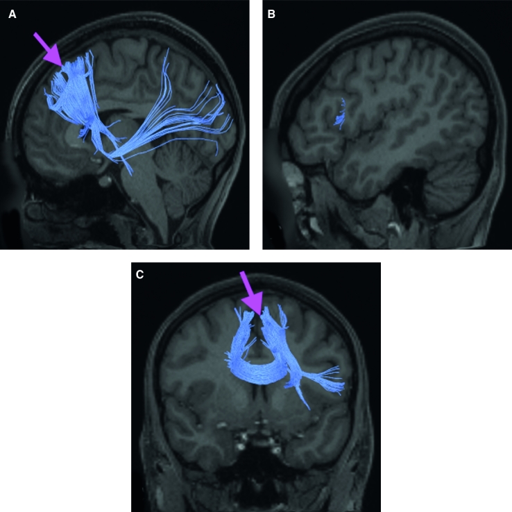
Area 8BM is connected to the contralateral hemisphere, frontal aslant tract, inferior front-occipital fasciculus, and thalamus. Contralateral connections course through the corpus callosum to end at 8BM and 9m. Frontal aslant tract fibers from 8BM project inferolaterally to end at area 44. Thalamic connections run inferior to 8BM and continue in the brainstem. Fibers with the inferior fronto-occipital fasciculus project inferior and posterior through the extreme/external capsule through the temporal lobe to end at parietal and occipital connections 7PC, V1, V2, and V3. Local short association bundles connect with 9m, d32, and SCEF (Figure 34).
FIGURE 34.
Structural connectivity of 8Bm in the left hemisphere, shown on T1-weighted MR images. Sagittal views of A, medial and B, lateral planes, and C, coronal view showing. Blue: white matter tracts of 8Bm demonstrating connections with the contralateral hemisphere, frontal aslant tract, inferior front-occipital fasciculus and thalamus.
What is known about its function?
Area 8Bm is involved in maintaining visuospatial information as well as coordinating and coding visuospatial information in terms of oculomotor and other body-centered coordinate systems.14
Area 9m
Where is it?
Area 9m (9 medial) is located in the anterior medial SFG.
What are its borders?
Area 9m shares borders with numerous areas. Its posterosuperior border includes areas 8BM and 8BL, its superior border includes areas 9a and area 9p. Its inferior and posterior border forms a wedge which is surrounded by areas p32, a24, p24, and d32 (from anterior to posterosuperior).
What is its functional connectivity?
Area 9m demonstrates functional connectivity to areas 9a, 9p, 10d, 8BL, 8AD, and 8AV in the dorsolateral frontal lobe, areas SFL, a24, 10r, 10v, and d32 in the medial frontal lobe, areas 44, 45, 47s, and 47l in the inferior frontal lobe, area AVI in the insula, areas TGd, TE1a, STSdp, STSva, and STSvp in the temporal lobe, areas PGi and PGs in the lateral parietal lobe, and areas 7m, POS1, 31pv, 31pd, 23d, v23ab, and d23ab in the medial parietal lobe (Figure 35).
FIGURE 35.
Functional connectivity of 9m demonstrated on an inflated left hemisphere. A, Lateral and medial views. B, Rostral and caudal views. C, Dorsal and ventral views. Parcellations with the strongest functional connectivity are shown in yellow. Pink arrows designate the parcellation of interest.
What are its white matter connections?
Area 9m is structurally connected to the contralateral hemisphere and cingulum. Connections to the contralateral hemisphere course through the corpus callosum to 9m, 8BL, and 8BM. Cingulum fibers project posteriorly, above the corpus callosum to end at precuneus areas 31pv, v23ab, and POS1. Local short association bundles connect with p32, 10d, and a24 (Figure 36).
FIGURE 36.
Structural connectivity of 9m in the left hemisphere, shown on T1-weighted MR images. A, Medial sagittal view and B, axial plane showing. Brown: white matter tracts of 9m demonstrating connections with the contralateral hemisphere and cingulum.
What is known about its function?
Area 9m is a newer parcellation of the pre-existing area 9, which is a major constituent of the dorsolateral prefrontal cortex. This region shows increased activity when monitoring multiple pieces of spatial information.14
Area 10r
Where is it?
Area 10r (10 rostral) is located in the anterior inferior portion of the medial SFG.
What are its borders?
Area 10r borders area 10d and 10pp anteriorly and inferiorly, respectively. Its superior boundary is area p32 and its posterior boundary is area s32.
What is its functional connectivity?
Area 10r demonstrates functional connectivity to areas 9p, 10d, and 8AD in the dorsolateral frontal lobe, areas 9m, a24, 10v, s32, and p32 in the medial frontal lobe, areas TGd, TE1a, STSva, PHA1, and the hippocampus in the temporal lobe, areas PGi and PGs in the lateral parietal lobe, and areas 7m, POS1, 31pv, 31pd, v23ab, and d23ab in the medial parietal lobe (Figure 37).
FIGURE 37.
Functional connectivity of 10r demonstrated on an inflated left hemisphere. A, Lateral and medial views. B, Rostral and caudal views. C, Dorsal and ventral views. Parcellations with the strongest functional connectivity are shown in yellow. Pink arrows designate the parcellation of interest.
What are its white matter connections?
Area 10r is connected to the contralateral hemisphere and cingulum. Connections to the contralateral hemisphere course through the genu of the corpus callosum to end at areas10r and 10v. Cingulum fibers project posteriorly from 10r to precuneus areas v23ab, POS1, and RSC. Local short association bundles connect with p32, 10d, and 10v (Figure 38). White matter tracts in the right hemisphere have less consistent connections with the cingulum.
FIGURE 38.
Structural connectivity of 10r in the left hemisphere, shown on T1-weighted MR images. A, Medial sagittal view and B, axial plane showing. Purple: white matter tracts of 10r demonstrating connections with the contralateral hemisphere and cingulum.
What is known about its function?
The rostral portion of area 10 (or area 10r) is a newer parcellation of the pre-existing area 10. Research on this particular region suggests that area 10r plays an important role in stimulus-oriented attention, indicating its importance in concentration and working memory.15
Area 10v
Where is it?
Area 10v is located in the depths of the inferior anterior most region of medial SFG.
What are its borders?
Area 10v shares a long inferior border with the OFC. Areas 10pp and 10d form its anteroinferior borders, area 10r is its superior neighbor. It has some border with area s32 superiorly. It terminates in the OFC posteriorly.
What is its functional connectivity?
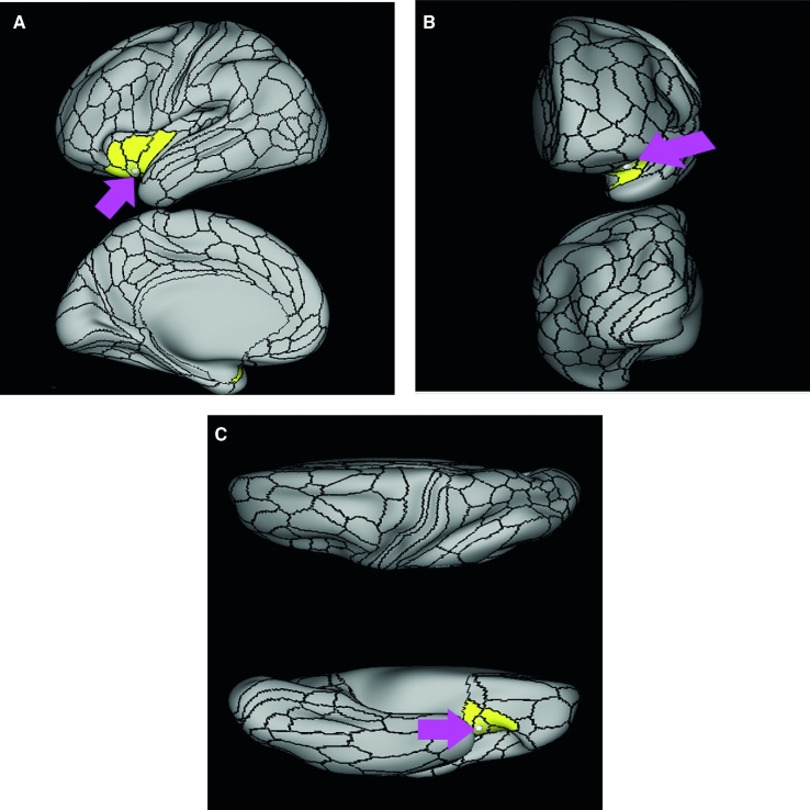
Area 10v demonstrates functional connectivity to areas 10d, 47l, and 8BL in the dorsolateral frontal lobe, areas 9m, 10r, and s32 in the medial frontal lobe, areas TGd, TE1a, STSva, and STSvp in the temporal lobe, areas PGi and PGs in the lateral parietal lobe, and areas 7m, POS1, 31pv, 31pd, and v23ab in the medial parietal lobe (Figure 39).
FIGURE 39.
Functional connectivity of 10v demonstrated on an inflated left hemisphere. A, Lateral and medial views. B, Rostral and caudal views. C, Dorsal and ventral views. Parcellations with the strongest functional connectivity are shown in yellow. Pink arrows designate the parcellation of interest.
What are its white matter connections?
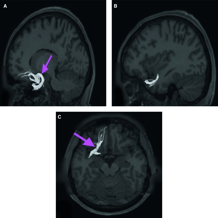
Area 10v is structurally connected to the contralateral hemisphere and the uncinate fasciculus. Connections with the uncinate fasciculus are not consistent across individuals. Contralateral connections course through the genu of the corpus callosum to end at areas 10v and 10d. Uncinate fasciculus fibers project through the limen insulae to temporal pole parcellation TGd (Figure 40).
FIGURE 40.
Structural connectivity of 10v in the left hemisphere, shown on T1-weighted MR images. Sagittal views of A, medial and B, lateral planes, and C, axial view showing. Red: white matter tracts of 10v demonstrating connections with the contralateral hemisphere and uncinate fasciculus.
What is known about its function?
Area 10v is a new ventral parcellation of the pre-existing area 10. This region is often associated with the ventromedial prefrontal cortex and is thought to play a role in behavioral decision making by integrating value appraisals from the OFC and ACC.16
ORBITOFRONTAL REGIONS
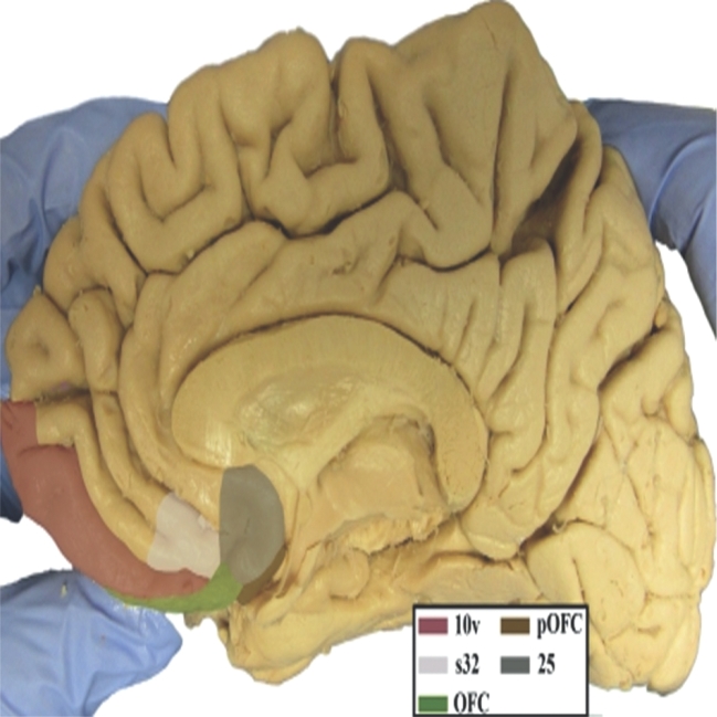
The orbitofrontal boundaries have been discussed in previous parts of this supplement, including discussions about areas 10 and 47 (and their subregions) which overlap with the edges of the orbitofrontal surface. The remaining orbitofrontal regions are discussed below. Of note, some of these regions were, admittedly, of limited power given limitations when imaging these areas. However, a standard nomenclature is beneficial in this area, as this region has generally been only roughly localized in the past into gyrus rectus and the orbitofrontal gyri. The anatomic location of these parcellations is shown on a cadaver brain in Figure 41. The combined tractography of the parcellations is shown in Figure 42.
FIGURE 41.
Anatomical location of medial frontal lobe parcellations shown on the right hemisphere of a cadaver brain. Medial view of the frontal lobe. Corresponding labels are shown in the lower right corner of each figure.
FIGURE 42.
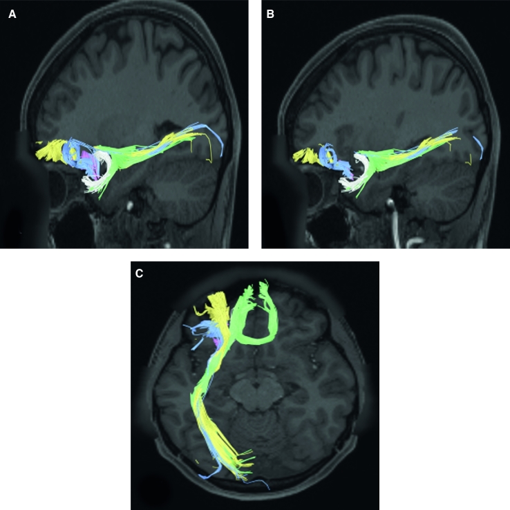
Combined structural connectivity of orbitofrontal region parcellations. Sagittal views of A, medial and B, lateral planes, and C, axial plane are shown. Tracks include OFC (green), pOFC (gray), 11l (yellow), 13l (pink), and 47m (blue).
Area OFC
Where is it?
Area OFC (orbitofrontal complex) is located in the gyrus rectus, the medial orbitofrontal cortices, and the intervening sulcus.
What are its borders?
Area OFC borders area 10pp anteriorly, areas 11l and 13l laterally, and area 10v on its superior medial bank. It abuts the pOFC posteriorly.
What is its functional connectivity?
Area OFC demonstrates functional connectivity to areas pOFC and 13L (Figure 43).
FIGURE 43.
Functional connectivity of OFC demonstrated on an inflated left hemisphere. A, Lateral and medial views. B, Rostral and caudal views. C, Dorsal and ventral views. Parcellations with the strongest functional connectivity are shown in yellow. Pink arrows designate the parcellation of interest.
What are its white matter connections?
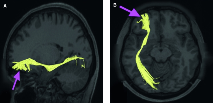
Area OFC is structurally connected to the contralateral hemisphere, the uncinate fasciculus, and inferior fronto-occipital fasciculus. Contralateral connections course through the genu of the corpus callosum to end at 10d, 10r, and 10v. Uncinate fasciculus fibers project through the limen insulae to temporal pole parcellation TGd. IFOF connections project posteriorly through the extreme/external capsule to occipital lobe area V1 (Figure 44).
FIGURE 44.
Structural connectivity of OFC in the left hemisphere, shown on T1-weighted MR images. Sagittal views of A, medial and B, lateral planes, and C, axial view showing. Green: white matter tracts of OFC demonstrating connections with the contralateral hemisphere, uncinate fasciculus, and inferior fronto-occipital fasciculus.
What is known about its function?
The Area OFC plays an integral role in the evaluation of rewards and punishment.17 Additionally, the OFC is suggested to be involved in self-regulation, behavioral inhibition, and emotional control by predicting the moment-to-moment value of stimuli, actions, and choices based on internal states.18
Area pOFC
Where is it?
Area pOFC (posterior orbitofrontal complex) is located in the posterior gyrus rectus and the medial posterior most portions of the orbitofrontal cortices.
What are its borders?
Area pOFC borders OFC anteriorly, areas 47s and Pir laterally, and area 25 superomedially. Its posterior border is with the anterior perforated substance.
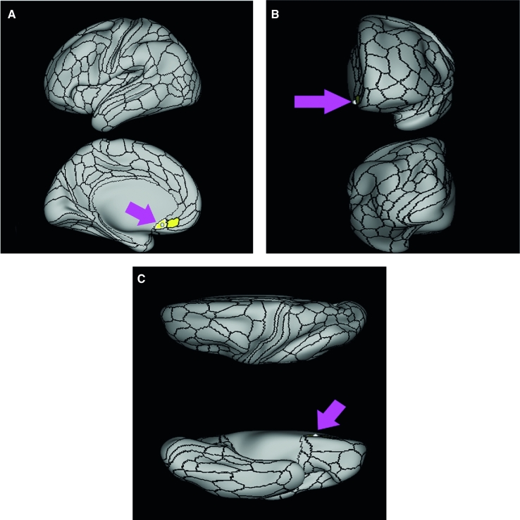
What is its functional connectivity?
Area pOFC demonstrates functional connectivity to the OFC (Figure 45).
FIGURE 45.
Functional connectivity of pOFC demonstrated on an inflated left hemisphere. A, Lateral and medial views. B, Rostral and caudal views. C, Dorsal and ventral views. Parcellations with the strongest functional connectivity are shown in yellow. Pink arrows designate the parcellation of interest.
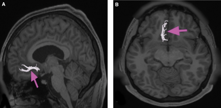
What are its white matter connections?
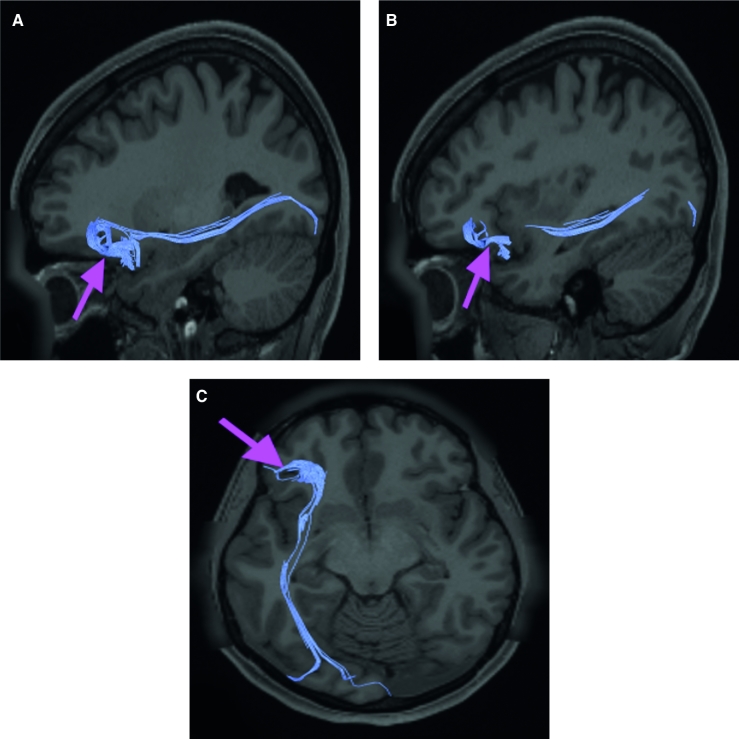
Area pOFC is connected to local parcellations. Short association bundles connect to the temporal pole through the insula to end at area TGd. There are anterior projections to OFC, 13l and PeEc (Figure 46)
FIGURE 46.
Structural connectivity of pOFC in the left hemisphere, shown on T1-weighted MR images. Sagittal views of A, medial and B, lateral planes, and C, axial view showing. Gray: white matter tracts of pOFC demonstrating connections with local parcellations.
What is known about its function?
This posterior parcellation of the OFC is thought to play a role in behavior and decision-making by evaluating sensory reinforcers, such as taste and odor.17
Area 11l
Where is it?
Area 11l is located in the anterior OFC.
What are its borders?
Area 11l borders area a10p anteriorly, area a47r laterally, areas 13l and 47m posteriorly, and OFC medially.
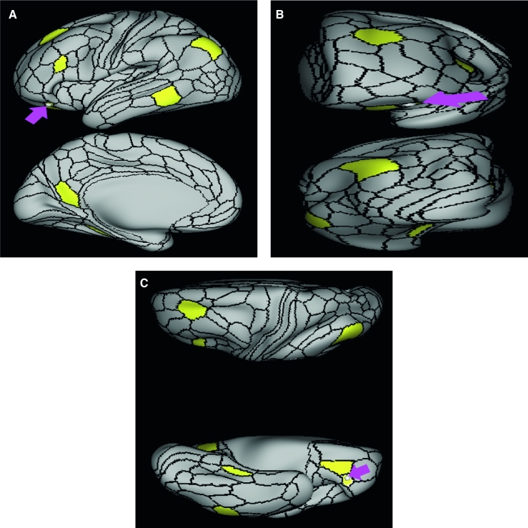
What is its functional connectivity?
Area 11l demonstrates functional connectivity to areas 46, a9-46v, p9-46v, and IFSa in the frontal lobe and IP2 in the parietal lobe (Figure 47).
FIGURE 47.
Functional connectivity of 11l demonstrated on an inflated left hemisphere. A, Lateral and medial views. B, Rostral and caudal views. C, Dorsal and ventral views. Parcellations with the strongest functional connectivity are shown in yellow. Pink arrows designate the parcellation of interest.
What are its white matter connections?
Area 11l is structurally connected to the occipital lobe through the inferior fronto-occipital fasciculus. In some individuals 11l also connects with the uncinate fasciculus but this tract is inconsistent. Inferior fronto-occipital fasciculus fibers project through the extreme/external to end at occipital lobe area V1 (Figure 48).
FIGURE 48.
Structural connectivity of 11l in the left hemisphere, shown on T1-weighted MR images. A, Sagittal and B, axial planes showing. Yellow: white matter tracts of 11l demonstrating connections with occipital lobe through the inferior fronto-occipital fasciculus.
What is known about its function?
Research on area 11l indicates its primary function is to receive, integrate, and modulate olfactory information to evaluate food-related reinforcers and analyze satiety and anticipation of food rewards.19,20
Area 13l
Where is it?
Area 13l is located in the posterior OFC.
What are its borders?
Area 13l borders area 11l anteriorly, areas 47s and 47m posteriorly and laterally, and OFC medially.
What is its functional connectivity?
Area 13l demonstrates functional connectivity to areas 47m and OFC (Figure 49).
FIGURE 49.
Functional connectivity of 13l demonstrated on an inflated left hemisphere. A, Lateral and medial views. B, Rostral and caudal views. C, Dorsal and ventral views. Parcellations with the strongest functional connectivity are shown in yellow. Pink arrows designate the parcellation of interest.
What are its white matter connections?
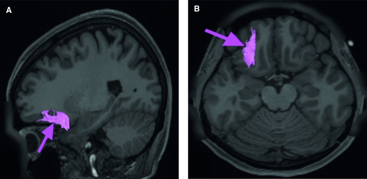
Area 13l is structurally connected to local parcellations. Short association bundles connect with 11l, 47m, and 47s (Figure 50).
FIGURE 50.
Structural connectivity of 13l in the left hemisphere, shown on T1-weighted MR images. A, Sagittal and B, axial planes showing. Pink: white matter tracts of 13l demonstrating connections with local parcellations.
What is known about its function?
Area 13l has been implicated as a secondary hub for olfactory, gustatory, visceral and food texture processing, integrating this information to assess satiety based on a comparison between food reward and current internal states.20-22
Area 47m
Where is it?
Area 47m (47 medial) is located in the posterior lateral OFC.
What are its borders?
Area 47m borders area 11l anteriorly, areas 47l and a47r laterally, area 47s posteriorly, and area 13l medially.
What is its functional connectivity?
Area 47m demonstrates functional connectivity to 13L in the orbitofrontal region, 8AD and IFSp in the frontal lobe, TE1p and PHA2 in the temporal lobe, and PGs and POS1 in the parietal lobe (Figure 51).
FIGURE 51.
Functional connectivity of 47m demonstrated on an inflated left hemisphere. A, Lateral and medial views. B, Rostral and caudal views. C, Dorsal and ventral views. Parcellations with the strongest functional connectivity are shown in yellow. Pink arrows designate the parcellation of interest.
What are its white matter connections?
Area 47m is structurally connected to local parcellations and the inferior fronto-occipital fasciculus. Fibers from the inferior fronto-occipital fasciculus project posteriorly through the extreme/external capsule to end at occipital lobe area V1. Local short association bundles connect with 13l, 47l, 47s, AAIC, and Pir (Figure 52).
FIGURE 52.
Structural connectivity of 47m in the left hemisphere, shown on T1-weighted MR images. Sagittal views of A, medial and B, lateral planes, and C, axial view showing. Blue: white matter tracts of 47m demonstrating connections with the IFOF and local parcellations.
What is known about its function?
Area 47m integrates emotional information, cognitive appraisals, and physiological internal states to aid in emotional regulation and assist in decision-making processes.23
DISCUSSION
A simplified scheme of prefrontal function views the prefrontal cortex as a 3-faced pyramid, with the dorsolateral face being responsible for working memory and executive function, the medial face being responsible for motivation, and the orbitofrontal face being responsible for emotional regulation. While very simplistic, there is some anatomic validity to considering the frontal lobe in this way: the dorsolateral prefrontal lobe largely communicates with the rest of the brain via the SLF/arcuate system as well as the IFOF; the medial frontal lobe is largely the realm of the cingulum; and the OFC largely communicates via the IFOF and uncinate. Interestingly, these 3 systems only seem to talk to each other through a series of local connections, and do not have substantial large interconnections between them. This chapter covers 2 of these areas, the medial frontal and orbitofrontal regions (the dorsolateral surface of the frontal lobe was covered in Chapter 2).
The Default Mode Network
The default mode network (DMN) was discovered during the analysis of the activity patterns of individuals not performing any specific task during functional imaging.24 It has been found to alternate its activity with the central executive network, a frontoparietal network largely thought to be involved in goal-directed behavior.25,26 The so-called salience network, which involves a more posterior medial frontal area and the insula, is thought to mediate the switching between these 2 networks.27 The DMN has been extensively studied given its putative role in a large number of psychological processes and its functional alteration in a wide number of diseases.28-33 Our group has found that its preservation is important in avoiding akinetic abulic states in surgery around the medial frontal lobe and corpus callosum.34
The main 3 components of the DMN are the anterior cingulate gyrus and adjacent frontal lobe, the PCC and RSC, and the lateral parietal lobes.35 Parts of the parahippocampal areas have also been included in some models of the network; however, this is not consistent between studies.36 Using this basic definition, and using functional connectivity patterns of the various parcellations, we would suggest that tentative members for inclusion as members of the DMN include the following parcellations:
Anterior cingulate regions: a24, p24, d32, 9M, 10r, 10v
Posterior cingulate regions: 31a, 31pv, 31pd, v23ab, d23ab, 23d, RSC, 7m, POS1, POS2
Lateral parietal lobe regions: PFm, PGs, PGi
Though further work is needed to compare published coordinate-based maps of resting state networks to the parcellation locations, these areas seem to be reasonable candidates for the purposes of discussion. Note that some areas activate a part of the distant network; for example, a32pr is functionally connected to RSC which is part of the DMN. Other areas, like a24pr, have functional connectivity with neighboring members that are part of the network, but do not demonstrate connectivity with distant parts of the network, suggesting that while they may interact with the network, they are not functionally part of it beyond any doubt.
The Middle Cingulate Gyrus and the Cingulum Bundle
The cingulum bundle is the white matter pathway linking the anterior and posterior cingulate components of the DMN. It is also the pathway by which the middle cingulate areas communicate with the PCC. The middle cingulate is primarily comprised of the “prime regions,” ie, a24pr, p24pr, a32pr, and p32pr. Our analysis suggests that these middle cingulate areas are physically highly interconnected with areas of the PCC. In many ways, they appear to be more broadly connected to the posterior cingulate region than the areas that are definitively in the DMN. However, they cannot be conclusively added to the network as they do not functionally correlate with the posterior cingulate areas to any appreciable degree, at least based on our thresholding method for functional connectivity.
Functional Segregation Within the DMN
The section on the posterior cingulate regions (Chapter 8) discusses the functional segregation of the DMN in more detail, including the concept of a dorsal PCC, which coordinates visuospatial body orientation,6,37-40 and a ventral PCC, which is involved in self-reflection, self-monitoring, and other internally motivated states.6,37-40 For the purposes of this discussion, we suggest that 31a, 23d, d23ab, and possibly 23c make up the dorsal PCC, and that 31pd, 31pv, and v23ab best describe the ventral PCC.
The PCC has been shown in graph theoretical analyses to have “rich club” topology,41 meaning these are highly interconnected areas which are connected to many other areas. Our own analysis suggests that there may be some basis for this beyond theory. First is the observation that all of the PCC areas are all functionally connected to other members of the PCC, effectively forming in a tight network. This means if you are part of the PCC, you talk to all other PCC members without exception, suggesting they are tightly linked. Second is the observation that all of the members of the PCC seem to have their own connections anteriorly, suggesting that there is no single hub or small set of hubs in the PCC and instead most of the PCC members are hubs in their own right. Thus, as a highly connected set of hubs, the PCC seems to qualitatively meet many of the criteria we would expect from a “rich club.”
Disentangling this anatomy is obviously complicated due to the numerous overlapping sets of connections. However, our own careful study of the structural connections suggests that there is a pattern in the connections of these PCC regions to their more anterior partners:
Dorsal PCC (31a, 23c, 23d, d23ab): These regions connect to other PCC members and to the middle cingulate parcellations. They do not connect to the anterior DMN members.
Ventral PCC (31pv, 31pd, v23ab, RSC): These regions connect to the anterior DMN members, the PCC members and also to the middle cingulate parcellations, making them more densely interconnected.
Given the suggested role of the ventral PCC in monitoring self-referential states, 6,37-40 it is reasonable to conclude that the ventral PCC regions and their connections with the anterior DMN components represent the DMN proper, and that the middle cingulate system represents a distinct but overlapping network running in parallel with the DMN.
The Orbitofrontal Regions
It is clear that the orbitofrontal regions play a critical role in emotional regulation,17,18 and should be preserved whenever possible in neurological surgery.42 However, study of this region has been limited by the complex nature of judgement and emotional regulation, which we all intuitively understand, but which defy easy quantification and definition. Further, complicating this problem is the difficulty with imaging-related artifacts caused by the skull base and nasal cavities in the anterior fossa.
The orbitofrontal regions are primarily connected to the rest of the brain through the IFOF and the uncinate pathways, which link them to the early visual areas and to TGd, respectively. Interestingly, while they are invariably linked to their neighboring frontal regions in the dorsolateral brain through local connections, there is no evidence that the orbitofrontal regions communicate with the dorsolateral or medial frontal systems through any form of large white matter tract, suggesting that these systems do not follow a small world model when communicating with each other.
Finally, in our opinion, the IFOF is a mysterious pathway, especially with its connections to this region. Why early visual areas need to talk directly to orbitofrontal regions is quite strange, and while this could play a role in feedback regulation of visual processing, we think this area needs further study. It may very well redefine the way we view “judgement.”
Disclosures
SynaptiveMedical assisted in the funding of all 18 chapters of this supplement. No other funding sources were utilized in the production or submission of this work.
Acknowledgments
Data were provided [in part] by the Human Connectome Project, WUMinn Consortium (Principal Investigators: David Van Essen and Kamil Ugurbil; 1U54MH091657) funded by the 16 NIH Institutes and Centers that support the NIH Blueprint for Neuroscience Research; and by the McDonnell Center for Systems Neuroscience at Washington University. We would also like to thank Brad Fernald, Haley Harris, and Alicia McNeely of Synaptive Medical for their assistance in constructing the network figures for Chapter 18 and for coordinating the completion and submission of this supplement.
REFERENCES
- 1. Sturm VE, Sollberger M, Seeley WW et al. Role of right pregenual anterior cingulate cortex in self-conscious emotional reactivity. Soc Cogn Affect Neurosci. 2013;8(4):468-474. [DOI] [PMC free article] [PubMed] [Google Scholar]
- 2. Devinsky O, Morrell MJ, Vogt BA. Contributions of anterior cingulate cortex to behaviour. Brain. 1995;118 (1):279-306. [DOI] [PubMed] [Google Scholar]
- 3. Drevets WC, Savitz J, Trimble M. The subgenual anterior cingulate cortex in mood disorders. CNS Spectr. 2008;13(08):663-681. [DOI] [PMC free article] [PubMed] [Google Scholar]
- 4. Beckmann M, Johansen-Berg H, Rushworth MF. Connectivity-based parcellation of human cingulate cortex and its relation to functional specialization. J Neurosci. 2009;29(4):1175-1190. [DOI] [PMC free article] [PubMed] [Google Scholar]
- 5. Palomero-Gallagher N, Mohlberg H, Zilles K, Vogt B. Cytology and receptor architecture of human anterior cingulate cortex. J Comp Neurol. 2008;508(6):906-926. [DOI] [PMC free article] [PubMed] [Google Scholar]
- 6. Glasser MF, Coalson TS, Robinson EC et al. A multi-modal parcellation of human cerebral cortex. Nature. 2016;536(7615):171-178. [DOI] [PMC free article] [PubMed] [Google Scholar]
- 7. Brodmann K. Vergleichende Lokalisationslehre der Grosshirnrinde in ihren Prinzipien dargestellt auf Grund des Zellenbaues. Springer: Barth; 1909. [Google Scholar]
- 8. Vogt BA, Nimchinsky EA, Vogt LJ, Hof PR. Human cingulate cortex: surface features, flat maps, and cytoarchitecture. J Comp Neurol. 1995;359(3):490-506. [DOI] [PubMed] [Google Scholar]
- 9. Vogt BA, Vogt L. Cytology of human dorsal midcingulate and supplementary motor cortices. J Chem Neuroanat. 2003;26(4):301-309. [DOI] [PubMed] [Google Scholar]
- 10. Gasquoine PG. Localization of function in anterior cingulate cortex: from psychosurgery to functional neuroimaging. Neurosci Biobehav Rev. 2013;37(3):340-348. [DOI] [PubMed] [Google Scholar]
- 11. Bush G, Vogt BA, Holmes J et al. Dorsal anterior cingulate cortex: a role in reward-based decision making. Proc Natl Acad Sci USA. 2002;99(1):523-528. [DOI] [PMC free article] [PubMed] [Google Scholar]
- 12. Bush G, Luu P, Posner MI. Cognitive and emotional influences in anterior cingulate cortex. Trends Cogn Sci. 2000;4(6):215-222. [DOI] [PubMed] [Google Scholar]
- 13. Stuphorn V. The role of supplementary eye field in goal-directed behavior. J Physiol Paris. 2015;109(1-3):118-128. [DOI] [PMC free article] [PubMed] [Google Scholar]
- 14. Petrides M, Pandya DN. Dorsolateral prefrontal cortex: comparative cytoarchitectonic analysis in the human and the macaque brain and corticocortical connection patterns. Eur J Neurosci. 1999;11(3):1011-1036. [DOI] [PubMed] [Google Scholar]
- 15. Burgess PW, Gilbert SJ, Dumontheil I. Function and localization within rostral prefrontal cortex (area 10). Philos Trans R Soc B: Biol Sci. 2007;362(1481):887-899. [DOI] [PMC free article] [PubMed] [Google Scholar]
- 16. Grabenhorst F, Rolls ET. Value, pleasure and choice in the ventral prefrontal cortex. Trends Cogn Sci. 2011;15(2):56-67. [DOI] [PubMed] [Google Scholar]
- 17. Kringelbach ML, Rolls ET. The functional neuroanatomy of the human orbitofrontal cortex: evidence from neuroimaging and neuropsychology. Prog Neurobiol. 2004;72(5):341-372. [DOI] [PubMed] [Google Scholar]
- 18. Rudebeck PH, Murray EA. The orbitofrontal oracle: cortical mechanisms for the prediction and evaluation of specific behavioral outcomes. Neuron. 2014;84(6):1143-1156. [DOI] [PMC free article] [PubMed] [Google Scholar]
- 19. Gottfried JA. What can an orbitofrontal cortex-endowed animal do with smells? Ann N Y Acad Sci. 2007;1121(1):102-120. [DOI] [PubMed] [Google Scholar]
- 20. Ongur D, Ferry AT, Price JL. Architectonic subdivision of the human orbital and medial prefrontal cortex. J Comp Neurol. 2003;460(3):425-449. [DOI] [PubMed] [Google Scholar]
- 21. Carmichael ST, Price JL. Sensory and premotor connections of the orbital and medial prefrontal cortex of macaque monkeys. J Comp Neurol. 1995;363(4):642-664. [DOI] [PubMed] [Google Scholar]
- 22. Rolls ET. Taste, olfactory and food texture reward processing in the brain and obesity. Int J Obes. 2011;35(4):550-561. [DOI] [PubMed] [Google Scholar]
- 23. Quirk GJ, Beer JS. Prefrontal involvement in the regulation of emotion: convergence of rat and human studies. Curr Opin Neurobiol. 2006;16(6):723-727. [DOI] [PubMed] [Google Scholar]
- 24. Raichle ME, MacLeod AM, Snyder AZ, Powers WJ, Gusnard DA, Shulman GL. A default mode of brain function. Proc Natl Acad Sci USA. 2001;98(2):676-682. [DOI] [PMC free article] [PubMed] [Google Scholar]
- 25. Chand GB, Dhamala M. Interactions among the brain default-mode, salience, and central-executive networks during perceptual decision-making of moving dots. Brain Connect. 2016;6(3):249-254. [DOI] [PubMed] [Google Scholar]
- 26. Chen AC, Oathes DJ, Chang C et al. Causal interactions between fronto-parietal central executive and default-mode networks in humans. Proc Natl Acad Sci USA. 2013;110(49):19944-19949. [DOI] [PMC free article] [PubMed] [Google Scholar]
- 27. Goulden N, Khusnulina A, Davis NJ et al. The salience network is responsible for switching between the default mode network and the central executive network: replication from DCM. Neuroimage. 2014;99:180-190. [DOI] [PubMed] [Google Scholar]
- 28. Shao J, Meng C, Tahmasian M et al. Common and distinct changes of default mode and salience network in schizophrenia and major depression. Brain Imaging Behav. 2018. [DOI] [PubMed] [Google Scholar]
- 29. Brakowski J, Spinelli S, Dorig N et al. Resting state brain network function in major depression - Depression symptomatology, antidepressant treatment effects, future research. J Psychiatr Res. 2017;92:147-159. [DOI] [PubMed] [Google Scholar]
- 30. Stephens JA, Salorio CF, Barber AD, Risen SR, Mostofsky SH, Suskauer SJ. Preliminary findings of altered functional connectivity of the default mode network linked to functional outcomes one year after pediatric traumatic brain injury. Dev Neurorehabil. 2017;1-8. [DOI] [PMC free article] [PubMed] [Google Scholar]
- 31. Wang C, Pan Y, Liu Y et al. Aberrant default mode network in amnestic mild cognitive impairment: a meta-analysis of independent component analysis studies. Neurol Sci. 2018;(5). [DOI] [PubMed] [Google Scholar]
- 32. Li M, Zheng G, Zheng Y et al. Alterations in resting-state functional connectivity of the default mode network in amnestic mild cognitive impairment: an fMRI study. BMC Med Imaging. 2017;17(1):919-931. [DOI] [PMC free article] [PubMed] [Google Scholar]
- 33. Padmanabhan A, Lynch CJ, Schaer M, Menon V. The default mode network in Autism. Biol Psychiatry Cogn Neurosci Neuroimaging. 2017;2(6):476-486. [DOI] [PMC free article] [PubMed] [Google Scholar]
- 34. Burks JD, Bonney PA, Conner AK et al. A method for safely resecting anterior butterfly gliomas: the surgical anatomy of the default mode network and the relevance of its preservation. J Neurosurg. 2017;126(6):1795-1811. [DOI] [PMC free article] [PubMed] [Google Scholar]
- 35. Harrison BJ, Pujol J, López-Solà M et al. Consistency and functional specialization in the default mode brain network. Proc Natl Acad Sci USA. 2008;105(28):9781-9786. [DOI] [PMC free article] [PubMed] [Google Scholar]
- 36. Ward AM, Schultz AP, Huijbers W, van Dijk KRA, Hedden T, Sperling RA. The parahippocampal gyrus links the default-mode cortical network with the medial temporal lobe memory system. Hum Brain Mapp. 2014;35(3):1061-1073. [DOI] [PMC free article] [PubMed] [Google Scholar]
- 37. Aggleton JP, Saunders RC, Wright NF, Vann SD. The origin of projections from the posterior cingulate and retrosplenial cortices to the anterior, medial dorsal and laterodorsal thalamic nuclei of macaque monkeys. Eur J Neurosci. 2014;39(1):107-123. [DOI] [PMC free article] [PubMed] [Google Scholar]
- 38. Bzdok D, Heeger A, Langner R et al. Subspecialization in the human posterior medial cortex. Neuroimage. 2015;106:55-71. [DOI] [PMC free article] [PubMed] [Google Scholar]
- 39. Leech R, Sharp DJ. The role of the posterior cingulate cortex in cognition and disease. Brain. 2014;137(1):12-32. [DOI] [PMC free article] [PubMed] [Google Scholar]
- 40. Vogt BA, Vogt L, Laureys S. Cytology and functionally correlated circuits of human posterior cingulate areas. Neuroimage. 2006;29(2):452-466. [DOI] [PMC free article] [PubMed] [Google Scholar]
- 41. van den Heuvel MP, Sporns O. Rich-Club organization of the human connectome. J Neurosci. 2011;31(44):15775-15786. [DOI] [PMC free article] [PubMed] [Google Scholar]
- 42. Burks JD, Conner AK, Bonney PA et al. Anatomy and white matter connections of the orbitofrontal gyrus. J Neurosurg. 2017;1-8. [DOI] [PubMed] [Google Scholar]


