Abstract
Objectives The objective of this study was to compare a bioceramic and a resin-based endodontic sealer with regard to extrusion and postoperative pain.
Materials and Methods Sixty-four patients requiring endodontic treatment of single-rooted maxillary teeth with necrotic pulps were included in this study. The root canal treatments were performed in a single visit using a size 40.06 single-file reciprocating system under 2.5% NaOCl irrigation. After irrigation with 17% ethylenediaminetetraacetic acid (EDTA) and 2.5% NaOCl, the canals were dried and randomly divided into two different groups ( n = 32) depending on the sealer used: resin-based group (RG) in which the canals were filled with the AH Plus, and the bioceramic group (BG) in which the canals were filled with the Sealer Plus BC. Ibuprofen (600 mg) was prescribed every 6 hours if the volunteers experienced pain. The patients registered their pain sensation in a visual analog scale (VAS) card, ranging from 0 to 10 at 24-hour, 48-hour, 72-hour, and 1-week intervals. Statistical analysis For statistical analysis, the level of significance was set at p < 0.05.
Results Sealer extrusion occurred in nine patients of the RG and in 19 patients of the BG ( p < 0.05). The average pain level at 24-hour and 48-hour intervals was, respectively, 1.46 ± 1.96 and 0.44 ± 0.86 for RG, and 1.21 ± 2.09 and 0.09 ± 0.38 for BG. There was no report of pain after 48 hours. The mean number of tablets taken for pain relief was 0.03 ± 0.17 for RG and 0.06 ± 0.24 for BG. No statistically significant difference was found with regard to pain level and intake of pain killer tablets ( p > 0.05).
Conclusions The BG sealer presented significantly more extrusion than the RG sealer. Sealer extrusion was not associated with pain. The average pain level and the mean number of tablets taken for pain relief were similar in both groups.
Keywords: root canal treatment, bioceramic, pain, reciprocating, resin-based sealer
Introduction
Root canal therapy (RCT) aims for the removal of vital and necrotic pulp tissue, and the tridimensional filling of root canal space. These steps should be achieved with the minimal postoperative discomfort. However, some factors such as trauma of the periapical tissues, bacterial extrusion, or remaining bacteria, might result in postoperative pain. 1
Different variables have been assessed with regard to postoperative pain such as the following: kinematics, 2 3 working length (WL) determination method, 4 foraminal enlargement, 5 and filling techniques. 6 The most common techniques require gutta-percha cone and sealer. Resin-based, zinc oxide eugenol-based, and calcium hydroxide are the sealers currently used. It seems that the extrusion of these root canal sealers does not impact the outcomes of teeth presenting pulp necrosis; however, sealer extrusion might lead to inconvenient results, mostly when the alveolar nerve is the target. 7 8 9
Resin-based sealers are known for their beneficial physical properties; on the other hand, there is a concern about its cytotoxic effects, pushing endodontics to seek a better sealer. 10 In an attempt to correct this drawback, bioceramic sealer (silicatum-based or hydraulic sealers) were recently launched. These sealers possess positive biological effects with low injury to vital tissues; 1 1 12 moreover, it has been recently hypothesized that they could benefit root integrity after root canal filling. 13 The solubility of these sealers remains a critical aspect of its properties. 11
Endodontic sealers should present an ideal flowability to fill all of the irregularities of the root canal system. Lack of flowability might prevent proper filling, while excessive flow rate could potentially increase the risk of extrusion. Filling material beyond the apex could increase the risk of pain or reach important anatomical landmarks. 14 While some studies presented bioceramic sealers to be more flowable than resin-based sealers, 15 a recent study showed discrepant results. 16 Under a clinical point of view, bioceramic sealers demonstrated good outcomes in nonsurgical root canal treatments. 17 A recent study showed that bioceramic sealers are similar to resin-based sealers with regard to postoperative pain in nonsurgical root canal retreatments when no extrusion was observed. 18 To the best of our knowledge, the extrusion rate and postoperative pain of both sealers in nonsurgical root canal treatment of teeth presenting with pulp necrosis is unknown.
Therefore, the aim of the present study was to compare the extrusion rate and the postoperative pain of a resin-based sealer (AH Plus, Dentsply-Sirona, Konstanz, Germany) and a bioceramic sealer (Sealer Plus BC; MKLife Medical and Dental Products, Porto Alegre, Brazil). The null hypotheses tested were that there was no difference between sealers with regard to extrusion rate, and there was also no difference in postoperative pain with both sealers.
Materials and Methods
Sample Size Calculation
This prospective, randomized clinical trial was approved by the Ethical Committee of Faculdade de Odontologia São Leopoldo Mandic (no. 2.270.666). Using the G* Power for Macintosh (Heinrich-Heine, Düsseldorf, Germany), the sample size was calculated based on a previous study that showed average pain level of 2.30 and 1.09 for respective experimental and control groups. 5 Aiming to achieve 80% power (α error of 0.05 and a power β of 0.95) and 95% confidence of difference between the groups, a minimum of 32 patients was set for each group.
Patient Selection
Sixty-four patients with the American Society of Anesthesiology (ASA) Class I or II who were non-smokers and diabetics, or who were pregnant, were invited for this research. Only patients with single-rooted, maxillary anterior teeth presenting straight canals, based on the radiograph evaluation, were selected. The inclusion criteria encompassed teeth with pulp necrosis, mature apices, and no signs of previously initiated RCT. Teeth with periodontal probe higher than 3 mm, radiographic aspect of root resorption, pain, swelling, or sinus tract were not included. Moreover, patients taking any analgesic, anti-inflammatory, or antibiotics, those allergic to Ibuprofen, or the ones who could not properly follow the instructions for filling the visual analog card (VAS) card were excluded.
After signing an informed consent, the patients were assigned in a parallel design and 1:1 allocation rate to one of the two groups ( n = 32). Sixty-four cards were printed and inserted in masked envelopes. Using a computer algorithm (random.org), these envelopes were randomly distributed. Each one of the envelopes indicated the volunteer for either the Sealer Plus BC (RG; MKLife Medical and Dental Products, Porto Alegre, Brazil) or bioceramic sealer group (BG). The volunteers were blinded to the group to which they were assigned, but the operator was aware of it. All of the procedures were the same except for the sealer and its insertion. In the RG, AH Plus was used, while in the BG, premixed Sealer Plus BC was chosen.
Treatment Protocol
After confirming the absence of pulp sensitivity in both a cold and an electric test, the volunteers were anesthetized with 3.6 mL 2% mepivacaine and 1:100,000 epinephrine. Thereafter, a rubber dam was placed and the pulp chamber was reached using a diamond bur, which was then copiously irrigated with 5 mL 2.5% sodium hypochlorite (NaOCl). The canal was gently explored with a size 10-K file; following the manufacturer’s instruction, only cases in which a size 20 K-file could reach the full WL were included. A size 40.06 single file reciprocating instrument (Reciproc, VDW, Munich, Germany) was used for root canal shaping in a VDW Silver motor (VDW) in the proper motion. The instrument was used in an in-and-out movement with, at the most, three movements under 3 to 4 mm of amplitude. After each insertion of the instrument, the canal was again irrigated with NaOCl, and foraminal patency was checked with a size 15-K file. After the preparation of the cervical and middle thirds, an electronic apex locator (EAL, NovaPex, Forum Technologies, Rishon Le-Zion, Israel) was used for WL determination. A periapical radiograph was used to confirm the WL 1 mm short of the apex.
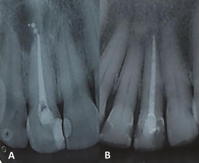
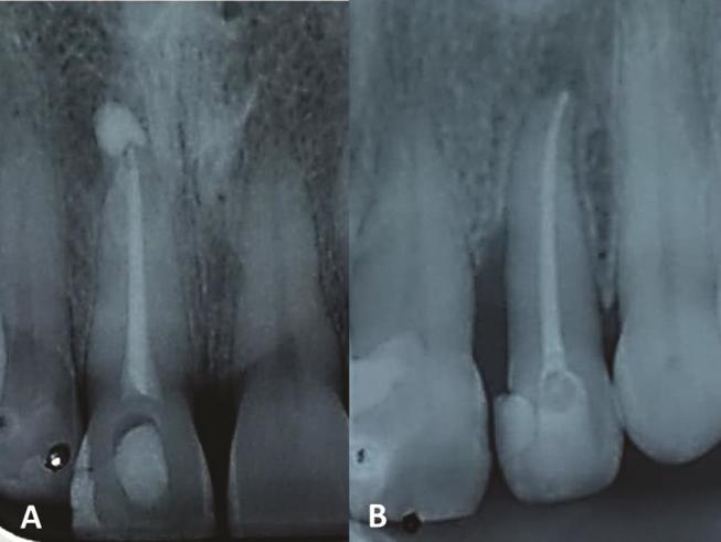
Each root canal was irrigated with the same volume of 2.5% NaOCl (40 mL) and 17% ethylenediaminetetraacetic acid (EDTA, 5 mL) followed by a final irrigation of 5 mL 2.5% NaOCl. A plastic device in reciprocating motion was used in three sessions of 20 seconds each to activate both EDTA and NaOCl final irrigation. 19 The canal was then dried with R40 paper points (VDW) and the same size gutta-percha cone was selected. After a radiograph was obtained, and when necessary, the gutta-percha tip was adjusted with a blade. At this point, one of the masked envelopes that had been previously randomized was selected, correspondent sealer was handled, and a single-cone technique filling was used. In the RG, the sealer was inserted with a lentulo; in the BG, the syringe and tip provided by the manufacturer was used. Then, the teeth were restored with a composite and the occlusion was checked. A final radiograph was obtained and the occurrence of sealer extrusion was registered by the operator ( Figs. 3 4 ). All root canal treatments were performed between January 2018 to August 2018 by the same experienced endodontist (BF).
Fig. 3.

Sample of treatments performed with AH Plus with ( A ) and without ( B ) extrusion.
Fig. 2.

Sample of treatments performed with BC Sealer with ( A ) and without ( B ) extrusion.
Pain Perception Assessment
After the RCT, the volunteers were requested to complete a VAS card with their pain perception ranging from 0 to 10. The 0 mark was used if the teeth remained asymptomatic, while the 10 mark was to be used for unbearable pain. Any other pain was to be registered according to the volunteers’ own perceptions. The pain sensation was registered at 24-hour, 48-hour, 72-hour, and 1-week intervals. Pain perception was classified as none, mild (1–2), moderate (3–7), or severe (8–10). The patients were oriented to take 600 mg Ibuprofen every 6 hours if they experienced any pain. The number of tablets taken for pain relief was also registered. The patients were to return their VAS cards after 1 week and contact the professional in case further appointments were necessary for pain relief.
Statistical Analysis
The chi-square test was used for assessment of differences in tooth type distribution, gender distribution, and occurrence of sealer extrusion between the groups. Pearson’s correlation analysis was applied to assess the correlation between extrusion and occurrence of pain ( p < 0.001). Results for pain level, mean age, and the number of tablets taken showed abnormal distribution; therefore, the Mann–Whitney U test was used for these differences between the two groups. All statistical analyses were performed using SAS version 9.3 (SAS Institute Inc, Cary, NC, United States). The test was performed at the level of significance of p < 0.05.
Results
All patients returned their VAS cards after 1 week. Overall, 24 male and 38 female patients ranging from 15 to 68 years old were enrolled. No statistically significant difference was found in the demographic distribution of the patients, as displayed in Table 1 ( p > 0.05).
Table 1. Demographic distribution of patients in both groups.
| RG | BG | p -Value | |
|---|---|---|---|
|
Mann-Whitney
U
test for statistical analysis of mean age; chi-square test for the gender, and tooth type distribution; 95% confidence interval.
*Significantlydifferent at the 0.05 level. | |||
| Mean age | 37.09 ± 13.10 | 38.5 ± 14.18 | >0.05 |
| Male | 14 | 12 | 0.6, x 2 = 0.25 |
| Female | 18 | 20 | |
| Central incisor | 21 | 14 | 0.08, x 2 = 3.1 |
| Lateral incisor | 6* | 14* | 0.03, x 2 = 4.6 |
| Canine | 5 | 4 | 0.9, x 2 = 0.006 |
BG presented a statistically significant amount of more extrusion (59.37%) than RG (28.12%) ( p < 0.05). Nonetheless, there was no correlation between extrusion of sealers and occurrence of pain for both BG (r = −0.071) and RG (r = 0.07).
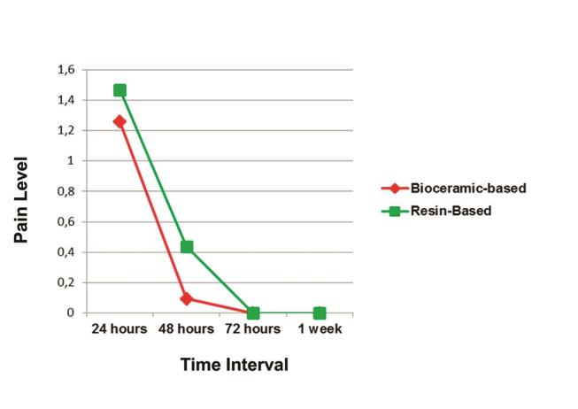
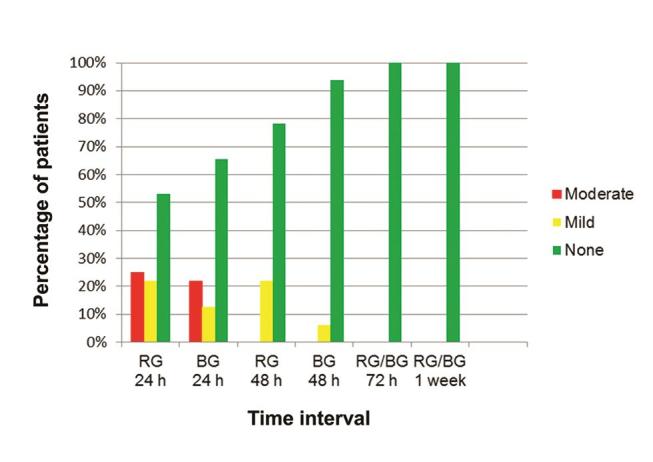
The average pain level in the RG was 1.46 ± 1.96 after 24 hours and 0.44 ± 0.86 after 48 hours; in the BG, it was 1.21 ± 2.09 after 24 hours and 0.09 ± 0.38 after 48 hours ( Fig. 1 ). With regard to pain intensity, in the RG, after 24 hours, 53.13% reported no pain, 21.88% mild pain, and 25% moderate pain; after 48 hours, 78.12% of the patients reported no pain and 21.78% reported mild pain. In the BG, after 24 hours, 65.63% reported no pain, 12.50% mild pain, and 21.88% moderate pain; after 48 hours, 93.65% of the patients reported no pain and 6.25% reported mild pain ( Fig. 2 ). There was no report of pain after 48 hours in either group, and there was no report of flare-up at any time interval.
Fig. 1.

Average pain level for RG and BG at all time intervals. BG, bioceramic group; RG, resin-based group.
Fig. 2.

Percentage of patients reporting none (0), mild (1–3) or moderate (4–7) pain at all time-intervals. There was no occurrence of severe (8–10) pain.
One patient in the RG and two patients in the BG took one tablet of Ibupofen in the first 24 hours; no patient required any medication after 24 hours. The mean number of tablets taken for pain relief was 0.03 ± 0.17 for RG and 0.06 ± 0.06 for BG. No statistically significant difference was found between the groups with regard to pain level and intake of pain killer tablets ( p > 0.05).
Discussion
Variations among the samples in randomized clinical trials might prevent the generation of meaningful results. Randomization resulted in groups with similar age and sex distribution, minimizing the effects of these factors on the results. 1 20 Aiming to standardize the samples as much as possible, only anterior maxillary teeth presenting straight, single-rooted teeth were used. 21 One might claim that the lateral incisor presents an apical curvature not usually present in central incisors or canines. To attenuate this possible variable, only teeth in which a size 20 K-file reached the estimated WL were included, thus enhancing standardization among the specimens. A recent study compared different unintentionally extruded endodontic sealers with regard to postoperative pain; however, that study included vital and necrotic pulps, different instrumentation systems, and single or multiple visits. 22 In the present study, all treatments of necrotic teeth were performed in single visits with the same single-file reciprocating protocol. A systematic review suggested that single-visit treatments might increase postoperative pain. 23 However, the protocol adopted herein was applied in a previous study, leading to a low-level of postoperative pain and analgesic intake. 5
Filling protocol is a key point when bioceramic sealers are used. The physical properties of sealers are subjected to damage in case of loss of humidity, which is promoted by excessive heating. 24 Nonetheless, recent studies showed acceptable properties for bioceramic sealers. 25 26 The present study adopted a single cone technique in both groups, therefore avoiding overheating of sealers. The only variable was that AH Plus was inserted in the canal with a lentulo, and the Sealer Plus BC was applied with the endodontic tip provided by the manufacturer. While different filling techniques might influence postoperative pain, the variance in using lentulo or an endodontic tip for inserting the sealer is unknown. 6
A proper flow rate allows the sealer to fill all irregularities within the root canal; meanwhile, excessive flowability increases the risk of sealer extrusion. Previous studies showed bioceramic sealers to present greater flowability than resin-based sealers. 15 27 Conversely, a recent study showed BC Sealer Plus presenting a lower flow rate than AH Plus. 16 It is worthwhile to mention that, in the aforementioned studies, the sealers were in accordance with the ISO 6876/2012 recommendations. Despite the findings of Mendes et al, 17 our results showed a higher rate of extrusion for the BC Sealer Plus (59.74%) when compared with the AH Plus sealer (28.13%). Therefore, our first hypothesis was rejected. A recent study showed a bioceramic sealer with an extrusion rate of 47.4%, even though a 90.9% success rate was achieved. 17 In spite of affecting neither postoperative pain in the present study nor long-term outcomes in Chybowsky et al, 17 sealer extrusion should not be considered harmless. There have been reports of accidents related to sealer extrusion, mainly affecting the mandibular molars and resulting in irreversible paresthesia. 8 28
The low-incidence of flare-ups and low-postoperative pain decreasing over time seems common in modern endodontics. 5 29 The present study had no report of flare-ups, and the majority (59.38%) of the patients reported no pain at any time interval regardless of the sealer used. Moderate pain occurred only in the first 48 hours, and medication intake only in the first 24 hours. Graunaite et al 18 did not report any case of flare-ups and showed 65% of patients to be pain free at all time intervals, with decreasing pain after an initial peak also occurred. That study assessed nonsurgical root canal retreatments in single-rooted teeth obturated with either a resin-based or a bioceramic sealer; however, no case of sealer extrusion occurred and the authors did not report that foraminal patency was achieved. In the present study, foraminal patency was achieved with a size 15-K file, therefore favoring the occurrence of unintentional sealer extrusion. Rodriguez-Lozano et al 30 showed bioceramic sealer to present more cytocompatibility than resin-based sealer; it was also found that cytotoxic effects decrease after 24 hours. Therefore, one might conclude that cytotoxic effects of sealers might impact pain perception. However, our findings showed pain occurring regardless of sealer extrusion, and no difference between the sealers. Therefore, possible cytotoxic effects found in vitro were not confirmed in our clinical findings.
In the present study, the operator could not be blinded with regard to the sealer used. Moreover, since the assessment of the sealer extrusion was performed by the same operator, these steps should be considered as limitations of the present study. Future studies should evaluate long-term outcomes of RCT with bioceramic and resin-based sealers. While the physical properties of AH Plus and different bioceramic sealers have been subject to several studies, the specific bioceramic sealer tested herein should undergo further scrutiny.
Our findings suggest that bioceramic sealer presented a significantly higher incidence of extrusion than the resin-based sealer. Sealer extrusion was not associated with pain. The average pain level and the mean number of tablets taken for pain relief were similar in both groups.
Footnotes
Conflict of Interest None declared.
References
- 1.Arias A, de la Macorra J C, Hidalgo J J, Azabal M. Predictive models of pain following root canal treatment: a prospective clinical study. Int Endod J. 2013;46(08):784–793. doi: 10.1111/iej.12059. [DOI] [PubMed] [Google Scholar]
- 2.Nekoofar M H, Sheykhrezae M S, Meraji N et al. Comparison of the effect of root canal preparation by using WaveOne and ProTaper on postoperative pain: a randomized clinical trial. J Endod. 2015;41(05):575–578. doi: 10.1016/j.joen.2014.12.026. [DOI] [PubMed] [Google Scholar]
- 3.Kherlakian D, Cunha R S, Ehrhardt I C, Zuolo ML, Kishen A, da Silveira Bueno CE. Comparison of the incidence of postoperative pain after using 2 reciprocating systems and a continuous rotary system: a prospective randomized clinical trial. J Endod. 2016;42(02):171–176. doi: 10.1016/j.joen.2015.10.011. [DOI] [PubMed] [Google Scholar]
- 4.Kara Tuncer A, Gerek M. Effect of working length measurement by electronic apex locator or digital radiography on postoperative pain: a randomized clinical trial. J Endod. 2014;40(01):38–41. doi: 10.1016/j.joen.2013.08.004. [DOI] [PubMed] [Google Scholar]
- 5.Cruz Junior J A, Coelho M S, Kato A S et al. The effect of foraminal enlargement of necrotic teeth with the reciproc system on postoperative pain: a prospective and randomized clinical trial. J Endod. 2016;42(01):8–11. doi: 10.1016/j.joen.2015.09.018. [DOI] [PubMed] [Google Scholar]
- 6.Alonso-Ezpeleta L O, Gasco-Garcia C, Castellanos-Cosano L, Martín-González J, López-Frías F J, Segura-Egea J J. Postoperative pain after one-visit root-canal treatment on teeth with vital pulps: comparison of three different obturation techniques. Med Oral Patol Oral Cir Bucal. 2012;17(04):e721–e727. doi: 10.4317/medoral.17898. [DOI] [PMC free article] [PubMed] [Google Scholar]
- 7.Szalma J, Soos B, Krajczar K, Lempel E. Piezosurgical management of sealer extrusion-associated mental nerve anaesthesia: a case report. Aust Endod J. 2018;45(02):274–280. doi: 10.1111/aej.12316. [DOI] [PubMed] [Google Scholar]
- 8.González-Martín M, Torres-Lagares D, Gutiérrez-Pérez J L, Segura-Egea J J. Inferior alveolar nerve paresthesia after overfilling of endodontic sealer into the mandibular canal. J Endod. 2010;36(08):1419–1421. doi: 10.1016/j.joen.2010.03.008. [DOI] [PubMed] [Google Scholar]
- 9.Ricucci D, Rôças I N, Alves F R, Loghin S, Siqueira J F.JrApically extruded sealers: fate and influence on treatment outcome J Endod 20164202243–249. [DOI] [PubMed] [Google Scholar]
- 10.Bouillaguet S, Wataha J C, Tay F R, Brackett M G, Lockwood P E. Initial in vitro biological response to contemporary endodontic sealers. J Endod. 2006;32(10):989–992. doi: 10.1016/j.joen.2006.05.006. [DOI] [PubMed] [Google Scholar]
- 11.Poggio C, Dagna A, Ceci M, Meravini M V, Colombo M, Pietrocola G. Solubility and pH of bioceramic root canal sealers: A comparative study. J Clin Exp Dent. 2017;9(10):e1189–e1194. doi: 10.4317/jced.54040. [DOI] [PMC free article] [PubMed] [Google Scholar]
- 12.Camps J, Jeanneau C, El Ayachi I, Laurent P, About I. Bioactivity of a calcium silicate-based endodontic cement (BioRoot RCS): Interactions with human periodontal ligament cells in vitro. J Endod. 2015;41(09):1469–1473. doi: 10.1016/j.joen.2015.04.011. [DOI] [PubMed] [Google Scholar]
- 13.Osiri S, Banomyong D, Sattabanasuk V, Yanpiset K. Root reinforcement after obturation with calcium silicate-based sealer and modified gutta-percha cone. J Endod. 2018;44(12):1843–1848. doi: 10.1016/j.joen.2018.08.011. [DOI] [PubMed] [Google Scholar]
- 14.Siqueira J F., Jr Microbial causes of endodontic flare-ups. Int Endod J. 2003;36(07):453–463. doi: 10.1046/j.1365-2591.2003.00671.x. [DOI] [PubMed] [Google Scholar]
- 15.Candeiro G T, Correia F C, Duarte M A, Ribeiro-Siqueira DC, Gavini G. Evaluation of radiopacity, pH, release of calcium ions, and flow of a bioceramic root canal sealer. J Endod. 2012;38(06):842–845. doi: 10.1016/j.joen.2012.02.029. [DOI] [PubMed] [Google Scholar]
- 16.Mendes A T, Silva P BD, Só B B et al. Evaluation of physicochemical properties of new calcium silicate-based sealer. Braz Dent J. 2018;29(06):536–540. doi: 10.1590/0103-6440201802088. [DOI] [PubMed] [Google Scholar]
- 17.Chybowski E A, Glickman G N, Patel Y, Fleury A, Solomon E, He J. Clinical outcome of non-surgical root canal treatment using a single-cone technique with Endosequence bioceramic sealer: A retrospective analysis. J Endod. 2018;44(06):941–945. doi: 10.1016/j.joen.2018.02.019. [DOI] [PubMed] [Google Scholar]
- 18.Graunaite I, Skucaite N, Lodiene G, Agentiene I, Machiulskiene V. Effect of resin-based and bioceramic root canal sealers on postoperative pain: A split-mouth randomized controlled trial. J Endod. 2018;44(05):689–693. doi: 10.1016/j.joen.2018.02.010. [DOI] [PubMed] [Google Scholar]
- 19.Kato A S, Cunha R S, da Silveira Bueno C E, Pelegrine R A, Fontana C E, de Martin A S. Investigation of the efficacy of passive ultrasonic irrigation versus irrigation with reciprocating activation: An environmental scanning electron microscopic study. J Endod. 2016;42(04):659–663. doi: 10.1016/j.joen.2016.01.016. [DOI] [PubMed] [Google Scholar]
- 20.Robinson M E, Riley JL I I, Myers C D et al. Gender role expectations of pain: relationship to sex differences in pain. J Pain. 2001;2(05):251–257. doi: 10.1054/jpai.2001.24551. [DOI] [PubMed] [Google Scholar]
- 21.Silva E J, Menaged K, Ajuz N, Monteiro MR, Coutinho-Filho TdeS. Postoperative pain after foraminal enlargement in anterior teeth with necrosis and apical periodontitis: a prospective and randomized clinical trial. J Endod. 2013;39(02):173–176. doi: 10.1016/j.joen.2012.11.013. [DOI] [PubMed] [Google Scholar]
- 22.Shashirekha G, Jena A, Pattanaik S, Rath J. Assessment of pain and dissolution of apically extruded sealers and their effect on the periradicular tissues. J Conserv Dent. 2018;21(05):546–550. doi: 10.4103/JCD.JCD_224_18. [DOI] [PMC free article] [PubMed] [Google Scholar]
- 23.Manfredi M, Figini L, Gagliani M, Lodi G. Single versus multiple visits for endodontic treatment of permanent teeth. Cochrane Database Syst Rev. 2016;12:CD005296. doi: 10.1002/14651858.CD005296.pub3. [DOI] [PMC free article] [PubMed] [Google Scholar]
- 24.Camilleri J. Sealers and warm gutta-percha obturation techniques. J Endod. 2015;41(01):72–78. doi: 10.1016/j.joen.2014.06.007. [DOI] [PubMed] [Google Scholar]
- 25.Prati C, Gandolfi M G. Calcium silicate bioactive cements: Biological perspectives and clinical applications. Dent Mater. 2015;31(04):351–370. doi: 10.1016/j.dental.2015.01.004. [DOI] [PubMed] [Google Scholar]
- 26.Silva Almeida L H, Moraes R R, Morgental R D, Pappen F G. Are premixed calcium silicate-based endodontic sealers comparable to conventional materials? A systematic review of in vitro studies. J Endod. 2017;43(04):527–535. doi: 10.1016/j.joen.2016.11.019. [DOI] [PubMed] [Google Scholar]
- 27.Zhou H M, Shen Y, Zheng W, Li L, Zheng Y F, Haapasalo M. Physical properties of 5 root canal sealers. J Endod. 2013;39(10):1281–1286. doi: 10.1016/j.joen.2013.06.012. [DOI] [PubMed] [Google Scholar]
- 28.Rosen E, Goldberger T, Taschieri S, Del Fabbro M, Corbella S, Tsesis I. The prognosis of altered sensation after extrusion of root canal filling materials: a systematic review of the literature. J Endod. 2016;42(06):873–879. doi: 10.1016/j.joen.2016.03.018. [DOI] [PubMed] [Google Scholar]
- 29.Alves V deO. Endodontic flare-ups: a prospective study. Oral Surg Oral Med Oral Pathol Oral Radiol Endod. 2010;110(05):e68–e72. doi: 10.1016/j.tripleo.2010.05.014. [DOI] [PubMed] [Google Scholar]
- 30.Rodríguez-Lozano F J, García-Bernal D, Oñate-Sánchez R E, Ortolani-Seltenerich P S, Forner L, Moraleda J M. Evaluation of cytocompatibility of calcium silicate-based endodontic sealers and their effects on the biological responses of mesenchymal dental stem cells. Int Endod J. 2017;50(01):67–76. doi: 10.1111/iej.12596. [DOI] [PubMed] [Google Scholar]


