Abstract
Purpose
While mutations in BRAF in metastatic colorectal cancer (mCRC) most commonly occur at the V600 amino acid, with the advent of next-generation sequencing, non-V600 BRAF mutations are increasingly identified in clinical practice. It is unclear whether these mutants, like BRAF V600E, confer resistance to anti-EGFR therapy.
Experimental Design
We conducted a multicenter pooled analysis of consecutive patients with non-V600 BRAF mutated mCRCs identified between 2010 and 2017. Non-V600 BRAF mutations were divided into functional classes based on signaling mechanism and kinase activity: activating and RAS-independent (class 2) or kinase-impaired and RAS-dependent (class 3).
Results
Forty patients with oncogenic non-V600 BRAF mutant mCRC received anti-EGFR antibody treatment (n=12 [30%] class 2, n=28 [70%] class 3). No significant differences in clinical characteristics were observed by mutation class. In contrast, while only one of 12 patients with class 2 BRAF mCRC responded, 14 of 28 patients with class 3 BRAF responded to anti-EGFR therapy (response rate, 8% and 50%, respectively, p=0.02). Specifically, in first- or second-line, one of six (17%) patients with class 2 and seven of nine (78%) patients with class 3 BRAF mutants responded (p=0.04). In third- or later-line, none of six patients with class 2 and seven of 19 (37%) patients with class 3 BRAF mutants responded (p=0.14).
Conclusions
Response to EGFR antibody treatment in mCRCs with class 2 BRAF mutants is rare, while a large portion of CRCs with class 3 BRAF mutants respond. CRC patients with class 3 BRAF mutations should be considered for anti-EGFR antibody treatment.
Keywords: BRAF non-V600 mutation, anti-EGFR therapy, colorectal cancer
Introduction
BRAF is a member of the RAF family of serine/threonine kinases and transduces signals in the mitogen-activated protein kinase (MAPK) pathway (1). It is directly activated by RAS, which induces the formation of RAF dimers that carry the MAPK signal downstream. Between 8% and 12% of metastatic colorectal cancer (mCRC) cases harbor a BRAF mutation (2–4). While mutations in BRAF most commonly occur at the V600 amino acid, up to a quarter of mutations in BRAF do not involve this residue, and, with next-generation sequencing, these mutants are increasingly identified in clinical practice (4–6).
BRAF mutations can be classified into three groups based on their biochemical and signaling mechanisms (7–9). Class 1 consists of BRAF V600 mutants, which exhibit high kinase activity and are RAS-independent because they can signal as monomers. Mutations outside of the V600 amino acid in BRAF are divided into class 2 and class 3. BRAF class 2 mutants are activating and RAS-independent; they dimerize and signal without RAS activation. Class 3 BRAF mutants exhibit low-kinase activity or are kinase-dead but activate the MAPK pathway through enhanced RAS binding and subsequent RAS-dependent CRAF activation (8–10). These distinct mechanisms of MAPK activation may affect clinical characteristics of BRAF mutant CRCs. While BRAF V600 mutant tumors are often right-sided, high grade, microsatellite instability (MSI)-high, and associated with a worse prognosis compared with BRAF wild-type (WT) tumors, BRAF non-V600 tumors are observed in younger patients with left-sided primaries and tend to have a favorable prognosis similar to BRAF WT tumors (5,11).
RAF inhibitors currently in use potently inhibit BRAF monomers but not dimers. They therefore are predicted to be useful for the treatment of BRAF V600 mutant tumors but not those driven by class 2 or class 3 mutations. This is the case in preclinical models and has, for the most part, been born out in clinical trials. Class 2 and 3 mutant driven tumors are ERK driven and predicted to be sensitive to MEK inhibition, but the effectiveness of the latter is limited by the narrow therapeutic index of these drugs. The data do offer the possibility that tumors driven by class 3 BRAF mutants in which RAS activation is dependent on receptor tyrosine kinase (RTK) signaling will be sensitive to inhibition of that RTK.
The obstacles to test this idea are the difficulty in identifying the dominant kinase driving the pathway in a particular tumor and in determining whether there is a single dominant driver at all. The epidermal growth factor receptor (EGFR) has been identified as important in the normal development and physiology of the colon and as important driver of a subset of colorectal cancers. We therefore undertook an international collaboration to assemble a large number of consecutive non-V600 BRAF mutant mCRC to determine whether colorectal cancers with non-V600 BRAF mutants would be sensitive to EGFR inhibition.
EGFR inhibitors represent an important regimen for mCRC associated with an overall survival (OS) benefit (12). Molecular markers have refined the population of patients treated with these agents. CRCs with activating RAS mutations, which constitutively signal downstream of EGFR, do not benefit from anti-EGFR monoclonal antibodies. Similarly, BRAF mutation may confer resistance to anti-EGFR therapy. Several retrospective studies and meta-analyses suggest that the presence of BRAF V600E is a negative predictor for response to these treatments (3,13–16). However, the effect of BRAF non-V600 mutations on response to targeted EGFR inhibition is largely unknown. Small studies and published case reports describe variable responses (17–19). We have now functionally characterized unknown BRAF mutants found in clinical practice and present an analysis of response of mCRC patients with non-V600 BRAF alterations to EGFR antibody therapy based on BRAF mutation functional class.
Methods
Study Design
To identify all mCRCs with non-V600 BRAF alterations sequenced between 2010 and 2017, we queried databases or searched the electronic records from prospective institutional sequencing of mCRC patients from the Japanese nationwide cancer genome screening project for gastrointestinal cancers (SCRUM-Japan GI-SCREEN), Memorial Sloan Kettering Cancer Center (MSK), the Biomarker Research for anti-EGFR monoclonal Antibodies by Comprehensive Cancer genomics (BREAC) study (18), Aichi Cancer Network, West Japan Oncology Group, and Massachusetts General Hospital Cancer Center. Data from consecutive patients with non-V600 BRAF mutant mCRC collected included: age at diagnosis, sex, primary tumor site, histology, stage, adjuvant chemotherapy status, RAS mutation status, MSI/ mismatch repair (MMR) protein status, last date of follow-up or date of death, and vital status. Primary site was designated as right-sided from the cecum to the distal transverse colon and left-sided from the distal transverse colon/splenic flexure (inclusive) to the rectum. Patients who received any systemic chemotherapy for metastatic disease were analyzed for OS, defined as the time from start of systemic chemotherapy for metastatic disease to death from any cause.
Efficacy of anti-EGFR antibody was assessed in patients with RAS WT tumors who received anti-EGFR antibody with or without chemotherapy. For these patients, information on prior treatments and clinical outcome of anti-EGFR antibody treatment were also collected. The efficacy endpoints were PFS, defined as the time from the start of anti-EGFR antibody treatment to disease progression or death from any cause; as well as OS; response rate, defined as the proportion of patients who had a complete or partial response to anti-EGFR-containing regimen. Response was assessed based on review of radiology reports and categorized according to the Response Evaluation Criteria in Solid Tumors (RECIST) version 1.1.
This study was approved by the respective institutional review boards in each participating center and complied with the Declaration of Helsinki. Each patient provided written informed consent.
Tumor Genotyping
Genomic DNA was extracted from formalin fixed paraffin embedded (FFPE) tissue obtained from biopsies or resections and sequenced with a multi-gene assay in a Clinical Laboratory Improvement Amendments- (CLIA) certified setting. For patients treated in Japan, sequencing was performed with either Oncomine® Cancer Research Panel (Life Technologies, Carlsbad, CA), Oncomine® Comprehensive Assay (Life Technologies), or GENOSEARCH™ BRAF or MEBGEN™ RASKET (MBL, Aichi, Japan). For the BREAC study, targeted capture sequencing was done with the Illumina HiSeq 2000 system (18). Tumor genomic analysis for MSK was performed with the mass-spectrometry based Sequenom assay (20) before 2015 and with MSK-IMPACT next-generation sequencing assay afterwards (21). Patients identified at Massachusetts General Hospital had tumor genomic analysis within a CLIA certified laboratory with SNaPshot, which utilizes next-generation sequencing and interrogates BRAF exons 11 and 15.
Circulating free DNA (cfDNA) Analysis
cfDNA analysis was performed with the Guardant360 commercial assay (Guardant Health) (22). This is a CLIA-certified targeted digital sequencing panel designed to detect single nucleotide variants (SNVs) and select insertions/deletions, amplifications, and fusions in 73 cancer genes.
Patient-derived Xenograft (PDX) Models
Patient derived tumor models were obtained from Crown Bioscience (San Diego, CA) or generated as previously described (23). The patient-derived tumor was implanted as subcutaneous xenografts into 6 weeks old NSG mice (Jackson Laboratories), and treatments started when tumors reached 100 mm3 volumes. Mice (at least 5/group) were randomized and dosed with vehicle or Cetuximab (50 mg/kg twice weekly intraperitoneal). Mice were observed daily throughout the treatment period for signs of morbidity/mortality, and body weights were recorded twice weekly. Tumors were measured twice weekly using calipers, and volume was calculated using the formula length × width2 × 0.52 (24). Cetuximab for animal experiments was purchased from the hospital pharmacy. All studies were performed in compliance with institutional guidelines under an Institutional Animal Care and Use Committee (IACUC) approved protocol. Investigators were not blinded when assessing the outcome of in vivo experiments.
Classification of BRAF Mutants in mCRC cases
The conditional RAS-knockout (“RAS-less”) cells, kindly provided by Mariano Barbacid (25), were used to classify unknown BRAF mutants. These HRAS−/−, NRAS−/−, KRASlox/lox-; RERTert/ert MEF cells were transfected to inducibly express wild-type BRAF or the BRAF mutants of interest (A33T, F24L, R558Q, L485F, and L525R) as previously described (9). Briefly, cells were grown in medium without 4-OHT (control cells expressing KRAS) or with 1 μM 4-OHT (cells with no RAS isoform expression) for one week. Expression of the BRAF proteins was induced by 10 ng/mL doxycycline treatment for 24 hours. Cells grown without 4-OHT were used to assay ERK activation and dimerization-dependence of the expressed BRAF mutants of interest; cells growth with 4-OHT to knock out the last RAS allele were used to investigate RAS dependence of the expressed BRAF mutants. Cells were collected, and whole cell lysates were prepared and examined by Western blot. RAS-GTP levels were determined using the active RAS pull-down assay (Thermo Fischer).
Statistical Analysis
Fisher’s exact test was used to compare clinicopathological features between BRAF mutational classes. For PFS and OS, survival curves according to BRAF mutational classification were estimated by the Kaplan-Meier method and were compared using log-rank test. A two-sided p < .05 was considered significant. All statistical analyses were performed using IBM SPSS statistics version 22.0 (IBM Corp, Armonk, NY).
Results
Response to EGFR inhibitors in preclinical patient-derived models
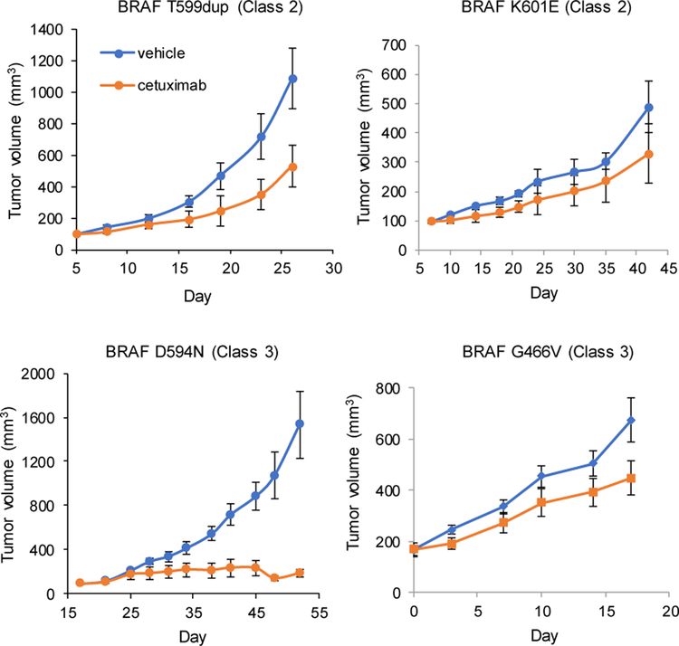
The effect of non-V600 BRAF mutants on clinical response to EGFR targeting antibodies in CRC is unknown. Given that mutant BRAF activates MAPK signaling downstream of EGFR, we hypothesized that CRCs with class 2 BRAF mutants may be refractory to anti-EGFR antibody treatment similar to those with class 1 BRAF V600 mutants. In contrast, class 3 BRAF mutants may amplify RAS signaling downstream of EGFR, and it has been proposed that these tumors may depend on EGFR activation for MAPK signaling (9). Therefore, we first analyzed patient-derived CRC models to evaluate response to EGFR antibodies. Five PDXs were generated from clinical samples of CRC, expanded in mice, and then treated with vehicle or the EGFR antibody cetuximab. The effect of cetuximab treatment in one PDX model was previously reported (9) and the other four PDX models are shown in Figure 1. Two of the PDXs were extremely sensitive to cetuximab treatment (Figure 1 and (9)), while the three others continued to grow despite cetuximab exposure, with no significant difference from vehicle treatment. Analysis by BRAF mutation class indicated that the two PDXs harboring class 2 BRAF mutants (K601E, T599dup) were resistant to cetuximab treatment, while the three PDXs harboring class 3 BRAF mutants (D594N, G466V) included two EGFR antibody sensitive tumors and a resistant tumor. Our preclinical models suggest that CRCs with BRAF mutations may respond differently to EGFR antibody treatment and response may vary by BRAF mutation class.
Fig. 1. Response to EGFR inhibitors in preclinical patient-derived models.
PDXs established from class 2 (T599dup, K601E) or class 3 (D594N, G466V) mutant BRAF were treated with either vehicle or cetuximab. Each treatment group consisted of 5 mice. Error bars indicate standard deviation.
Classification of BRAF mutants in mCRC cases
Based on our preclinical observations, we sought to evaluate the effect of non-V600 BRAF mutants on clinical response to EGFR antibodies in patients. We assembled an international collaboration to identify consecutive CRC patients with non-V600 BRAF alterations seen in the clinic. A total of 153 patients with non-V600 BRAF mutant mCRC were identified (Supplementary Fig. S1). Four patients with concurrent class 1 BRAF mutations and 31 patients without chemotherapy treatment for metastatic disease were excluded from further analysis. In the total population of 118 patients, 41 received anti-EGFR antibody treatment.
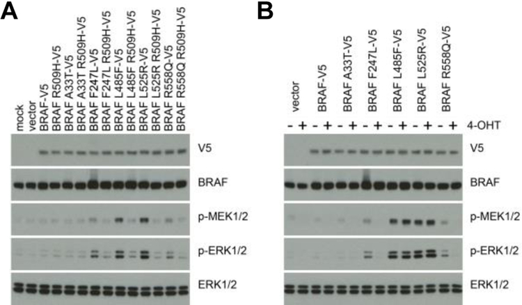
We first classified the BRAF mutants in the 118 cases into class 2 or class 3 based on published studies of the biochemical effects of the BRAF alterations (9,18,26–29). We sought to evaluate the effect of EGFR antibodies in all patients exposed to these agents by BRAF mutation class. Three patients had non-V600 BRAF mutations that had not been previously characterized (A33T, F247L, R558Q), and two cases had BRAF mutations (L485F and L525R) that had not been fully characterized. To classify these mutants, we first expressed each of these BRAF mutants in mouse embryonic fibroblasts (MEFs) to assess their ability to activate downstream proteins phospho-ERK and phospho-MEK (Fig. 2A). A change in ERK signaling was not detectable in cells when WT BRAF was overexpressed. Expression of BRAF A33T also had no effect on ERK signaling. BRAF F247L and R558Q led to modest activation of ERK signaling, and BRAF L485F and L525R both strongly activated ERK signaling, compared to WT BRAF. We next evaluated if dimerization is required for these mutants to activate the MEK/ERK cascade by introducing the R509H mutation into BRAF. It has been reported that this mutation impairs BRAF dimerization (30). The activation of ERK signaling seen with the BRAF mutants F247L, R558Q, L485F, and L525R was abrogated by the R509H mutation, indicating that all these mutants require dimerization to signal. Furthermore, we determined RAS independence by expressing each of these BRAF mutants in the RAS-less cells (9). In these cells, the only RAS allele (kras) was removed by 4-hydroxytamoxifen (4-OHT) induced Cre expression. Then, the dependence of ERK signaling of the mutants on RAS signaling was assayed (Fig. 2B). Absence of RAS expression abrogated ERK activation by BRAF F247L and R558Q but had no effect on ERK activation by BRAF L485F and L525R. Based on these data, BRAF A33T was considered to have no effect on ERK signaling, BRAF F247L and R558Q were classified as class 3 mutants, and BRAF L525R and L485F were classified as class 2 mutants.
Fig. 2. Classification of BRAF mutants.
(A) Mouse embryonic fibroblasts (MEFs) were transfected to express the V5-tagged, indicated BRAF proteins or the same BRAF proteins together with R509H mutation. Cells were collected to assay expression of activated pathway intermediates. (B) Expression of the indicated BRAF proteins was induced in the conditional “RAS-less” MEFs after pre-treatment with 4-hydroxytamoxifen (4-OHT) to knock out the last RAS allele. Cells were collected to assay expression of activated pathway intermediates.
Concurrent MAPK pathway alterations
The 118 cases in our series thus consisted of 32 class 2 BRAF mutant tumors (27%), 72 class 3 BRAF mutant cases (61%), 13 uncharacterized BRAF mutants, and a mutant with no effect on ERK signaling (Supplementary Table S1). Elevated induction of ERK output by class 3 BRAF mutants requires adequate RAS activity, which could result from RTK activation or mutations upstream in the pathway (e.g., RAS). In class 3 BRAF mutant epithelial tumors such as non-small cell lung cancer and CRC, we have shown that RAS activation often relies on RTK signaling. By contrast, in melanoma, class 3 BRAF mutants almost always coexist with mutants that activate RAS signaling, usually inactivating mutations of NF1 (9). In our larger dataset of non-V600 BRAF mutant CRCs, concurrent RAS hotspot mutations were detected in 31 patients and were enriched in tumors with class 3 BRAF mutants versus those with class 2 BRAF mutants (25% versus 6.3%, respectively; p = 0.03) (Supplementary Table S2). Furthermore, 20 of the 53 class 3 BRAF mutant CRCs co-occurred with another MAPK pathway alteration, including RAS, NF1, and MAP2K1 alterations, and concurrent MAPK pathway alterations were significantly more common in the class 3 BRAF mutant CRCs compared with class 2 BRAF mutant ones (20/53 versus 4/29, p = 0.025) (Supplementary Fig. S2). These results indicate that about a third of class 3 BRAF mutant CRCs coexist with a MAPK alteration expected to confer resistance to EGFR inhibitors and that the remaining about two-thirds may be sensitive to RTK inhibition.
Clinical characteristics and survival
Clinical characteristics of the 118 patients are summarized in Supplementary Table S2. There was a preponderance of male patients in the entire cohort and also within the class 2 and class 3 BRAF mutant CRC cases. A larger portion of class 3 BRAF mutant CRCs originated in the rectum, but this trend did not reach statistical significance. The large majority of cases exhibited conventional histology, not mucinous. Additionally, most of the non-V600 BRAF mutations occurred in microsatellite stable tumors; one class 2 BRAF mutant occurred in a microsatellite instable tumor and one frameshift alteration of unknown significance occurred in a microsatellite instable tumor. The novel BRAF alteration A33T that did not affect ERK signaling occurred in a microsatellite instable tumor.
Median OS for the patients with non-V600 BRAF mutant mCRC was 40.2 months (95% confidence interval [CI], 30.4 to 50.0 months) (Supplementary Fig. S3A). Patients with concurrent RAS tumor mutation appeared to have a poorer prognosis compared to patients with RAS WT tumors, but this difference did not reach statistical significance (median OS 29.0 months versus 44.2 months, respectively; p = 0.07) (Supplementary Fig. S3B). Among patients with RAS WT tumors, those with class 2 BRAF mutant mCRC had a trend towards shorter survival compared to those with class 3 BRAF mutant tumors (median OS 26.0 versus 44.8 months, respectively; p = 0.21) (Supplementary Fig. S3C).
Efficacy of anti-EGFR antibody treatment
Clinical characteristics for the 40 class 2 or class 3 BRAF mutant mCRC patients treated with anti-EGFR antibody are summarized in Supplementary Table S3; no significant differences were observed by BRAF mutation class. The regimen received, tumor BRAF mutation, and best response are listed in Table 1. We found clear differences in response to anti-EGFR therapy by mutational class. The response rate to anti-EGFR containing-regimen in class 2 and 3 BRAF mutants was 8% and 50%, respectively (p = 0.02) (Table 2). In the subgroup analysis according to line of therapy and sidedness, there was also trend towards higher response in class 3 patients. In the first-line or second-line setting, about half of patients had a response to treatment containing an EGFR antibody, including seven of nine patients with class 3 BRAF mutant mCRC and one of six patients with class 2 BRAF mutant mCRC (response rate, 78% and 17%, respectively, p = 0.04). Notably, two patients with class 3 BRAF mutation were able to undergo resection after response to chemotherapy. In third- or later-line, no patients with class 2 BRAF mutant mCRC responded to EGFR inhibitors, while seven of 19 (37%) patients with class 3 BRAF mutant mCRC responded (p = 0.14). More than half of the left-sided class 3 BRAF mutant tumors responded to anti-EGFR therapy whereas only two of 7 right-sided class 3 tumors achieved response (Supplementary Table S4).
Table 1.
Response details for patients treated with anti-EGFR antibody therapy.
| Class | BRAF | Line | Regimen | Response | PFS |
|---|---|---|---|---|---|
| 2 | G469V | 1 | FOLFOX+Pmab | PR | 18.9 |
| 2 | T599_V600TinsT | 1 | FOLFOX+Pmab | PD | 1.2 |
| 2 | G469A | 2 | Irinotecan+Pmab | SD | 4.0* |
| 2 | G469A | 2 | Irinotecan+Cmab | SD | 3.5* |
| 2 | L597R | 2 | Irinotecan+Pmab | PD | 3.3 |
| 2 | L597R | 2 | FOLFIRI+Pmab | SD | 16.5 |
| 2 | G469A | 3 | FOLFIRI**+Cmab | SD | 4.4 |
| 2 | G469A | 3 | Cmab | SD | 2.8 |
| 2 | G469V | 3 | Irinotecan**+Cmab | SD | 10.9 |
| 2 | L485F | 3 | Irinotecan**+Cmab | SD | 6.3 |
| 2 | L525R | 3 | Cmab | SD | 4 |
| 2 | K601E | 3 | Pmab | PD | 1.4 |
| Class | BRAF | Line | Regimen | Response | PFS |
| 3 | N581I | 1 | Irinotecan+Cmab | SD | 10.3* |
| 3 | N581S | 1 | FOLFIRI+Cmab | PR | 16 |
| 3 | N581T | 1 | FOLFOX+Pmab | PR | 1.4* |
| 3 | D594G | 1 | FOLFOX+Pmab | SD | 7.0* |
| 3 | D594G | 1 | FOLFOX+Pmab | PR | 2.8* |
| 3 | D594G | 1 | FOLFOX+Pmab | PR | 8.3* |
| 3 | F595L | 1 | FOLFOX+Pmab | PR | 10.2* |
| 3 | G466E | 2 | Irinotecan**+Pmab | PR | 8.3* |
| 3 | D594G | 2 | FOLFIRI+Cmab | PR | 3.2 |
| 3 | G466E | 3 | Irinotecan**+Pmab | PR | 8.3 |
| 3 | G466V | 3 | Irinotecan**+Pmab | PR | 6.1 |
| 3 | Q524L | 3 | Pmab | PD | 2.3 |
| 3 | D594G | 3 | Irinotecan**+Pmab | NE | 1.8* |
| 3 | D594G | 3 | Irinotecan**+Pmab | SD | 1.8* |
| 3 | D594G | 3 | FOLFIRI**+Cmab | SD | 3.6 |
| 3 | D594G | 3 | Irinotecan**+Pmab | PD | 2.8 |
| 3 | D594G | 3 | FOLFIRI**+Cmab | PD | 2.4 |
| 3 | D594G | 3 | FOLFIRI**+Pmab | SD | 2.6* |
| 3 | D594G | 3 | FOLFIRI**+Cmab | SD | 3.7 |
| 3 | D594G | 3 | Irinotecan**+Cmab | SD | 6.6 |
| 3 | D594G | 3 | Irinotecan**+Pmab | SD | 2.3 |
| 3 | D594G | 3 | Irinotecan**+Cmab | SD | 2.2* |
| 3 | D594G | 3 | Irinotecan**+Pmab | PR | 6.7* |
| 3 | D594N | 3 | Irinotecan**+Pmab | PR | 14.9 |
| 3 | D594N | 3 | 5-FU***+Cmab | PD | 1.6 |
| 3 | D594N | 5 | Irinotecan**+Pmab | PR | 14.7* |
| 3 | F247L | 3 | FOLFIRI**+Cmab | CR | 12.6 |
| 3 | R558Q | 4 | Irinotecan**+Cmab | CR | 37.7* |
CR, complete response; NE, not evaluable; PD, progressive disease; PR, partial response; SD, stable disease; Cmab, Cetuximab; Pmab, Panitumumab.
censored
Irinotecan refractory
5-FU refractory
Table 2.
Response to anti-EGFR antibody in patients based on BRAF classification.
| Class 2 | Class 3 | P | |||
|---|---|---|---|---|---|
| No. of patients | No. Responded (%) | No. of patients | No. Responded (%) | ||
| 1st or 2nd line | 6 | 1 | 9 | 7 | 0.04 |
| (17) | (78) | ||||
| 3rd or later line | 6 | 0 | 19 | 7 | 0.14 |
| (37) | |||||
| Total | 12 | 1 | 28 | 14 | 0.02 |
| (8) | (50) | ||||
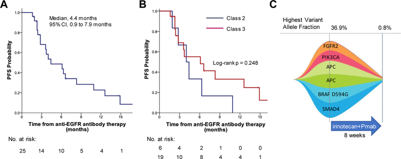
Progression free survival (PFS) for patients who received EGFR-inhibitor containing therapy at third- or later-line was 4.4 months (95% CI, 0.9–7.9 months) (Fig. 3A). In the subgroup analysis, the median duration of response to EGFR antibody therapy was 4.0 months (95% CI, 2.1 to 5.9 months) for class 2 BRAF mutant mCRC and 6.1 months (95% CI, 0.8 to 11.4 months) for class 3 BRAF mutant mCRC patients (p = 0.25) (Fig. 3B).
Fig. 3. Survival of patients with non-V600 BRAF alterations.
(A) PFS in a total of 25 patients treated with anti-EGFR antibody at the third- or later-line, or (B) stratified by BRAF mutation class. (C) Measurements of cell tumor DNA before and during treatment with panitumumab and irinotecan. Numbers for highest allelic fraction correspond to BRAF D594G mutant allelic fraction detected in blood.
In a patient with BRAF D594G mutant (class 3) mCRC who responded to panitumumab and irinotecan after prior irinotecan, we were able to collect plasma for cfDNA analysis before treatment and at 8 weeks. cfDNA analysis indicated that the BRAF mutation had the highest variant allelic frequency in blood at the start of treatment of 36.9%, suggesting it is a truncal mutation and not a subclonal event (Fig. 3C). After 8 weeks of treatment, the frequency of BRAF D594G decreased to 0.8%, indicating that the BRAF mutant clone had decreased with EGFR inhibitor treatment, rather than a separate BRAF WT clone responding. The variant allelic frequency of all the circulating tumor DNA detectable in blood in this patient decreased with the panitumumab/irinotecan treatment, supporting the benefit of EGFR inhibitor treatment in this patient with a non-V600 BRAF mutation.
Class 2 mutations can cause secondary resistance to EGFR inhibitors
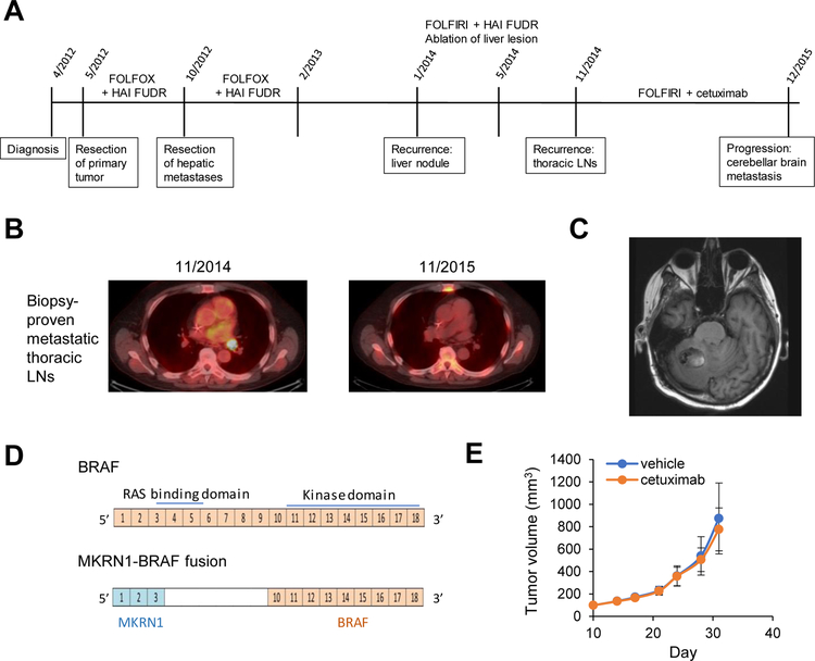
The patient with BRAF F247L mCRC achieved a long-lasting response to EGFR antibody-containing therapy of over a year. At resistance, a new, additional class 2 BRAF mutation was detected. Specifically, the patient received third-line FOLFIRI and cetuximab for biopsy proven metastatic thoracic lymphadenopathy (Clinical course summarized in Fig. 4A). Treatment led to complete regression of the hypermetabolic thoracic nodes (Fig. 4B), but the patient developed a brain metastasis after a year of treatment (Fig. 4C). The brain metastasis was removed and sequencing identified a new MKRN1-BRAF fusion (Fig. 4D), a class 2 activating alteration that no longer includes the RAS binding domain of BRAF. This fusion does not require RAS binding to dimerize. Sequencing of both the primary colon cancer and the thoracic lymph node biopsy specimen did not identify this fusion confirming that this alteration was acquired in the resistant brain metastasis. We were able to generate a PDX from the cerebellar metastasis. Treatment of the PDX with vehicle or cetuximab (5 mice per group) indicated that cetuximab did not inhibit tumor growth (Fig. 4E). These data suggest that the acquired class 2 BRAF alteration caused resistance to cetuximab treatment and further supports the limited efficacy of EGFR inhibitors against class 2 BRAF mutants.
Fig. 4. Class 2 mutations can cause secondary resistance to EGFR inhibitors.
(A) Timeline of patient’s treatment history, (B) Representative images from PET/CT showing biopsy-proven, hypermetabolic metastatic thoracic lymphadenopathy at the start of FOLFIRI/cetuximab treatment and after one year of treatment, (C) Representative MRI image showing new cerebellar metastasis, (D) Schema of the MKRN1-BRAF fusion identified on sequencing the cerebellar metastasis, (E) Growth curve of mice bearing the PDX with MKRN1-BRAF fusion treated with vehicle or cetuximab. Five mice were treated in each group and standard deviations are indicated with error bars.
Discussion
In this study, we find in both preclinical models and in our large clinical series that response to EGFR antibody treatment in CRCs with class 2 BRAF mutants is rare, while a large portion of CRCs with class 3 BRAF mutants respond to EGFR antibody treatment. This is the first demonstration of the sensitivity of tumors driven by RAS- or RAF-dependent activation of ERK signaling to RTK inhibition. Moreover, we have defined a biomarker (Class 3 BRAF mutation) that allows identification of patients who are candidates for this therapy.
Our data indicate that CRC patients with class 3 BRAF mutations should not be excluded from EGFR antibody treatment. Anti-EGFR antibody containing treatment decreased the frequency of BRAF D594G circulating tumor DNA, indicating that the BRAF mutant clone responded to the treatment, rather than a separate BRAF WT clone responding. When anti-EGFR antibody treatment was used with first-line chemotherapy, seven of nine patients treated achieved responses, and two of them were able to undergo surgical resection. Consistent with our data, Cremolini et al found that three of four patients with either BRAF 594 or 596 mutation achieved partial response with upfront chemotherapy plus cetuximab (31). Together these data indicate that RAS activation in many class 3 BRAF mutant mCRC is driven by EGFR and targeting this RTK leads to regression of these tumors.
It has recently been appreciated that within RAS WT tumors, patients with a left-sided colon primary benefit from anti-EGFR antibodies, and those with right-sided primaries obtain limited benefit (32–34). Consistent with this observation, we found that left-sided tumors were significantly more sensitive to EGFR antibody treatment. Given that class 3 BRAF mutants require RAS activation to effectively amplify MAPK signaling, our data suggest that EGFR, which is primarily activated in the left-colon, often provides the activation of RAS for MAPK pathway activation and tumorigenesis in class 3 BRAF mutant tumors.
Not all the class 3 BRAF CRC patients responded to anti-EGFR therapy. In addition to RAS mutations, activation of other RTKs leads to resistance to anti-EGFR therapy (35,36). By identifying RTKs playing dominant roles in RAS activation, class 3 BRAF mutant tumors may be better targeted by treating with the matched RTK inhibitor with or without a MEK inhibitor. Despite this, our data suggest that in a large portion of class 3 BRAF mutants, the single dominant driver is EGFR, which can be effectively targeted in the clinic.
None of the class 2 BRAF mutants responded to anti-EGFR antibodies in treatment beyond the first-line setting. In line with this, cetuximab did not cause regression in our PDX models. Additionally, we found that a CRC patient acquired a class 2 BRAF alteration, the MKRN1-BRAF fusion, at resistance to cetuximab, further suggesting that class 2 mutants cause resistance. Similarly, BRAF fusions have been reported as a mechanism of acquired resistance to EGFR inhibition in EGFR mutant non-small cell lung cancer (NSCLC) (37). While class 2 BRAF mutants thus appear to rarely respond to EGFR inhibitors, we note that occasional responses have been described (19), though these responses appear short-lived. There may be a range of ERK activation with class 2 BRAF mutants and, in our series, we see a low, but non-zero, rate of concurrent RAS mutation in the class 2 BRAF mutant tumors. These data suggest that in some class 2 BRAF mutants, while mutant BRAF activates ERK, an additional contribution to ERK activation can come from upstream signaling. Consistent with this understanding, in class 2 BRAF mutant NSCLC lines, EGFR inhibition partially suppressed MAPK signaling and cell growth, but not enough to induce tumor regression (38). Thus, while a subset of class 2 BRAF mutant CRC may respond to EGFR inhibitors, these cases are rare and EGFR inhibitor treatment may not sufficiently suppress ERK signaling for durable clinical benefit.
Class 2 BRAF tumors are resistant to approved BRAF drugs designed to inhibit mutant V600. Newer RAF inhibitors may have activity against these mutants. Type II RAF inhibitors can bind to the DFG-out inactive conformation of BRAF and induce RAF dimerization but inhibit the kinase activity of the dimer (8,39). Type II inhibitors demonstrated preclinical efficacy in xenograft models from BRAF class 2 mutant NSCLC cell lines (38). MEK inhibitors may also be effective, however, the effects of these drugs are attenuated by re-activation of receptors, such as EGFR. Combined inhibition of BRAF and MEK with or without EGFR could be an effective strategy for class 2 BRAF mutants and is currently being investigated in clinical trials (UMIN000031857; ).
While our study is the largest series to interrogate the efficacy of anti-EGFR antibodies in patients with BRAF non-V600 mutations, clear limitations of our analysis are the small number of BRAF class 2 and 3 mutated CRC patients, in line with the low frequency of these populations, and the retrospective nature of our cohort. Because non-V600 BRAF alterations occur in about 2% of mCRCs, it would be difficult to perform a prospective study; in fact, in BRAF V600E mCRC, which is the most frequent BRAF alteration in CRC, the consensus that this genomic alteration is associated with resistance to EGFR antibody treatment comes from meta-analyses. A randomized study of EGFR antibodies has not been conducted for this specific population and the subset analyses of randomized controlled trials that included BRAF V600E mCRC did not have sufficient numbers to demonstrate statistically significant differences in response rate (40,41). Yet despite this, clinically, assessing BRAF V600E status is very important in deciding whether to give EGFR antibody treatment alone (42–44). We now show that non-V600 BRAF alterations do not clinically represent a uniform group and response to EGFR antibody treatment should be considered by BRAF mutation class. Prospective analysis would therefore require very large patient numbers and would not be feasible. We also acknowledge that we could not directly assess the efficacy of anti-EGFR antibodies monotherapy, since most of the patients were treated with chemotherapy. This may contribute to the discordance between the similar PFS intervals for the two BRAF mutation classes and the substantially different response rates observed. Additionally, many patients underwent mutational analysis after progression through standard chemotherapy in order to enroll on clinical trials, which could have led to a selection bias resulting in better OS in our cohort.
In conclusion, our results indicate that many class 3, but not class 2, BRAF mutant-driven colorectal cancers are driven by EGFR and can be sensitive to its inhibition. These data provide clinical guidance on the application of EGFR antibodies in patients with CRC with non-V600 BRAF mutations. CRC patients with class 3 BRAF mutations should be considered for anti-EGFR antibody treatment. The lack of efficacy seen in class 2 BRAF mutant tumors suggests that these patients may not benefit from anti-EGFR therapy and highlights the importance to develop new targeted approaches for this patient population.
Supplementary Material
Statement of Translational Relevance.
BRAF mutations can be classified into three groups based on their biochemical properties: class 1 mutants (i.e. V600) exhibit high kinase activity and signal as RAS-independent monomers; class 2 mutants are activating and signal as RAS-independent dimers; and class 3 mutants have impaired kinase activity and depend on RAS to activate signaling. In metastatic colorectal cancer (mCRC), BRAF mutations consist of 2/3 class 1 and 1/3 class 2 and 3 alterations. The class 2 and 3 BRAF mutants do not respond to approved RAF inhibitors. Class 3 mutants, however, may be sensitive to inhibition of receptors activating RAS, such as EGFR in CRC. Here, we have assembled a large series of non-V600 BRAF mutant mCRC treated with EGFR targeting antibodies to test this hypothesis. We show that patients with class 3 BRAF mutations commonly respond to anti-EGFR treatment, while mCRC patients with class 2 BRAF mutations are unlikely to respond.
Acknowledgements
Supported by Grants-in-Aid for Scientific Research (16K07164) and Fund for the Promotion of Joint International Research (15KK0303) from Japan Society for the Promotion of Science (H.Ebi), and Takeda Science Foundation (H.Ebi), Grant-in-Aid to Project for Cancer Research and Therapeutics Evolution (P-CREATE) from the Japan Agency for Medical Research and Development (H. Ebi, 19cm0106513h0004), MSK Cancer Center core grant P30 CA 008748 and R01 CA233736 (N. Rosen., R. Yaeger.).
Conflict of Interest Statement:
R.Y. has received research funding from Array BioPharma, Genentech, GlaxoSmithKline, and Novartis and has served as an advisory board member for GlaxoSmithKline. D.K received honoraria from Eli Lilly, Takeda, Chugai, and Merck biopharma. H.T. has received research funding from Daiichi Sankyo and Takeda and received honoraria from Takeda, Taiho, Merck Biopharma, and Chugai. T.Y has received research grant from Novartis Pharma K.K., MSD K.K., Sumitomo Dainippon Pharma Co. Ltd., Chugai, Sanofi, Daiichi Sankyo, Parexel, and Ono. N.R. is on the scientific advisory board (SAB) of Chugai, BeiGene, Fortress Biotech, Daiichi-Sankyo, AstraZeneca, F-Prime, Zai Lab, Arvinas, and Array BioPharma; and he is a past SAB member of Millennium-Takeda, Kadmon, Kura Oncology, and Araxes. N.R. is also a consultant to Novartis Biomed, Boehringer Ingelheim, Tarveda, Foresite Capital, Array BioPharma, and Revolution Medicines; and in recent years has also consulted with Eli Lilly, Merrimack, Kura Oncology, Araxes, and Kadmon. N.R. owns equity in BeiGene, Zai Lab, Fortress Biotech, Kura Oncology, Araxes, Kadmon, and Effector. N.R. collaborates with Plexxikon; he receives research support from Chugai. R.B.C. is a consultant/advisory board member for Amgen, Array Biopharma, Astex Pharmaceuticals, Avidity Biosciences, BMS, Chugai, Fog Pharma, Genentech, LOXO, Merrimack, N-of-one, Novartis, nRichDx, Roche, Roivant, Shire, Spectrum Pharmaceuticals, Symphogen, Taiho, and Warp Drive Bio; holds equity in Avidity Biosciences and nRichDx; and has received research funding from Asana, AstraZeneca, and Sanofi. The remaining authors have nothing to disclose.
References
- 1.Holderfield M, Deuker MM, McCormick F, McMahon M. Targeting RAF kinases for cancer therapy: BRAF-mutated melanoma and beyond. Nat Rev Cancer 2014;14(7):455–67 10.1038/nrc3760. [DOI] [PMC free article] [PubMed] [Google Scholar]
- 2.Davies H, Bignell GR, Cox C, Stephens P, Edkins S, Clegg S, et al. Mutations of the BRAF gene in human cancer. Nature 2002;417(6892):949–54 10.1038/nature00766. [DOI] [PubMed] [Google Scholar]
- 3.Sanz-Garcia E, Argiles G, Elez E, Tabernero J. BRAF mutant colorectal cancer: prognosis, treatment, and new perspectives. Ann Oncol 2017;28(11):2648–57 10.1093/annonc/mdx401. [DOI] [PubMed] [Google Scholar]
- 4.Yaeger R, Chatila WK, Lipsyc MD, Hechtman JF, Cercek A, Sanchez-Vega F, et al. Clinical Sequencing Defines the Genomic Landscape of Metastatic Colorectal Cancer. Cancer Cell 2018;33(1):125–36 10.1016/j.ccell.2017.12.004. [DOI] [PMC free article] [PubMed] [Google Scholar]
- 5.Jones JC, Renfro LA, Al-Shamsi HO, Schrock AB, Rankin A, Zhang BY, et al. (Non-V600) BRAF Mutations Define a Clinically Distinct Molecular Subtype of Metastatic Colorectal Cancer. J Clin Oncol 2017;35(23):2624–30 10.1200/JCO.2016.71.4394. [DOI] [PMC free article] [PubMed] [Google Scholar]
- 6.Yaeger R, Corcoran RB. Targeting Alterations in the RAF-MEK Pathway. Cancer Discov 2019;9(3):329–41 10.1158/2159-8290.CD-18-1321. [DOI] [PMC free article] [PubMed] [Google Scholar]
- 7.Wan PT, Garnett MJ, Roe SM, Lee S, Niculescu-Duvaz D, Good VM, et al. Mechanism of activation of the RAF-ERK signaling pathway by oncogenic mutations of B-RAF. Cell 2004;116(6):855–67. [DOI] [PubMed] [Google Scholar]
- 8.Yao Z, Torres NM, Tao A, Gao Y, Luo L, Li Q, et al. BRAF Mutants Evade ERK-Dependent Feedback by Different Mechanisms that Determine Their Sensitivity to Pharmacologic Inhibition. Cancer Cell 2015;28(3):370–83 10.1016/j.ccell.2015.08.001. [DOI] [PMC free article] [PubMed] [Google Scholar]
- 9.Yao Z, Yaeger R, Rodrik-Outmezguine VS, Tao A, Torres NM, Chang MT, et al. Tumours with class 3 BRAF mutants are sensitive to the inhibition of activated RAS. Nature 2017;548(7666):234–8 10.1038/nature23291. [DOI] [PMC free article] [PubMed] [Google Scholar]
- 10.Heidorn SJ, Milagre C, Whittaker S, Nourry A, Niculescu-Duvas I, Dhomen N, et al. Kinase-dead BRAF and oncogenic RAS cooperate to drive tumor progression through CRAF. Cell 2010;140(2):209–21 10.1016/j.cell.2009.12.040. [DOI] [PMC free article] [PubMed] [Google Scholar]
- 11.Roth AD, Tejpar S, Delorenzi M, Yan P, Fiocca R, Klingbiel D, et al. Prognostic role of KRAS and BRAF in stage II and III resected colon cancer: results of the translational study on the PETACC-3, EORTC 40993, SAKK 60–00 trial. J Clin Oncol 2010;28(3):466–74 10.1200/JCO.2009.23.3452. [DOI] [PubMed] [Google Scholar]
- 12.Jonker DJ, O’Callaghan CJ, Karapetis CS, Zalcberg JR, Tu D, Au HJ, et al. Cetuximab for the treatment of colorectal cancer. N Engl J Med 2007;357(20):2040–8 10.1056/NEJMoa071834. [DOI] [PubMed] [Google Scholar]
- 13.Seymour MT, Brown SR, Middleton G, Maughan T, Richman S, Gwyther S, et al. Panitumumab and irinotecan versus irinotecan alone for patients with KRAS wild-type, fluorouracil-resistant advanced colorectal cancer (PICCOLO): a prospectively stratified randomised trial. Lancet Oncol 2013;14(8):749–59 10.1016/S1470-2045(13)70163-3. [DOI] [PMC free article] [PubMed] [Google Scholar]
- 14.Rowland A, Dias MM, Wiese MD, Kichenadasse G, McKinnon RA, Karapetis CS, et al. Meta-analysis of BRAF mutation as a predictive biomarker of benefit from anti-EGFR monoclonal antibody therapy for RAS wild-type metastatic colorectal cancer. Br J Cancer 2015;112(12):1888–94 10.1038/bjc.2015.173. [DOI] [PMC free article] [PubMed] [Google Scholar]
- 15.Pietrantonio F, Petrelli F, Coinu A, Di Bartolomeo M, Borgonovo K, Maggi C, et al. Predictive role of BRAF mutations in patients with advanced colorectal cancer receiving cetuximab and panitumumab: a meta-analysis. Eur J Cancer 2015;51(5):587–94 10.1016/j.ejca.2015.01.054. [DOI] [PubMed] [Google Scholar]
- 16.van Brummelen EMJ, de Boer A, Beijnen JH, Schellens JHM. BRAF Mutations as Predictive Biomarker for Response to Anti-EGFR Monoclonal Antibodies. Oncologist 2017;22(7):864–72 10.1634/theoncologist.2017-0031. [DOI] [PMC free article] [PubMed] [Google Scholar]
- 17.De Roock W, Claes B, Bernasconi D, De Schutter J, Biesmans B, Fountzilas G, et al. Effects of KRAS, BRAF, NRAS, and PIK3CA mutations on the efficacy of cetuximab plus chemotherapy in chemotherapy-refractory metastatic colorectal cancer: a retrospective consortium analysis. Lancet Oncol 2010;11(8):753–62 10.1016/S1470-2045(10)70130-3. [DOI] [PubMed] [Google Scholar]
- 18.Shinozaki E, Yoshino T, Yamazaki K, Muro K, Yamaguchi K, Nishina T, et al. Clinical significance of BRAF non-V600E mutations on the therapeutic effects of anti-EGFR monoclonal antibody treatment in patients with pretreated metastatic colorectal cancer: the Biomarker Research for anti-EGFR monoclonal Antibodies by Comprehensive Cancer genomics (BREAC) study. Br J Cancer 2017;117(10):1450–8 10.1038/bjc.2017.308. [DOI] [PMC free article] [PubMed] [Google Scholar]
- 19.Wang Y, Jones JC, Kipp BR, Grothey A. Activity of EGFR Antibody in Non-V600 BRAF Mutant Metastatic Colorectal Cancer. Ann Oncol 2018. 10.1093/annonc/mdy477. [DOI] [PubMed]
- 20.Arcila M, Lau C, Nafa K, Ladanyi M. Detection of KRAS and BRAF mutations in colorectal carcinoma roles for high-sensitivity locked nucleic acid-PCR sequencing and broad-spectrum mass spectrometry genotyping. J Mol Diagn 2011;13(1):64–73 10.1016/j.jmoldx.2010.11.005. [DOI] [PMC free article] [PubMed] [Google Scholar]
- 21.Cheng DT, Mitchell TN, Zehir A, Shah RH, Benayed R, Syed A, et al. Memorial Sloan Kettering-Integrated Mutation Profiling of Actionable Cancer Targets (MSK-IMPACT): A Hybridization Capture-Based Next-Generation Sequencing Clinical Assay for Solid Tumor Molecular Oncology. J Mol Diagn 2015;17(3):251–64 10.1016/j.jmoldx.2014.12.006. [DOI] [PMC free article] [PubMed] [Google Scholar]
- 22.Lanman RB, Mortimer SA, Zill OA, Sebisanovic D, Lopez R, Blau S, et al. Analytical and Clinical Validation of a Digital Sequencing Panel for Quantitative, Highly Accurate Evaluation of Cell-Free Circulating Tumor DNA. PLoS One 2015;10(10):e0140712 10.1371/journal.pone.0140712. [DOI] [PMC free article] [PubMed] [Google Scholar]
- 23.Yaeger R, Yao Z, Hyman DM, Hechtman JF, Vakiani E, Zhao H, et al. Mechanisms of Acquired Resistance to BRAF V600E Inhibition in Colon Cancers Converge on RAF Dimerization and Are Sensitive to Its Inhibition. Cancer Res 2017;77(23):6513–23 10.1158/0008-5472.CAN-17-0768. [DOI] [PMC free article] [PubMed] [Google Scholar]
- 24.Solit DB, Garraway LA, Pratilas CA, Sawai A, Getz G, Basso A, et al. BRAF mutation predicts sensitivity to MEK inhibition. Nature 2006;439(7074):358–62 10.1038/nature04304. [DOI] [PMC free article] [PubMed] [Google Scholar]
- 25.Drosten M, Sum EY, Lechuga CG, Simon-Carrasco L, Jacob HK, Garcia-Medina R, et al. Loss of p53 induces cell proliferation via Ras-independent activation of the Raf/Mek/Erk signaling pathway. Proc Natl Acad Sci U S A 2014;111(42):15155–60 10.1073/pnas.1417549111. [DOI] [PMC free article] [PubMed] [Google Scholar]
- 26.Foster SA, Whalen DM, Ozen A, Wongchenko MJ, Yin J, Yen I, et al. Activation Mechanism of Oncogenic Deletion Mutations in BRAF, EGFR, and HER2. Cancer Cell 2016;29(4):477–93 10.1016/j.ccell.2016.02.010. [DOI] [PubMed] [Google Scholar]
- 27.Rodriguez-Viciana P, Tetsu O, Tidyman WE, Estep AL, Conger BA, Cruz MS, et al. Germline mutations in genes within the MAPK pathway cause cardio-facio-cutaneous syndrome. Science 2006;311(5765):1287–90 10.1126/science.1124642. [DOI] [PubMed] [Google Scholar]
- 28.Agianian B, Gavathiotis E. Current Insights of BRAF Inhibitors in Cancer. J Med Chem 2018;61(14):5775–93 10.1021/acs.jmedchem.7b01306. [DOI] [PubMed] [Google Scholar]
- 29.Ng PK, Li J, Jeong KJ, Shao S, Chen H, Tsang YH, et al. Systematic Functional Annotation of Somatic Mutations in Cancer. Cancer Cell 2018;33(3):450–62 10.1016/j.ccell.2018.01.021. [DOI] [PMC free article] [PubMed] [Google Scholar]
- 30.Poulikakos PI, Zhang C, Bollag G, Shokat KM, Rosen N. RAF inhibitors transactivate RAF dimers and ERK signalling in cells with wild-type BRAF. Nature 2010;464(7287):427–30 10.1038/nature08902. [DOI] [PMC free article] [PubMed] [Google Scholar]
- 31.Cremolini C, Di Bartolomeo M, Amatu A, Antoniotti C, Moretto R, Berenato R, et al. BRAF codons 594 and 596 mutations identify a new molecular subtype of metastatic colorectal cancer at favorable prognosis. Ann Oncol 2015;26(10):2092–7 10.1093/annonc/mdv290. [DOI] [PubMed] [Google Scholar]
- 32.Tejpar S, Stintzing S, Ciardiello F, Tabernero J, Van Cutsem E, Beier F, et al. Prognostic and Predictive Relevance of Primary Tumor Location in Patients With RAS Wild-Type Metastatic Colorectal Cancer: Retrospective Analyses of the CRYSTAL and FIRE-3 Trials. JAMA Oncol 2016. 10.1001/jamaoncol.2016.3797. [DOI] [PMC free article] [PubMed]
- 33.Boeckx N, Koukakis R, Op de Beeck K, Rolfo C, Van Camp G, Siena S, et al. Primary tumor sidedness has an impact on prognosis and treatment outcome in metastatic colorectal cancer: results from two randomized first-line panitumumab studies. Ann Oncol 2017;28(8):1862–8 10.1093/annonc/mdx119. [DOI] [PMC free article] [PubMed] [Google Scholar]
- 34.Arnold D, Lueza B, Douillard JY, Peeters M, Lenz HJ, Venook A, et al. Prognostic and predictive value of primary tumour side in patients with RAS wild-type metastatic colorectal cancer treated with chemotherapy and EGFR directed antibodies in six randomized trials. Ann Oncol 2017;28(8):1713–29 10.1093/annonc/mdx175. [DOI] [PMC free article] [PubMed] [Google Scholar]
- 35.Bertotti A, Papp E, Jones S, Adleff V, Anagnostou V, Lupo B, et al. The genomic landscape of response to EGFR blockade in colorectal cancer. Nature 2015;526(7572):263–7 10.1038/nature14969. [DOI] [PMC free article] [PubMed] [Google Scholar]
- 36.Bardelli A, Corso S, Bertotti A, Hobor S, Valtorta E, Siravegna G, et al. Amplification of the MET receptor drives resistance to anti-EGFR therapies in colorectal cancer. Cancer Discov 2013;3(6):658–73 10.1158/2159-8290.CD-12-0558. [DOI] [PMC free article] [PubMed] [Google Scholar]
- 37.Yu HA, Suzawa K, Jordan E, Zehir A, Ni A, Kim R, et al. Concurrent Alterations in EGFR-Mutant Lung Cancers Associated with Resistance to EGFR Kinase Inhibitors and Characterization of MTOR as a Mediator of Resistance. Clin Cancer Res 2018;24(13):3108–18 10.1158/1078-0432.CCR-17-2961. [DOI] [PMC free article] [PubMed] [Google Scholar]
- 38.Kotani H, Adachi Y, Kitai H, Tomida S, Bando H, Faber AC, et al. Distinct dependencies on receptor tyrosine kinases in the regulation of MAPK signaling between BRAF V600E and non-V600E mutant lung cancers. Oncogene 2018;37(13):1775–87 10.1038/s41388-017-0035-9. [DOI] [PubMed] [Google Scholar]
- 39.Karoulia Z, Gavathiotis E, Poulikakos PI. New perspectives for targeting RAF kinase in human cancer. Nat Rev Cancer 2017;17(11):676–91 10.1038/nrc.2017.79. [DOI] [PMC free article] [PubMed] [Google Scholar]
- 40.Bokemeyer C, Van Cutsem E, Rougier P, Ciardiello F, Heeger S, Schlichting M, et al. Addition of cetuximab to chemotherapy as first-line treatment for KRAS wild-type metastatic colorectal cancer: pooled analysis of the CRYSTAL and OPUS randomised clinical trials. Eur J Cancer 2012;48(10):1466–75 10.1016/j.ejca.2012.02.057. [DOI] [PubMed] [Google Scholar]
- 41.Karapetis CS, Jonker D, Daneshmand M, Hanson JE, O’Callaghan CJ, Marginean C, et al. PIK3CA, BRAF, and PTEN status and benefit from cetuximab in the treatment of advanced colorectal cancer--results from NCIC CTG/AGITG CO.17. Clin Cancer Res 2014;20(3):744–53 10.1158/1078-0432.CCR-13-0606. [DOI] [PubMed] [Google Scholar]
- 42.Van Cutsem E, Cervantes A, Adam R, Sobrero A, Van Krieken JH, Aderka D, et al. ESMO consensus guidelines for the management of patients with metastatic colorectal cancer. Ann Oncol 2016;27(8):1386–422 10.1093/annonc/mdw235. [DOI] [PubMed] [Google Scholar]
- 43.Yoshino T, Arnold D, Taniguchi H, Pentheroudakis G, Yamazaki K, Xu RH, et al. Pan-Asian adapted ESMO consensus guidelines for the management of patients with metastatic colorectal cancer: a JSMO-ESMO initiative endorsed by CSCO, KACO, MOS, SSO and TOS. Ann Oncol 2018;29(1):44–70 10.1093/annonc/mdx738. [DOI] [PubMed] [Google Scholar]
- 44.Benson AB, Venook AP, Al-Hawary MM, Cederquist L, Chen YJ, Ciombor KK, et al. NCCN Guidelines Insights: Colon Cancer, Version 2.2018. J Natl Compr Canc Netw 2018;16(4):359–69 10.6004/jnccn.2018.0021. [DOI] [PMC free article] [PubMed] [Google Scholar]
Associated Data
This section collects any data citations, data availability statements, or supplementary materials included in this article.


