Abstract
Backgrounds/Aims
Residual gallbladder mucosa left after subtotal/partial cholecystectomy is prone to develop recurrent lithiasis and become symptomatic, which mandates surgical removal.
Methods
We retrospectively evaluated the patients with residual gallbladder referred to us from January 2011 to December 2017. Based on MRCP we classified calot's anatomy to – type I where cystic duct was seen and type II where sessile GB stump was seen.
Results
21 patients with median age 38 years and M:F::1:9.5, had undergone cholecystectomy (3 months-20 years) prior, presented with recurrent biliary pain. 3 had jaundice (CBD stone, Mirizzi and biliary stricture), 1 had pancreatitis and one had malignancy of the GB. Imaging revealed type I anatomy in 14 (67%) and type II in 7 (33%). All underwent completion cholecystectomy – open in 18 and laparoscopic in 3 (one converted to open). Additional procedure was required in 5 patients – CBD exploration in 2 (10%) and one each Hepatico-jejunostomy, extended cholecystectomy and splenectomy. Median hospital stay was 1 day. There was no mortality and 10% morbidity. One patient with malignancy died at 2 years, two died of unrelated cause, one developed incisional hernia and the remaining were well at a median follow up of 29 months.
Conclusions
Residual GB lithiasis should be suspected if there are recurrent symptoms after cholecystectomy. MRCP based proposed classification can guide the management strategy. Completion cholecystectomy is curative.
Keywords: Gall bladder, Cholecystectomy, Residual, Cystic duct, Remnant, Recurrent
INTRODUCTION
Cholecystectomy is a widely performed procedure throughout the globe. Difficult dissection and dense adhesions in calots triangle due to inflammatory process as a result of stone disease can put the bile duct at risk of injury. Various strategies have been devised to minimize the risk of bile duct injuries.1,2 One of them is performing partial or subtotal cholecystectomy (STC). It not only avoids the difficult calots dissection in the setting of inflammation but also keeps the dissection away from bile duct and hepatic artery.1,2,3,4,5 However, such a strategy has been reported to increase the risk of bile leak.3,4,5,6 More so, it also leaves some amount of the gall bladder (GB) attached to the biliary tree. This diseased portion of this remnant gall bladder is prone to develop recurrent stones.3,4,5,6,7,8,9,10
A large series has reported 0.4% incidence of subtotal cholecystectomies being performed.1 However, the natural history of the residual gall bladder stump after partial cholecystectomy is not clearly understood. About 10% develop symptoms and seek medical advice.1,3,5,6 There is a paucity of literature regarding the management and outcome of recurrent lithiasis in the gall bladder stump after cholecystectomy.7,8,9,10,11 The present report focuses on symptomatic residual gall bladder lithiasis after a safe subtotal cholecystectomy.
MATERIALS AND METHODS
From January 2011 to December 2017, 21 consecutive patients referred to us with symptomatic residual gall bladder stone after previous cholecystectomy, were reviewed retrospectively at Postgraduate Institute of Medical Education and Research, Chandigarh, a tertiary care centre in north India. The hospital records and charts were reviewed for demographic data, clinical, radiological and operative details. Post operative outcome was recorded. Patients with incomplete information and records were excluded. All patients were followed up till March 2018.
Management protocol
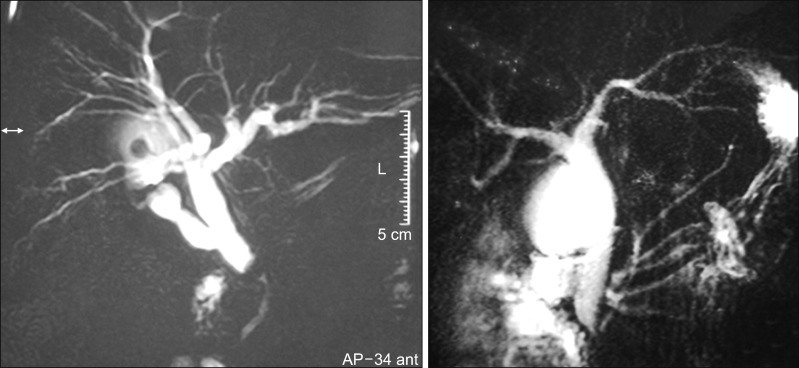
All patients underwent detail evaluation of symptom along with biochemical, hematological and coagulation parameters. The diagnosis was initially established on ultrasonogram of the abdomen. All the patients underwent magnetic resonance cholangio-pancreatography (MRCP) for evaluation of biliary anatomy (Fig. 1). Based on MRCP findings, we classified anatomy as:
Fig. 1. MRCP to show type I (left) and type II (right) Calot's anatomy. Note the presence of cystic duct in type IA. This patient had concomitant choledocholithiasis.
Type I Calot's anatomy: cystic duct was seen below the pouch of residual gall bladder. This was further categorized to
IA when gall bladder pouch was present
IB when only a long cystic duct stump was present without any residual gall bladder
Type II Calot's anatomy: sessile gall bladder with total obliteration of plane between medial GB wall and bile duct.
In case of other associated findings, additional imaging was carried out as required.
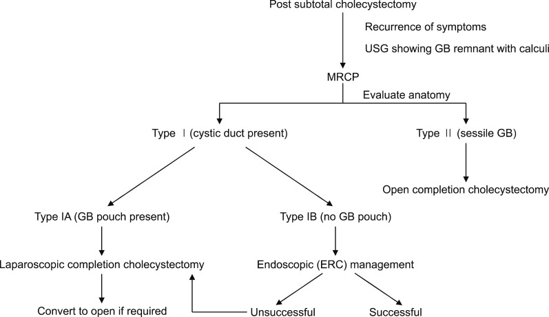
Further plan of management was based on the algorithm as described in the flow chart (Fig. 2).
Fig. 2. Flow chart to show the algorithm of management.
Surgical procedure
Adhesiolysis was done from lateral to medial side so as to visualize the residual gall bladder. Due care was taken to separate colon and duodenum. Dissection was carried out by blunt and sharp dissection using short burst of monopolar diathermy and by bipolar current. Small size and large stone in residual GB, made it difficult to grasp in some patients, so the dissection was proceeded by removing stones through a cholecystostomy.
Type I anatomy: underwent completion cholecystectomy; calot's triangle could be dissected to visualize cystic duct and artery, which were safely ligated and divided. Gall bladder was separated from the cystic plate in retrograde fashion.
Type II anatomy: underwent redo subtotal cholecystectomy; there were dense adhesions between the gall bladder and common bile duct. Antegrade dissection of gall bladder from cystic plate was done and removing about 90% of the GB leaving a very short stump on the cystic duct, which was closed with fine interrupted absorbable sutures.
RESULTS
Demographic & clinical data
The median age was 38 years with a sex ratio M:F of 1:9.5. Index cholecystectomy was done for symptomatic cholelithiasis in all the patients. Two patients had concomitant bile duct stones which were cleared prior to cholecystectomy, one patient had undergone distal gastrectomy done for gastric cancer previously prior to cholecystectomy. Cholecystectomy was performed by open procedure in 7 and laparoscopically in 14, however 3 of them were converted to an open. Two of these patients were referred to us immediately after cholecystectomy for the management of biliary fistula. Both had bile leak from the closed stump of GB and were successfully managed conservatively and were followed up for the development of symptoms. Operative details were available for 11 patients and only three of them had mentioned regarding subtotal cholecystectomy.
Median interval between index cholecystectomy and recurrence of symptoms was 3 years (3 months to 20 years). Recurrence of of biliary pain was seen in all the patients. Three patients had jaundice and one had pancreatitis. One patient had carcinoma in the residual GB stump, which developed 4 years after the index operation. Two patients with bile leak developed symptoms 8 months and 2 years after initial operation.
Imaging findings
Ultrasonogram revealed a small pouch of gall bladder in 20 and long cystic stump with stone in one, stones were seen in 20 and malignancy was seen in one. In addition one patient had evidence of extrahepatic portal vein obstruction. and splenomegaly.
On MRCP; type I anatomy was seen in 14 (67%) (IA in 13 & IB in 1) and type II in 7 (33%). 20 patients had a GB stump with a pouch and one had a long cystic duct with calculus.
Three patients with jaundice had: Bismuth type II biliary stricture in one, common duct stone in one and Mirizzi's syndrome in another.
18 FDG PET CT performed in patient with malignancy in GB stump, showed a 25 mm mass lesion with an SUV uptake of 11.5.
Endoscopic retrograde cholangiography was attempted in patient with cystic duct stone (type IB) but was not successful and the patient was managed surgically.
Operative findings
Type I anatomy: completion cholecystectomy was performed in 14 patients – open approach in 11 and laparoscopic approach in 3 out of which one was converted to open.
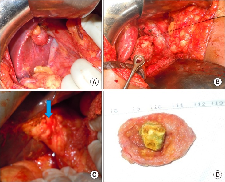
Three other patients required additional procedure – one with extrahepatic portal vein obstruction underwent subtotal cholecystectomy and splenectomy (had hypersplenism) while another patient with mass in GB underwent completion extended cholecystectomy and the third one with common duct stone (CBD) was planned for endoscopic clearance. However, during stone retrieval, the dormia basket got impacted in the CBD and patient was taken up for emergent open cholecystectomy and bile duct exploration in the same sitting (Fig. 3).
Fig. 3. Operative picture to show (A) type I anatomy with gall bladder pouch, (B) Calot's triangle could be dissected and completion cholecystectomy was performed, (C) type II anatomy, small gall bladder with obliterated Calot's triangle (arrow), (D) small sized Gall bladder with a single large stone occupying the lumen.
Type II anatomy: redo subtotal cholecystectomy was done in all the 7 patients as an open procedure. Additional procedure was required in two patients – one with mirizzi syndrome was managed with subtotal cholecystectomy and T tube drainage and one with concomitant biliary stricture underwent Roux en Y hepaticojejunostomy.
Histopathology was chronic cholecystitis in 16, xanthgranulomatous cholecystitis in 4 and adenocarcinoma (T2N1) in one. The median hospital stay was 1 day (0–6 days). Three had superficial surgical site infection and one (5%) had deep space infection which required image guided drainage and rest other made an uneventful recovery. One patient with carcinoma died after 2 years of recurrent disease and two others died of an unrelated cause. Remaining 18 patients are symptom free at a median follow up of 29 months (6 months to 6 years). One patient developed incisional hernia 18 months after the surgery (Table 1).
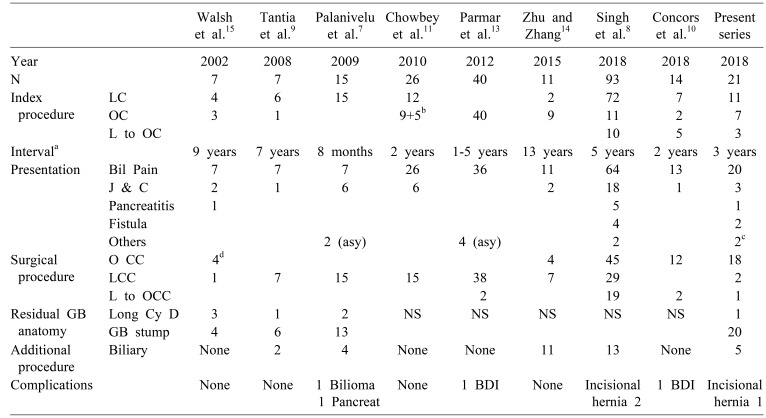
Table 1. Comparative analysis of the published series on Residual GB after cholecystectomy.
LC, Laparoscopic cholecystectomy; OC, Open cholecystectomy; L to OC, Laparoscopic converted to open cholecystectomy; LCC, Laparoscopic completion cholecystectomy; OCC, Open completion cholecystectomy; L to OCC, Laparoscopic converted to open completion cholecystectomy; J&C, Jaundice and cholangitis; BDI, Bile duct injury; Asy, asymptomatic; Cy D, cystic duct; GB, gallbladder
aInterval between indes cholecystectomy and second surgery
bPatients underwent cholecystostomy
c1 had malignancy and another had biliary stricture
dTwo patients with cystic duct calculi managed endoscopically
DISCUSSION
The present study has emphasized the importance preoperative imaging to classify calot's anatomy while planning re-operation in patients with symptomatic residual gall bladder. If a cystic duct stump is seen classified as type I anatomy, then complete re-excision of the gall bladder should be contemplated, however, if again a sessile gall bladder is seen classified as type II anatomy, then cholecystectomy leaving a small stump of GB on bile duct wall should be undertaken. More so, the present report is the first of its kind to describe de-novo malignancy in the residual GB stump.
Subtotal cholecystectomy is being performed as a damage control operation for a difficult gall bladder. The incidence reported from large series varies from 0.4% to 3%.1,3,5,6,7,8,12 Although many reports have shown the safety of this procedure,1,2,12 but only a few have described the short and long term complications associated with it.3,5,6 We observed a 10% incidence of biliary fistula in the entire cohort. Jara et al.5 in a study of 22 STC, reported 9% incidence of biliary fistula. A study of 191 subtotal cholecystectomies reported 11% incidence of biliary fistula.6 On the contrary, another large retrospective study of 93 cases of residual gall bladder reported biliary fistula in 4%.8 We found a residual stump of GB in 20 patients; this indirectly suggests prior reconstituting type subtotal cholecystectomy. Leaving a part of diseased GB stump is more likely to give symptoms when compared to long cystic duct stump. Others have also found frequent need for interventions when reconstituting subtotal cholecystectomy was done when compared to fenestrating type.6
We reported the recurrence of symptoms after index operation after 3 years. Most of the studies have reported a median time interval in many years after the index operation.8,9,10,11,13,14,15 On the contrary, Palanivelu et al.7 reported a mean of 8 months. This probably might be due to the fact that majority of the STC were performed by them and they could have been more aggressive in follow up. Recurrent symptoms may occur as early as 3 months to as late as 40 years.7,8,9,10,11,13,14,15 Early recurrence of symptoms is probably related to incomplete removal of stones from GB.
Though STC is considered as a safe procedure, we observed the occurrence of concomitant biliary stricture in one patient where type II anatomy was found subsequently. Patient was symptomatic for stricture and residual GB was an incidental finding. Other studies describing the management of residual gall bladder lithiasis did not report any case of biliary stricture.7,8,9,10,11,13,14,15 However, a review of 15 studies on partial cholecystectomy reported one case of biliary stricture.12 Another recent study on 191 patients of STC reported one case of biliary stricture following reconstituiting type cholecystectomy.6 The development of stricture was a delayed event suggesting diathermy injury as the probable mechanism underlying the stricture formation. This can happen as an overzealous attempt to cauterize residual GB mucosa to prevent malignancy in future. The occurrence of fistula after STC is higher, but it does not lead on to stricture formation as the dissection is usually away from the bile duct. We did not observe any stricture in both the patients with bile leaks. Others also did not report any stricture as a consequence of bile leak.3,5,6,8 In view of a higher incidence of bile leak, use of omentum over the STC stump has been advocated.16
The present series has reported 5% incidence of malignancy in the residual GB stump. Other reports from the west did not find any case of malignancy in the GB stump.10,14,15 This is probably related to a lower incidence of carcinoma GB in those regions. India being an endemic region for the occurrence of GB cancer, the residual at risk GB mucosa left after STC mandates a close follow up for the development of de novo neoplasm. Even studies from India with considerably long follow up after STC did not report any case of malignancy.7 Large Indian series on the clinical profile of residual GB did not report any case of malignancy.8,11,13 This probable might be due to a very small residual mucosa at risk left after STC. On the contrary, Do et al.17 has reported a case of cystic duct adenocarcinoma 10 years after index cholecystectomy where a long cystic duct stump was left. Occurrence of de-novo malignancy, in the present study indicate the need for surveillance of residual GB mucosa where complete excision was not possible. Diathermy destruction of the residual mucosa can mitigate the risk however, caution should be exercised as an overzealous attempt can lead on to stricture as was observed in the present study.
Although a few studies report successful laparoscopic re-excision,7,9,11,13 but other have reported open procedures in majority.8,10,15 Singh et al.8 in a large study on residual GB reported conversion to open procedure in 19 of the 48 patients in whom laparoscopy was attempted. Another recent study reported conversion to an open procedure in both the patients where laparoscopy was attempted.10 One of them sustained major biliary injury. Here in lies the importance of imaging based classification in the present study. Whereas, type I anatomy can undergo complete re-excision and laparoscopic approach can be safe in his situation, on the contrary, type II anatomy should be directly subjected to open procedure. One should be prepared to do subtotal cholecystectomy again in this setting. The proposed algorithm also sub-classify type I into two types based on the presence or absence of GB pouch. In the absence of GB pouch, there is a role of endoscopic management. Walsh et al reported successful endoscopic management of 2 patient with cystic duct calculi.15
Two series have reported iatrogenic bile duct injury10,13 and one has a report of post operative bile collection.7 Despite the fact, that reoperation is carried out by an experienced team, the difficult anatomy, puts the bile duct at risk of injury. Getting the preoperative information of calot's anatomy can circumvent this problem. We believe that patients with type II anatomy should be more prone to biliary injury particularly, if complete removal is attempted. No mortality has been reported so far.
Concluding, residual gall bladder can be a significant problem after subtotal cholecystectomy. Only the ones symptomatic need treatment and asymptomatic ones should be kept under surveillance. It is better to classify the calot's anatomy and plan re-operation either by laparoscopic or open approach. Utmost care should be taken to prevent biliary injury.
References
- 1.Kim Y, Wima K, Jung AD, Martin GE, Dhar VK, Shah SA. Laparoscopic subtotal cholecystectomy compared to total cholecystectomy: a matched national analysis. J Surg Res. 2017;218:316–321. doi: 10.1016/j.jss.2017.06.047. [DOI] [PubMed] [Google Scholar]
- 2.Dissanaike S. A step-by-step guide to laparoscopic subtotal fenestrating cholecystectomy: a damage control approach to the difficult gallbladder. J Am Coll Surg. 2016;223:e15–e18. doi: 10.1016/j.jamcollsurg.2016.05.006. [DOI] [PubMed] [Google Scholar]
- 3.Shin M, Choi N, Yoo Y, Kim Y, Kim S, Mun S. Clinical outcomes of subtotal cholecystectomy performed for difficult cholecystectomy. Ann Surg Treat Res. 2016;91:226–232. doi: 10.4174/astr.2016.91.5.226. [DOI] [PMC free article] [PubMed] [Google Scholar]
- 4.Nakajima J, Sasaki A, Obuchi T, Baba S, Nitta H, Wakabayashi G. Laparoscopic subtotal cholecystectomy for severe cholecystitis. Surg Today. 2009;39:870–875. doi: 10.1007/s00595-008-3975-4. [DOI] [PubMed] [Google Scholar]
- 5.Jara G, Rosciano J, Barrios W, Vegas L, Rodríguez O, Sánchez R, et al. Laparoscopic subtotal cholecystectomy: a surgical alternative to reduce complications in complex cases. Cir Esp. 2017;95:465–470. doi: 10.1016/j.ciresp.2017.07.013. [DOI] [PubMed] [Google Scholar]
- 6.van Dijk AH, Donkervoort SC, Lameris W, de Vries E, Eijsbouts QAJ, Vrouenraets BC, et al. Short- and long-term outcomes after a reconstituting and fenestrating subtotal cholecystectomy. J Am Coll Surg. 2017;225:371–379. doi: 10.1016/j.jamcollsurg.2017.05.016. [DOI] [PubMed] [Google Scholar]
- 7.Palanivelu C, Rangarajan M, Jategaonkar PA, Madankumar MV, Anand NV. Laparoscopic management of remnant cystic duct calculi: a retrospective study. Ann R Coll Surg Engl. 2009;91:25–29. doi: 10.1308/003588409X358980. [DOI] [PMC free article] [PubMed] [Google Scholar]
- 8.Singh A, Kapoor A, Singh RK, Prakash A, Behari A, Kumar A, et al. Management of residual gall bladder: a 15-year experience from a north Indian tertiary care centre. Ann Hepatobiliary Pancreat Surg. 2018;22:36–41. doi: 10.14701/ahbps.2018.22.1.36. [DOI] [PMC free article] [PubMed] [Google Scholar]
- 9.Tantia O, Jain M, Khanna S, Sen B. Post cholecystectomy syndrome: role of cystic duct stump and re-intervention by laparoscopic surgery. J Minim Access Surg. 2008;4:71–75. doi: 10.4103/0972-9941.43090. [DOI] [PMC free article] [PubMed] [Google Scholar]
- 10.Concors SJ, Kirkland ML, Schuricht AL, Dempsey DT, Morris JB, Vollmer CM, et al. Resection of gallbladder remnants after subtotal cholecystectomy: presentation and management. HPB (Oxford) 2018;20:1062–1066. doi: 10.1016/j.hpb.2018.05.005. [DOI] [PubMed] [Google Scholar]
- 11.Chowbey P, Soni V, Sharma A, Khullar R, Baijal M. Residual gallstone disease - laparoscopic management. Indian J Surg. 2010;72:220–225. doi: 10.1007/s12262-010-0058-8. [DOI] [PMC free article] [PubMed] [Google Scholar]
- 12.Henneman D, da Costa DW, Vrouenraets BC, van Wagensveld BA, Lagarde SM. Laparoscopic partial cholecystectomy for the difficult gallbladder: a systematic review. Surg Endosc. 2013;27:351–358. doi: 10.1007/s00464-012-2458-2. [DOI] [PubMed] [Google Scholar]
- 13.Parmar AK, Khandelwal RG, Mathew MJ, Reddy PK. Laparoscopic completion cholecystectomy: a retrospective study of 40 cases. Asian J Endosc Surg. 2013;6:96–99. doi: 10.1111/ases.12012. [DOI] [PubMed] [Google Scholar]
- 14.Zhu JG, Zhang ZT. Laparoscopic remnant cholecystectomy and transcystic common bile duct exploration for gallbladder/cystic duct remnant with stones and choledocholithiasis after cholecystectomy. J Laparoendosc Adv Surg Tech A. 2015;25:7–11. doi: 10.1089/lap.2014.0186. [DOI] [PubMed] [Google Scholar]
- 15.Walsh RM, Chung RS, Grundfest-Broniatowski S. Incomplete excision of the gallbladder during laparoscopic cholecystectomy. Surg Endosc. 1995;9:67–70. doi: 10.1007/BF00187890. [DOI] [PubMed] [Google Scholar]
- 16.Matsui Y, Hirooka S, Kotsuka M, Yamaki S, Yamamoto T, Kosaka H, et al. Use of a piece of free omentum to prevent bile leakage after subtotal cholecystectomy. Surgery. 2018;164:419–423. doi: 10.1016/j.surg.2018.04.022. [DOI] [PubMed] [Google Scholar]
- 17.Do JH, Choi YS, Ze EY. Adenocarcinoma developed from remnant cystic duct after cholecystectomy. BMC Gastroenterol. 2014;14:175. doi: 10.1186/1471-230X-14-175. [DOI] [PMC free article] [PubMed] [Google Scholar]


