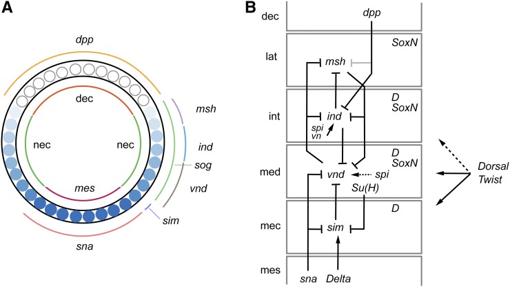
Figure 3.
Dorsal–ventral (D–V) patterning and neural identity genes. (A) Cross-section of a blastoderm embryo showing major cell types, gradient of Dorsal protein, and expression of D–V patterning genes (ventral is bottom). Inside shows the distribution of the three main cell types: mesoderm (mes), neuroectoderm (nec), and dorsal ectoderm (dec). The blue circles represents blastoderm nuclei and indicate the levels of Dorsal protein with dark shades equivalent to high levels of nuclear protein. The domains of expression of D–V patterning genes are shown on the outside. Adapted from Hong et al. 2008, copyright (2008) National Academy of Sciences. (B) Genetic interactions and expression patterns occurring in the different neuroectodermal domains that promote neural precursor identity. Neuroectodermal domains are lateral (lat), intermediate (int), and medial (med) neuroectoderm, and mesectoderm (mec). Also shown are dorsal ectoderm (dec) and mesoderm (mes). dpp is a stronger repressor of ind expression (dark) than msh expression (gray). Maintenance of vnd expression by spi signaling is indicated by a dashed arrow. Dorsal-Twist regulation: solid lines indicate regulation by both TFs and dotted line indicates regulation by only Dorsal. Dichaete (D) and SoxN are shown in their columns of expression.

