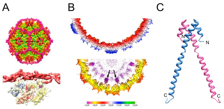
Figure 3.
Size redirection by P4 and type 1 SaPIs. (A) Isosurface representation of the P4 procapsid reconstruction (top) showing the dodecahedral cage made by Sid (magenta). The bottom image shows the Sid density (red) as it interacts with the CP hexamer underneath (yellow surface; three copies of CP are modeled into the density). (B) Slice through the three-dimensional reconstructions of 80α (top) and SaPI1 (bottom) procapsids, scaled radially according to the color bar (radius in Å). The internal protrusions corresponding to CpmB (purple) are indicated by arrows. (C) Ribbon representation of the SaPI1 CpmB dimer, made as a composite between the CpmB NMR structure (PDB ID: 2L8T) and the SaPI1 cryo-EM structure (PDB ID: 6B23). N- and C-termini are labeled.

