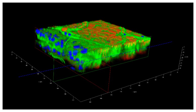
Figure 5.
Three-dimensional confocal microscopy image of a midgut preparation, which had been soaked for 2 h in a suspension containing 5 nm (1.99 × 10⁹ nanoparticles/mL, labeled with FITC) and 50 nm (1.99 × 10⁹ nanoparticles/mL, labeled with Cy3) gold-nanoparticles. The midgut was dissected from a female mosquito at 72 h post-oral acquisition of a proteinmeal consisting of a 20% BSA solution. The dashed blue line depicts the zone of the midgut lumen, the horizontal green and red lines the location of the midgut surfaces. The image was captured using an inverted spectral confocal microscope (TCP SP8 MP, Leica Microsystems). Blue = nuclei stained with DAPI; green = FITC labeled 5 nm gold-nanoparticles; red = Cy3 labeled 50 nm gold-nanoparticles. Irregular line structures (green) represent tracheae.

