Figure 6.
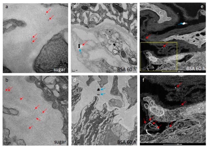
Ultrastructural (TEM) detection of 5 nm gold-nanoparticles (red arrows) and 50 nm gold-nanoparticles (blue arrows) within strands of the midgut BL (a–d). Midguts were obtained from sugar-fed (a,b) and proteinmeal-fed Ae. aegypti (c–f; at 60 h pf of a 20% BSA solution). Images were captured using a JEOL JEM 1400 transmission electron microscope. (e,f) Detection of 5 nm (red arrows) and 50 nm (blue arrow) gold-nanoparticles associated with the midgut BL by scanning transmission electron microscopy (STEM) to enhance visibility of the nanoparticles. Images were generated using a ThermoFisher Tecnai F30 Twin 300kV TEM/STEM operated at 200 kV in high angle annular dark field (HAADF) image mode.

