Abstract
Investigation of the physiologic role of the A3 adenosine receptor has been facilitated by the availability of selective agonists and antagonists. Selective agonists include IB-MECA and the 2-chloro derivative Cl-IB-MECA. Selective antagonists have been identified and designed with the aid of molecular modeling among various nonpurine classes of heterocycles: flavonoids, 1,4-dihydropyridine derivatives, triazoloquinazolines, isoquinolines, and a triazolonaphthyridine. The dihydropyridine 3-ethyl 5-benzyl 2-methyl-6-phenyl-4-phenylethynyl-1,4-(±)-dihydropyridine-3,5-dicarboxylate (MRS 1191) is 1,300-fold selective for human A3 (Ki of 31 nM) vs. A1/A2A adenosine receptors and also 28-fold A3 selective in rat tissue (Ki of 1.42 mM). 9-Chloro-2-(2-furyl)-5-phenylacetylamino[1,2,4]-triazolo[1,5-c]quinazoline (MRS 1220) is useful as an A3 selective antagonist only in human tissue, with a Ki value of 0.65 nM. The pyridine derivative 5-propyl 2-ethyl-4-propyl-3-(ethylsulfanylcarbonyl)-6-phenylpyridine-5-carboxylate (MRS 1523) is a selective antagonist of both rat and human A3 receptors, with Ki values of 113 and 19 nM, respectively. Paradoxical effects of A3 agonists in the brain, heart and other tissues indicate that acute activation of A3 receptors at greater than 10 mM concentrations acts as a lethal input to cells, whereas low, nanomolar concentrations of A3 receptor agonists protect against apoptosis or ischemic damage. Adenosine A3 receptor agonists, antagonists, or both, may be useful in treating inflammatory conditions.
Keywords: purines, A3 receptors, cell viability, dihydropyridines, pyridines, molecular modeling
INTRODUCTION
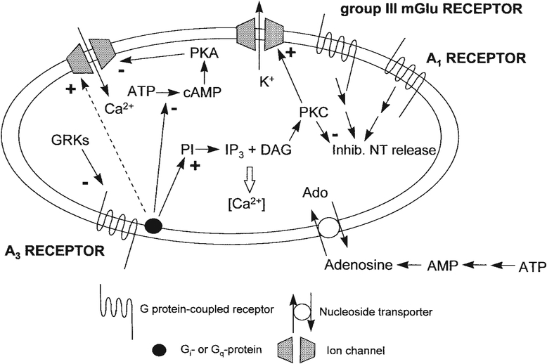
Most tissues contain one or more of the four known adenosine receptor subtypes, A1, A2A, A2B, and A3 [Linden, 1994; Olah and Stiles, 1995], consistent with the ubiquitous role of adenosine in maintaining homeostasis, especially in conditions of stress or ischemia. The A1 and A2A adenosine receptors, pharmacologically well characterized mainly through the use of selective ligands, generally have a protective role, i.e., in decreasing energy demand and increasing energy supply, respectively. A1 receptor activation inhibits the release of potentially damaging excitatory neurotransmitters (Fig. 1). Mice in which the A2A receptors have been knocked out have been bred [Ledent et al., 1997], and observations with these viable animals point to regulatory differences in the cardiovascular and central nervous systems. A2B receptors are the only subtype for which there are not yet highly selective agonists and antagonists, although SAR studies have been carried out in functional assays at this subtype [de Zwart et al., 1998]. Development of selective agonists and antagonists for the A3 receptor has made possible pharmacologic studies of this novel receptor. By virtue of effects on apoptosis, adenosine A3 receptors may play a critical role in human disease states, such as neurodegeneration, cancer, and inflammation. Adenosine A3 receptor antagonists may be useful in treating asthma. The acute administration of an A3 antagonist or the chronic administration of an A3 agonist appears to protect brain cells in a global ischemia model and, thus, may be potential therapeutic approaches for preventing stroke damage. In the heart, because A3 receptor activation protects both in a preconditioning model and during prolonged ischemic, selective agonists may be of great clinical importance.
Fig. 1.
Processes resulting from activation of A3 adenosine receptors. The dashed line indicates activity only at micromolar concentrations of IB-MECA [Kohno et al., 1996; Casavola et al., 1998]. Presynaptic hippocampal A3 receptor activation induces inhibition of the effects of A1 receptors [Dunwiddie et al., 1997] and a PKC-dependent inhibition of mGlu receptors [Macek et al., 1998]. In CA3 pyramidal neurons, potentiated calcium current through a protein kinase A (PKA)-dependent mechanism [Fleming and Mogul, 1997]. In cardiac myocytes, A3 receptor activation is proposed to induce PKC-dependent activation of ATP-sensitive K+ channels, which results in cardioprotection [Stambaugh et al., 1997]. PI, phosphatidyl inositol; PKC, protein kinase C; DAG, diacylglycerol; NT, neurotransmitter; GRKs, G-protein-coupled receptor kinases.
The distribution of A3 adenosine receptors is species dependent and in the human occurs in the lungs, liver, heart, kidneys, brain, and testes [Linden, 1994]. The only primary human tissue in which high density radioligand binding to A3 adenosine receptors has been demonstrated is eosinophils, suggesting a role of this subtype in inflammatory diseases. In functional studies, A3 adenosine receptors have been detected in the central nervous system, in both neurons [Dunwiddie et al., 1997] and astrocytes [Abbracchio et al., 1997b], although the density in the brain is low and relatively diffusely distributed. On the basis of studies that used selective agonists and antagonists, it has been proposed that modulating A3 adenosine receptors may provide new therapeutic approaches for treating inflammatory and neurodegenerative diseases, asthma, and cardiac ischemia [Beaven et al., 1994; Liu et al., 1994; von Lubitz et al., 1994; Strickler et al., 1996; Tracey et al., 1997; Knight et al., 1997; Stambaugh et al., 1997; Walker et al., 1997]. A3 knockout mice are also being studied [Salvatore and Jacobson, 1996], and preliminary information indicates that the homozygous knockout is nonlethal.
SELECTIVE A3 ADENOSINE RECEPTORS AGONISTS
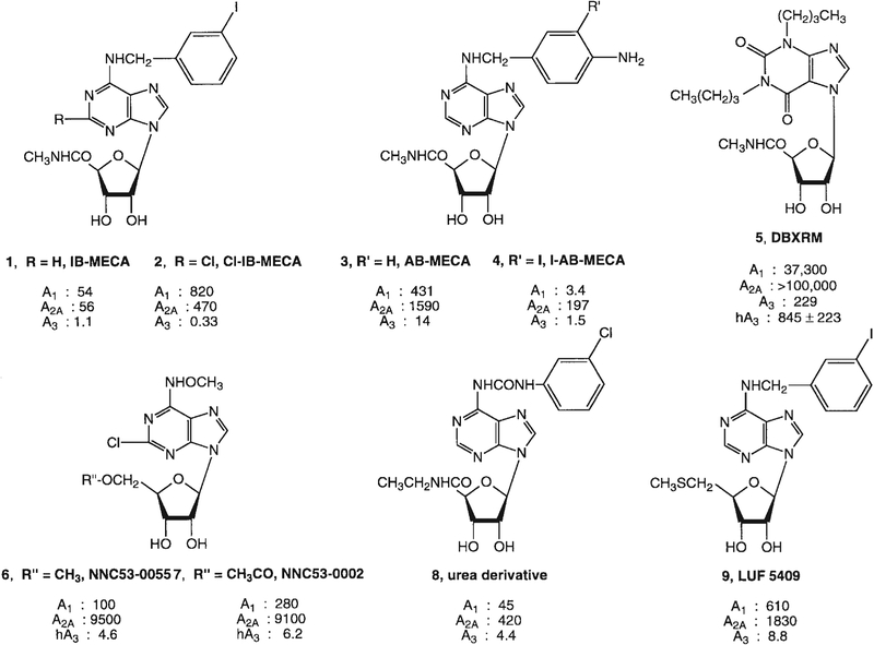
All of the currently synthesized adenosine agonists with moderate to high selectivity for the A3 receptor subtype contain modifications at two sites on the adenosine structure, the N6- and 5′-positions [Jacobson et al., 1995]. The monosubstituted N6-(3-iodobenzyl)adenosine is only slightly A3 selective, whereas the corresponding 5′-uronamide derivatives are more highly selective. For example, N6-(3-iodobenzyl)-adenosine-5′-N- methyluronamide (IB-MECA, 1, Fig. 2) was the first highly potent and selective A3 agonist, both in vitro, in species as diverse as human [Jacobson et al., 1997], dog [Auchampach et al., 1997b], and chick [Stambaugh et al., 1997], and in vivo [Jacobson et al., 1995; von Lubitz et al., 1994]. It is approximately 50-fold selective in binding assays for rat A3 vs. either rat A1 or rat A2A receptors. Substitution at the 2-position of adenosine in combination with modifications at N6 and 5′-positions further enhanced A3 affinity and selectivity. Thus, 2-chloro-N6-(3-iodobenzyl)-adenosine-5′-N-methyluronamide (2, Cl-IB-MECA) [Jacobson et al., 1995] displayed a Ki value of 0.33 nM at A3 receptors and is selective for rat A3 vs. A1 and A2A receptors by 2500- and 1400-fold, respectively. Although highly selective, Cl-IB-MECA at micromolar concentrations has been shown to activate A2A receptors in human neutrophils [Hannon et al., 1998]. Thus, for the range of A3 receptor effects that have been demonstrated only at micromolar concentrations of A3 agonists (Table 1), it is imperative to compare results with agonists of selectivity for A1 and A2A adenosine receptors and where feasible to test antagonism by A3 receptor selective antagonists
Fig. 2.
Structures of highly potent A3 adenosine receptor agonists. Ki values in receptor binding at rat (unless indicated) A1/A2A/A3 receptors (nM) are shown. h, human.
TABLE 1.
Effects of Potent A3 Receptor Agonists
| Parameter | Cl-IB-MECA (~EC50 nM) |
|---|---|
| In vitro | |
| Inhibition of chemotaxis (eosinophils) | 0.1 |
| Cardioprotection | 1–10 |
| Stimulation of phospholipase C (striatum) | 50 |
| Inhibition of adenylyl cyclase | 60 |
| Astroglial cell changes (morphology, Bcl-XL) | 100 |
| Antagonism of A1-mediated inhibition of neurotransmission Elevation of [Ca2+]i | 500 |
|
70 |
|
2000 |
|
10,000 |
| Necrosis (cerebellum), apoptosis (cardiac, inflamm. system), and cell growth arrest (recomb. receptor in CHO cells) | 10,000 |
| Locomotor depression | |
| Histamine release | |
| Cerebroprotection—chronic administration (NOS ⇓, MAP2 ⇑) |
N6-(4-Aminobenzyl)-adenosine-5′-N-methyluronamide (AB-MECA, 3) was prepared as a precursor for radioiodination, such that an iodo substituent directed to the 3-position would be expected to enhance affinity at A3 receptors. Although AB-MECA is a moderately A3-selective agonist, the resulting radioligand [125I]I-AB-MECA [Olah et al., 1994], 4, although a high-affinity (Kd of 0.59 nM) probe for A3 receptors, is not as A3 selective as IB-MECA [Shearman and Weaver, 1997]. Thus, in brain autoradiographic studies, [125I]I-AB-MECA also bound to A1 and A2A subtypes [Shearman and Weaver, 1997].
In an attempt initially directed toward the derivatization of xanthines as A3 receptor antagonists by forming 7-riboside derivatives, DBXRM (5, Fig. 2) was found to be 140-fold selective in binding to rat A3- vs. A1 adenosine receptors. However, DBXRM proved to be an agonist at recombinant rat A3 receptors [Kim et al., 1994; Park et al., 1998]. A 3′-deoxy derivative of DBXRM was found to be an antagonist at A1 and partial agonist at A3 adenosine receptors. Thus, it is possible for the same compound to stimulate one adenosine receptor subtype (A3) and block another subtype (A1) within the same species [Park et al., 1998]. Full agonists, such as Cl-IB-MECA or I-AB-MECA, were more potent and effective than the partial agonist DBXRM in causing desensitization of rat A3 receptors, as indicated by loss of [35S]GTPγ[S] binding to RBL-2H3 cell membranes.
Knutsen and coworkers have developed hydroxylamino derivatives such as 6 and 7 as A3-selective agonists [Knutsen et al., 1998]. This series emphasized the flexibility of substitution at the 5′-position with amide, chloromethyl, vinyl, ester, and isoxazole groups. Baraldi and coworkers [Baraldi et al., 1998] have developed urea derivatives such as 8 and related amides as A3 selective agonists. IJzerman and coworkers [van Tilburg et al., 1998] have reported that substitution with a methylthio group at the 5′-position of N6-benzyladenosine analogues results in partial agonists such as 9.
SELECTIVE A3 ADENOSINE RECEPTOR ANTAGONISTS
The low affinity of xanthines, the classic antagonists of A1, A2A, and A2B subtypes, at rat A3 adenosine receptors is striking [Linden, 1994; Jacobson et al., 1995; Olah and Stiles, 1995]. At human, dog, and sheep A3 adenosine receptors [Linden et al., 1993; Salvatore et al., 1993; Auchampach et al., 1997a], certain xanthines are of intermediate potency as antagonists; however, highly A3 selective xanthines have not yet been identified. The species differences in antagonist affinity and the low degree of homology between human and rat receptor sequences (72%) suggest the existence of two subtypes of A3 adenosine receptors, although this needs to be further investigated.
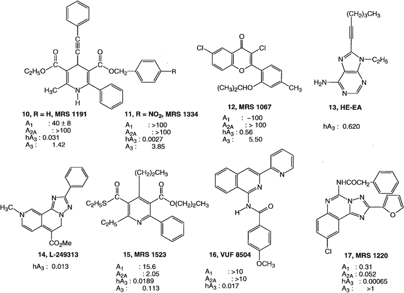
A3 adenosine receptor antagonists, which have been introduced only recently [Jacobson et al., 1996; Karton et al., 1996; Kim et al., 1996; Jiang et al., 1997a], were previously hypothesized [Beaven et al., 1994; von Lubitz et al., 1994] to act as potential antiasthmatic [Olah and Stiles, 1995], anti-inflammatory, or cerebroprotective agents. The need for selective antagonists is critical, especially in light of the fact that most effects of high concentrations of A3 agonists (Table 1) have not unequivocally been ascribed to activation of A3 receptors. The dramatic species differences in antagonist affinity and in A3 receptor responses makes the extrapolation of studies in rodents to the potential treatment of human disease more challenging. Thus, it is desirable to obtain antagonists that are A3 selective across species [Li et al., 1998] for preclinical studies. Promising leads for selective antagonists for human A3 receptors have appeared among nonxanthine heterocycles (Fig. 3), including flavonoids, 1,4-dihydropyridine derivatives, triazoloquinazolines, isoquinolines, and a triazolonaphthyridine. Our initial screening of chemically diverse substances as potential antagonists, consisted of single-point displacement of [125I]I-AB-MECA binding at human A3 receptors expressed in HEK-293 cells. 1,4-Dihydropyridines (DHPs) [Jiang et al., 1997a], which act as potent l-type calcium channel antagonists, were found to have micromolar affinity at this subtype. Common DHP drugs typically bound to various adenosine receptor subtypes either nonselectively, as for example, nifedipine, with a Ki value of 8.3 μM at A3 receptors, or in some cases with selectivity for the A3 vs. other adenosine receptor subtypes, as for example, S-niguldipine, with a Ki value of 2.8 μM. At human A2B receptors, such 1,4-DHPs are essentially inactive [Dunwiddie et al., 1997]. Careful structural optimization of the 1,4-DHP core as a template for adenosine antagonists then ensued. Key features that boost affinity at adenosine (especially A3) receptors and completely deselect for affinity at l-type Ca2+-channels (Ki < 100 μM) are separation at the 4-position of the typical phenyl ring substituent by a two-carbon unsaturated chain (vinyl or acetylene) and at the 6-position substitution of methyl with a bulky phenyl group. Small cycloalkyl groups at the 6-position of 4-phenylethynyl-DHPs were also favorable for high affinity at human A3 adenosine receptors.
Fig. 3.
Structures of selective A3 adenosine receptor antagonists. Ki values in receptor binding at rat (unless indicated) A1/A2A/A3 receptors (micromolar) are shown. h, human.
Among those DHPs binding to A3 receptors selectively and with high affinity were a trisubstituted 1,4-dihydro-6-phenylpyridine analogue, 3-ethyl 5-benzyl 2-methyl-6-phenyl-4-phenylethynyl-1,4-(±)-dihydropyridine-3,5-dicarboxylate (MRS 1191, Fig. 3, 10) [Jiang et al., 1997]. The enhancement of affinity in MRS 1334, 11, apparently corresponded to the presence of the electron-withdrawing p-nitro group on the benzyl ring. These racemic DHPs await optical resolution before radioligands may be synthesized; however, side-by-side comparison of previously known DHP enantiomers shows that the stereoselectivity at A3 receptors favors the R-isomer, the opposite of the stereoselectivity at l-type calcium channels. Even in rat tissue, MRS 1191 was a moderately selective antagonist, e.g., it bound with 28-fold higher affinity for A3 (Ki of 1.42 μM) vs. A1 receptors [Jacobson et al., 1997]. In chick ventricular myocyte cultures [Stambaugh et al., 1997] and in the CA1 region of the rat hippocampus [Dunwiddie et al., 1997], 10 μM MRS 1191 selectively antagonized the A3 subtype in the presence of A1 and A2 receptors.
Flavones and flavonols, which are naturally occurring phenolic derivatives, provided another structural lead for development of A3 antagonists [Karton et al., 1996]. The affinity of common phytochemicals at adenosine receptors suggests that a wide range of natural substances in the human diet may potentially antagonize the effects of endogenous adenosine, including those mediated by means of the A3 subtype. The flavonoid class has been chemically optimized in the form of MRS 1067 (12) [Karton et al., 1996], which is 200-fold selective for human A3 vs. A1 adenosine receptors. Other high-affinity A3-selective antagonists that have been recently reported include a triazolonaphthyridine (l-249313, 14) [Jacobson et al., 1996], a series of isoquinoline derivatives such as VUF 8504 (16) [Van Muijlwijk-Koezen et al., 1998], and a derivative (MRS 1220, 17) of the triazoloquinazoline CGS 15943, a nonselective adenosine antagonist [Kim et al., 1996]. Related to the parent compound CGS 15943 simply through acylation at the N5-amino position with a phenylacetyl group, MRS 1220 is the antagonist of highest affinity (Ki 0.65 nM) at human A3 receptors currently reported. In rat tissue, the selectivity of MRS1220 shifts to A2A >> A3 receptors.
Binding of MRS 1067, MRS 1191, and MRS 1220 at human A3 receptors was shown to be competitive by Scatchard analysis vs. binding of [125I]I-AB-MECA [Jacobson et al., 1997]. Antagonism was demonstrated in functional assays consisting of agonist-induced inhibition of adenylate cyclase and the stimulation of binding of [35S]GTPγ[S] to the associated G-proteins. MRS 1220 and MRS 1191, with KB values of 1.7 and 92 nM, respectively, proved to be highly selective for human A3 receptor vs. human A1 receptor-mediated effects on adenylate cyclase.
Recently, we have explored pyridine derivatives, prepared from 1,4-DHPs through oxidation, as A3 receptor antagonists [Li et al., 1998]. Certain 3,5-diacyl-2,4-dialkyl-6-phenylpyridine derivatives displayed nanomolar affinity in radioligand binding at recombinant human A3 receptors and were also considerably selective in binding to recombinant rat A3 receptors. The 4-Pr derivative, MRS 1523, 15, was selective and highly potent at both human and rat A3 receptors (Ki values of 18.9 and 113 nM, respectively). Key modifications of the SAR in the pyridine series included a thioester at the 3-acyl substituents and a small alkyl group at the 4-position. As for the 4-substituted 1,4-DHPs, a 6-phenyl group was required for optimal A3 selectivity. Unlike the DHPs, a 5-position benzyl ester in the pyridine series decreased affinity at adenosine receptors.
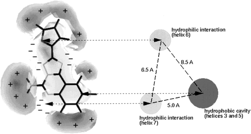
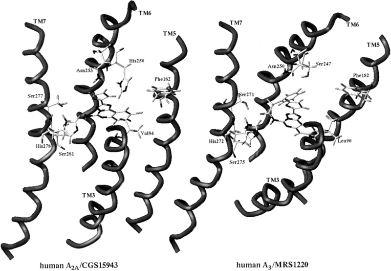
A general pharmacophore model for antagonist binding to the human A3 receptor has been constructed [Moro et al., 1998b]. A combination of ab initio quantum mechanical calculations, electrostatic potential map comparison, and the steric and electrostatic alignment (SEAL) method led to a general pharmacophore map that was based on adenines, xanthines, triazoloquinazolines, flavonoids, thiazolopyridines, 6-phenyl-1,4-DHPs, and 6-phenyl-pyridines as A3 adenosine receptor antagonists. According to the proposed pharmacophore map (Fig. 4), recognition of all antagonists at a common region inside the receptor binding site and, consequently, a common electrostatic potential profile is possible. To help interpret these results, a rhodopsin-based model of the human A3 receptor (Fig. 5) was built, and the triazoloquinazoline reference ligand CGS 15953 (9-chloro-2-(2-furyl)[1,2,4]triazolo[1,5-c]quinazolin-5-amine) could be docked in the putative binding site. The model of the ligand-receptor complex was derived and refined by using our recently introduced cross-docking procedure [Moro et al., 1998a], which simulates the reorganization of the native receptor structure induced by a ligand. All other ligands could be docked according to the overlay with respect to CGS 15953 as a template, obtained through the SEAL approach. The proposed interactions between the ligands and specific helical domains of the human A3 receptor are shown in Figure 5. A major structural difference between the hypothetical binding sites among adenosine receptor subtypes is that the A3 receptor does not contain the histidine residue in TM6 common to all A1 (His251 in hA1 receptor) and A2 (His250 in hA2A and His251 in hA2B receptors) subtypes. This histidine residue has been shown to participate in both agonist and antagonist recognition to A2A receptors [Jiang et al., 1997b; Kim et al., 1995]. In the A3 receptor, this histidine in TM6 is replaced with a serine residue (Ser275 in hA3 receptors). This replacement reduces the steric hindrance in this region of the binding cavity. In our model, substituents at the N5-position of CGS 15943 are located close to Ser275 (TM6) (see Fig. 5). According to this model, the binding region of the receptor surrounding the α-carbon of the acetyl group of MRS1220 is not sterically restricted, and large substituents could enhance the A3 binding affinity. In fact, we found that phenylacetyl (MRS 1220) and diphenylacetyl (MRS 1406) derivatives are among the most potent antagonists at the human A3 receptor (Ki values of 0.65 nM and 0.59 nM, respectively). As shown in Figure 5, other important residues for the ligand binding are the following: Ser271 and Ser275 (TM7), close to the N5-HR-position of CGS 15943; Asn250 (TM6), close to the oxygen of the furan ring of CGS 15943; Leu90 (TM3), Phe182 (TM5), and Ile186 (TM5) around the chlorophenyl moiety of CGS 15943.
Fig. 4.
Common pharmacophore model for binding of antagonists at the human A3-adenosine receptor. The isopotential surface of CGS 15943 (+ region = 5 kcal/mol; – region = −5 kcal/mol) and three points of interaction between transmembrane helical domains of the receptor protein and shared features of antagonist molecules (e.g., Fig. 3) are shown.
Fig. 5.
Model of the ligand (high-affinity triazoloquinazoline antagonists shown) binding site of human A2A and A3 adenosine receptors. CGS 15943 is shown bound to the A2A receptor, and the corresponding N5-phenylacetyl derivative MRS 1220 is shown bound to the A3 receptor. According to this model, the absence of a key aromatic residue found in A1 and A2A receptors (His 250, TM6) in the A3 receptor allows for the introduction of the added bulky phenylacetyl group of MRS 1220 (or similarly a diphenylacetyl group in MRS 1406, not shown).
BIOLOGICAL EFFECTS OF A3 RECEPTOR AGONISTS
In general, there is a strikingly large potency differential among various functional activities of A3 selective agonists (Table 1), i.e., the same agonists may act functionally in the low nanomolar range (consistent with their affinity in competitive binding assays) for some functional responses, whereas in other activities, even within the same species, micromolar concentrations of the agonists are needed. Although for many receptors, the measured affinity is typically lower than EC50 values in functional assays, the wide range of these values for A3 adenosine receptors, i.e., spanning ≥ 4 orders of magnitude, is unusual. The role of spare receptors in this phenomenon has not been explored.
Effector mechanisms.
In addition to a unique structure-activity profile for agonists and particularly for antagonists, activation of the A3 receptor has a characteristic second messenger profile (Fig. 1), in that it has been shown to stimulate directly phospholipases C [Abbracchio et al., 1995; Olah and Stiles, 1995] and D [Ali et al., 1996] and to inhibit adenylate cyclase [Olah and Stiles, 1995]. Rat adenosine A3 receptors can interact with Giα2, Giα3, and to a lesser extent Gq [Palmer et al., 1995]. Recombinant A3 adenosine receptors undergo agonist-induced desensitization, the mechanism of which involves phosphorylation of the C-terminal segment of the receptor by G protein receptor coupled kinases such as GRK 2, 3, and 5 [Palmer et al., 1996].
The effects of A3 agonists on intracellular calcium are complex. In HL-60 cells, activation of A3 receptors by ≥10 μM Cl-IB-MECA results in influx of Ca2+ and release from intracellular stores [Kohno et al., 1996]. Similar concentrations of IB-MECA in rat cardiac myocytes cause Ca2+ release in the absence of extracellular Ca2+ [Shneyvays et al., 1998]. In RBL-2H3 mast cells, the potency of adenosine agonists in raising [Ca2+]i but not IP1 levels parallels A3 receptor affinity [Shin et al., 1996]. In frog A6 kidney cells, a commonly used model of the mammalian collecting duct, micromolar concentrations of Cl-IB-MECA applied to the apical membrane cause an influx of Ca2+ but not a release from intracellular stores [Casavola et al., 1998].
Chloride channels are also activated by the A3 adenosine receptors. In the human eye, A3 adenosine receptor agonists stimulate chloride channels of nonpigmented ciliary epithelial cells [Mitchell et al., 1999].
Protective versus lethal effects of A3 receptor activation.
With the recent availability of selective agonists and antagonists, both protective and lethal effects of A3 adenosine receptor activation have been discovered (Table 2). The concentration of endogenous adenosine required for half-occupancy of A1 and A2A adenosine receptors is in the range of 10−8 to 10−7 M [Jacobson et al., 1995; Olah and Stiles, 1995] concentrations that might be achieved in the basal, resting state of an organ. The Ki value of adenosine in binding to the rat A3 receptor has not been determined directly, but rather estimated to be 10−6 M [Jacobson et al., 1995]; activation of this subtype may require a relatively high concentration of adenosine, such as would occur during hypoxic stress and other cellular damage. Thus, the pathophysiologic role of A3 receptors may be very different from the role of A1 and A2A subtypes, in that it would act as an endogenous regulator only under conditions of more severe challenge. The varied effects of A3 receptor agonists, in vitro and in vivo, seem to be dual and opposite, i.e., either cytoprotective or cytotoxic, depending on the level of receptor activation and the paradigm studied. The mechanisms involved in these opposite effects are not yet fully understood.
TABLE 2.
Cytoprotective vs. Lethal Effects of A3 Receptor Agonists and Antagonists
In the heart, both A1 and A3 adenosine receptor agonists appear to protect cardiac myocytes [Liu et al., 1994; Strickler et al., 1996; Stambaugh et al., 1997; Tracey et al., 1997; Auchampach et al., 1997b]; but the latter evoke a longer window of protection [Liang and Jacobson, 1998] and do not cause the hypotension and hypothermia associated with agonists for the other adenosine receptor s. A3 receptors occur on ventricular but not atrial cardiac myocytes [Strickler et al., 1996]. There are protective effects of A3 receptor activation in heart cells, administered both before [Strickler et al., 1996; Tracey et al., 1997] and during [Stambaugh et al., 1997] an ischemic episode. IB-MECA also protects against myocardial stunning in conscious rabbits [Auchampach et al., 1997b]. In cultured chick cardiac myocytes, a brief prior exposure to nanomolar concentrations of the A3 receptor agonist Cl-IB-MECA protected cells from damage induced by subsequent hypoxia [Liu et al., 1994; Strickler et al., 1996], thus simulating the protection afforded by a brief hypoxic period, a phenomenon termed “preconditioning.” Activation by endogenous adenosine of both adenosine A1 and A3 receptors is thought to mediate preconditioning. The protective potential was prolonged up to 45 min after exposure to the A3 agonist [Liang and Jacobson, 1998]. Because the culture consisted only of almost exclusively ventricular myocytes, this was not an indirect effect of activation of mast cells. Thus, an A3 agonist at low concentration is potentially useful therapeutically as a cardioprotective agent, having fewer in vivo side effects than other (e.g., A1 selective) adenosine agonists [Jacobson et al., 1996; Liu et al., 1994].
Although nanomolar concentrations of selective A3 agonists tend to protect cells, ≥10 μM concentrations are often toxic (Table 2), causing apoptosis, as in rat cardiac myocytes [Shneyvays et al., 1998]. In a variety of human cell lines of the immune system, A3 agonists at such high concentrations often prove lethal (Table 1). Apoptosis, with the characteristic DNA fragmentation, has been shown to occur in human leukemia HL-60 cells, MCF-7 breast cancer cells, and in human blood eosinophils in response to high concentrations of A3 selective agonists [Kohno et al., 1996; Yao et al., 1997]. A positive mediator of apoptosis, bak, is up-regulated under these conditions [Yao et al., 1997]. Clarification of the need for such high doses of agonists, thousands of fold higher than the Ki values at A3 receptors, has awaited the introduction of selective A3 receptor antagonists, which are now available for the human A3 receptor.
In certain cultured cell lines, antagonists alone, representing three diverse chemical classes, caused apoptosis (programmed cell death, see below) [Yao et al., 1997], suggesting that there may exist a tonic state of low-level activation of the A3 receptor which has a protective role. If indeed a tonic A3 receptor activation exists, the apoptotic effects of A3 antagonists may simply be explained on the basis of a blockade of a protective action induced by endogenous adenosine. To explain how ≥10 μM concentrations of agonist alone may induce rather than prevent apoptosis, one could propose differential activation of different second messengers by the same receptor at low and high doses (Fig. 1). Such hypotheses will require further investigation, which would be greatly aided by the development of a high-affinity antagonist radioligand for the A3 receptor. The low density of A3 receptors has also made study difficult.
A3 adenosine receptor ligands have been shown to be protective in cerebral ischemia models in gerbils [von Lubitz et al., 1994]. The first cytoprotective effects of an A3 agonist were shown after its chronic administration in gerbils in a model of stroke. In an in vivo gerbil model of global ischemia, the acute administration of IB-MECA during the ischemia exacerbated histologic and functional damage, i.e., clearly worsened the postocclusive outcome [von Lubitz et al., 1994]. However, chronic preadministration of the same agonist over several weeks had a highly neuroprotective, postischemic effect. In the same gerbil model, acute administration of the A3 antagonist MRS1191 was protective [von Lubitz et al., 1997].
Several possible explanations for the damaging effects of acute A3 activation during ischemia have been offered. It may involve detrimental effects observed on cerebral blood flow [von Lubitz et al., 1994] or conceivably release of a cytotoxic agent. Alternately, the effects may be by means of neuronal A3 receptors, which may regulate other receptors. For example, acute activation of presynaptic hippocampal A3 receptors antagonizes the action of metabotropic glutamate receptors, thus, resulting in enhanced glutamate release [Macek et al., 1998]. Dunwiddie et al. [1997] found that A3 activation counteracts protective effects of A1 receptor activation at the hippocampal synapse; i.e., the depression of excitatory transmission elicited by A1 agonists is blunted by selective A3 agonists. In contrast, Fleming and Mogul [1997] have shown that A3 receptor activation increases cellular excitability in these neurons through a pathway independent of A1 receptors. Activation of A3 receptors in isolated CA3 pyramidal neurons from the guinea pig hippocampus by a low concentration of a selective agonist was also found to potentiate a calcium current through a PKA-dependent/PKC-independent mechanism.
In primary astroglial cell cultures, effects of selective A3 agonists are also biphasic, with 100–200 nM protecting against cell death and inducing differentiation, whereas ≥10 μM concentrations increased cell death [Abbracchio et al., 1997a]. In human ADF cells of astroglial lineage, 100 nM Cl-IB-MECA caused a marked reorganization of the cytoskeleton, with appearance of stress fibers and numerous cell protrusions (which became enriched in the antiapoptotic protein Bcl-XL), accompanied by induction of the expression of Rho, a small GTP-binding protein possibly related to cytoskeletal changes [Abbracchio et al., 1997b]. 10 μM Cl-IB-MECA was lethal to cultured rat cerebellar granule neurons, and the toxic effects of glutamate were also augmented [Sei et al., 1997].
Several years ago, a commentary by Beaven et al. [1994] suggested that a then hypothetical A3 receptor antagonist could be a useful antiasthmatic drug. The acute activation of A3 receptors in rodents leads to release of histamine and other mediators from mast cells, which also results in hypotension [Fozard et al., 1996]. In microcirculation of the hamster cheek pouch, activation of A3 receptors results in vasoconstriction, which also occurs through activation of mast cells [Shepherd et al., 1996]. Walker et al. [1997] postulated a role for A3 receptors in lung inflammation, because adenosine leads to exaggerated airway narrowing in individuals with inflammatory airway disorders. Evidence was found that in humans the A3 receptor gene expression is localized to inflammatory cells (eosinophils, but not mast cells) and that gene expression is up-regulated in airway inflammation. Cl-IB-MECA was found to inhibit eosinophil migration without affecting adhesion receptors CD18 and selectin or assembly of F-actin, and this effect was blocked by l-294,313 [Knight et al., 1997]. Based on this effect, it is not known whether an A3 agonist or antagonist would be more useful in treating asthma, because eosinophil activation could theoretically either augment (by means of migration to site) or counteract (by means of migration away from site) inflammation. However, other experiments suggest the utility of an antagonist. For example, Meade et al. [1996] found that in the BDE rat model of airway disease, A3 agonists induced bronchospasm by means of mast-type cells. Although aerosol challenge of antigen-immunized rabbits with the nonselective agonist APNEA did not elicit dose-dependent changes in either airways resistance or dynamic compliance [Ali et al., 1997], it was found that the agonists IB-MECA and Cl-IB-MECA caused bronchoconstriction. Selective activation of A3 receptors appears to inhibit human neutrophil degranulation, suggesting the anti-inflammatory potential of A3 adenosine agonists in neutrophil-mediated tissue injury [Bouma et al., 1997].
There may be an involvement of A3 receptors in cancer. Activation of A3 receptors reduced cytotoxic lymphocyte adhesion to tumor cells [Mackenzie et al., 1994].
REFERENCES
- Abbracchio MP, Brambilla R, Ceruti S, Kim HO, von Lubitz D, Jacobson KA, Cattabeni F. 1995. G protein-dependent activation of phospholipase C by adenosine A3 receptors in rat brain. Mol Pharmacol 48:1038–1045. [PubMed] [Google Scholar]
- Abbracchio MP, Ceruti S, Brambilla R, Franceschi C, Malorni W, Jacobson KA, Von Lubitz D, Cattabeni F. 1997a. Modulation of apoptosis by adenosine in the central nervous system: a possible role for the A3 receptor. Pathophysiological significance and therapeutic implications for neurodegenerative disorders. Ann New York Acad Sci 825:11–22. [DOI] [PMC free article] [PubMed] [Google Scholar]
- Abbracchio MP, Rainaldi G, Giammarioli AM, Ceruti S, Brambilla R, Cattabeni F, Barbieri D, Franceschi C, Jacobson KA, Malorni W. 1997b. The A3 adenosine receptor mediates cell spreading, reorganization of actin cytoskeleton, and distribution of Bcl- XL: Studies in human astroglioma cells. Biochem Biophys Res Commun 241:297–304. [DOI] [PMC free article] [PubMed] [Google Scholar]
- Ali H, Choi OH, Fraundorfer PF, Yamada K, Gonzaga HMS, Beaven MA. 1996. Sustained activation of phospholipase D via adenosine A3 receptors is associated with enhancement of antigen- and Ca2+- ionophore-induced secretion in a rat mast cell line. J Pharmacol Exp Ther 276:837–845. [PubMed] [Google Scholar]
- Ali S, Jacobson KA, Mustafa SJ. 1997. Adenosine A3 receptor in the airways of allergic rabbits. FASEB J 11:A346. [Google Scholar]
- Auchampach JA, Jin XW, Wan TC, Caughey GH, Linden J. 1997a. Canine mast cell adenosine receptors: cloning and expression of the A3 receptor and evidence that degranulation is mediated by the A2B receptor. Mol Pharmacol 52:846–860. [DOI] [PubMed] [Google Scholar]
- Auchampach JA, Rizvi A, Qiu YM, Tang XL, Maldonado C, Teschner S, Bolli R. 1997b. Selective activation of A3 adenosine receptors with N-6-(3-iodobenzyl)adenosine-5′-N-methyluronamide protects against myocardial stunning and infarction without hemodynamic changes in conscious rabbits. Circ Res 80:800–809. [DOI] [PubMed] [Google Scholar]
- Baraldi PG, Cacciari B, Pineda de las Infantas MJ, Romagnoli R, Spalluto G, Volpini R, Costanzi S, Vittori S, Cristalli G, Melman N, Park K-S, Ji X-d, Jacobson KA. 1998. Synthesis and biological activity of a new series of N6-arylcarbamoyl-, 2-(ar)alkynyl-N6-arylcarbamoyl, and N6-carboxamido- derivatives of adenosine-5′-N-ethyluronamide (NECA) as A1 and A3 adenosine receptor agonists. J Med Chem 41:3174–3185. [DOI] [PMC free article] [PubMed] [Google Scholar]
- Beaven MA, Ramkumar V, Ali H. 1994. Adenosine-A3 receptors in mast-cells. Trends Pharmacol Sci 15:13–14. [DOI] [PubMed] [Google Scholar]
- Bouma MG, Jeunhomme T, Boyle DL, Dentener MA, Voitenok NN, vandenWildenberg F, Buurman WA. 1997. Adenosine inhibits neutrophil degranulation in activated human whole blood: involvement of adenosine A2 and A3 receptors? J Immunol 158: 5400–5408. [PubMed] [Google Scholar]
- Casavola V, Di Sole F, Guerra L, Debellis L, Reshkin SJ, Jacobson KA. 1998. Adenosine A3 receptor activation increases cytosolic calcium concentration via calcium influx in A6 cells. Drug Dev Res 43:62. [Google Scholar]
- de Zwart M., Link R, von Frijtag Drabbe Künzel JK, Cristalli G, Jacobson KA, Townsend-Nicholson A, IJzerman AP. 1998. A screening of adenosine analogues on the human adenosine A2B receptor as part of a search for potent and selective agonists. Nucleosides Nucleotides 17:969–986. [DOI] [PMC free article] [PubMed] [Google Scholar]
- Dunwiddie TV, Diao LH, Kim HO, Jiang JL, Jacobson KA. 1997. Activation of hippocampal adenosine A(3) receptors produces a desensitization of A(1) receptor-mediated responses in rat hippocampus. J Neurosci 17:607–614. [DOI] [PMC free article] [PubMed] [Google Scholar]
- Fleming KM, Mogul DJ. 1997. Adenosine A3 receptors potentiate hippocampal calcium current by a PKA-dependent/PKC-independent pathway. Neuropharmacology 36:353–362. [DOI] [PubMed] [Google Scholar]
- Fozard JR, Pfannkuche HJ, Schuurman HJ. 1996. Mast cell degranulation following adenosine A3 receptor activation in rats. Eur J Pharmacol 298:293–297. [DOI] [PubMed] [Google Scholar]
- Hannon JP, Bray-French KM, Fozard JR. 1998. Further pharmacological characterization of the adenosine receptor subtype mediating the oxidative burst in human isolated neutrophils. Drug Dev Res 43:214–224. [Google Scholar]
- Jacobson KA, Suzuki F. 1996. Recent development in selective agonists and antagonists acting at purine and pyrimidine receptors. Drug Dev Res 39:289–300. [DOI] [PMC free article] [PubMed] [Google Scholar]
- Jacobson KA, Kim HO, Siddiqi SM, Olah ME, Stiles G, von Lubitz DKJE. 1995. A3 adenosine receptors: design of selective ligands and therapeutic prospects. Drugs Future 20:689–699. [DOI] [PMC free article] [PubMed] [Google Scholar]
- Jacobson KA, Park KS, Jiang JL, Kim YC, Olah ME, Stiles GL, Ji XD. 1997. Pharmacological characterization of novel A3 adenosine receptor -selective antagonists. Neuropharmacology 36:1157–1165. [DOI] [PMC free article] [PubMed] [Google Scholar]
- Jacobson MA, Chakravarty PK, Johnson RG, Norton R. 1996. Novel non-xanthine selective A3 adenosine receptor antagonists. Drug Dev Res 37:131. [Google Scholar]
- Jiang JL, Van Rhee AM, Chang L, Patchornik A, Ji XD, Evans P, Melman N, Jacobson KA. 1997a. Structure-activity relationships of 4-(phenylethynyl)-6-phenyl-1,4-dihydropyridines as highly selective A3 adenosine receptor antagonists. J Med Chem 40:2596–2608. [DOI] [PMC free article] [PubMed] [Google Scholar]
- Jiang JL, Lee BX, Glashofer M, Van Rhee AM, Jacobson KA. 1997b. Mutagenesis reveals structure-activity parallels between human A2A-adenosine receptors and biogenic-amine G protein-coupled receptors. J Med Chem 40:2588–2595. [DOI] [PMC free article] [PubMed] [Google Scholar]
- Karton Y, Jiang JL, Ji XD, Melman N, Olah ME, Stiles GL, Jacobson KA. 1996. Synthesis and biological activities of flavonoid derivatives as A3 adenosine receptor antagonists. J Med Chem 39:2293–2301. [DOI] [PMC free article] [PubMed] [Google Scholar]
- Kim HO, Ji X-d, Melman N, Olah ME, Stiles GL, Jacobson KA. 1994. Selective ligands for rat A3-adenosine receptors: structure-activity relationships of 1,3-dialkylxanthine-7-riboside derivatives. J Med Chem 37:4020–4030. [DOI] [PMC free article] [PubMed] [Google Scholar]
- Kim JH, Wess J, Van Rhee AM, Schöneberg T, Jacobson KA. 1995. Site-directed mutagenesis identifies residues involved in ligand recognition in the human A2A adenosine receptor. J Biol Chem 270:13987–13997. [DOI] [PMC free article] [PubMed] [Google Scholar]
- Kim YC, Ji XD, Jacobson KA. 1996. Derivatives of the triazoloquinazoline adenosine antagonist. CGS15943) are selective for the human A3 receptor subtype. J Med Chem 39:4142–4148. [DOI] [PMC free article] [PubMed] [Google Scholar]
- Knight D, Zheng XY, Rocchini C, Jacobson M, Bai T, Walker B. 1997. Adenosine A3 receptor stimulation inhibits migration of human eosinophils. J Leukoc Biol 62:465–468. [DOI] [PubMed] [Google Scholar]
- Knutsen LJS, Sheardown MJ, Roberts SM, Morgensen JP, Olsen UB, Thomsen C, Bowler AN. 1998. Adenosine A1 and A3 selective N-alkoxypurines as novel cytokine modulators and neuroprotectants. Drug Dev Res 45:214–221. [Google Scholar]
- Kohno Y, Sei Y, Koshiba M, Kim HO, Jacobson KA. 1996. Induction of apoptosis in HL-60 human promyelocytic leukemia cells by adenosine A3 receptor agonists. Biochem Biophys Res Commun 219:904–910. [DOI] [PMC free article] [PubMed] [Google Scholar]
- Ledent C, Vaugeois JM, Schiffmann SN, Pedrazzini T, El Yacoubi M, Vanderhaeghen JJ, Costentin J, Heath JK, Vassart G, Parmentier M. 1997. Aggressiveness, hypoalgesia and high blood pressure in mice lacking the adenosine A2a receptor. Nature 388:674–678. [DOI] [PubMed] [Google Scholar]
- Li A-H, Moro S, Melman N, Ji X-d, Jacobson KA. 1998) Structure activity relationships of 3,5-diacyl-2,4-dialkylpyridine derivatives as selective A3 adenosine receptor antagonists. J Med Chem 41:3186–3201. [DOI] [PMC free article] [PubMed] [Google Scholar]
- Liang BT, Jacobson KA. 1998. A physiological role of the adenosine A3 receptor: sustained cardioprotection. Proc Natl Acad Sci USA 95:6995–6999. [DOI] [PMC free article] [PubMed] [Google Scholar]
- Linden J. 1994. Cloned adenosine A3 receptors: pharmacological properties, species differences and receptor functions. Trends Pharmacol Sci 15:298–306. [DOI] [PubMed] [Google Scholar]
- Linden J, Taylor HE, Robeva AS, Tucker AL, Stehle JH, Rivkees SA, Fink JS, Reppert SM. 1993. Molecular-cloning and functional expression of a sheep-A3 adenosine receptor with widespread tissue distribution. Mol Pharmacol 44:524–532. [PubMed] [Google Scholar]
- Liu GS, Richards SC, Olsson RA, Mullane KH, Walsh RS, Downey JM. 1994. Evidence that the adenosine A3 receptor may mediate the protection afforded by preconditioning in the isolated rabbit heart. Cardiovasc Res 28:1057–1061. [DOI] [PubMed] [Google Scholar]
- Macek TA, Schaffhauser H, Conn PJ. 1998. Protein kinase C and A3 adenosine receptor activation inhibit presynaptic metabotropic glutamate receptor (mGluR) function and uncouple mGluRs from GTP-binding proteins. J Neurosci 18:6138–6146. [DOI] [PMC free article] [PubMed] [Google Scholar]
- Mackenzie WM, Hoskin DW, Blay J. 1994. Adenosine inhibits the adhesion of anti-Cd3-activated killer lymphocytes to adenocarcinoma cells through an A3 receptor. Cancer Res 54:3521–3526. [PubMed] [Google Scholar]
- Meade CJ, Mierau J, Leon I, Ensinger HA. 1996. In vivo role of the adenosine A3 receptor: N-6–2-(4-aminophenyl)ethyladenosine induces bronchospasm in BDE rats by a neurally mediated mechanism involving cells resembling mast cells. J Pharmacol Exp Ther 279:1148–1156. [PubMed] [Google Scholar]
- Mitchell CH, Peterson-Yantorno K, Carré DC, McGlinn AM, Coca-Prados M, Stone RA, Civan MM. 1999. A3 adenosine receptors regulate Cl-channels of nonpigmented ciliary epithelial cells Amer J Physiol, in press. [DOI] [PubMed] [Google Scholar]
- Moro S, Guo DP, Camaioni E, Boyer JL, Harden TK, Jacobson KA. 1998a. Human P2Y1 receptor: molecular modeling and site-directed mutagenesis as tools to identify agonist and antagonist recognition sites. J Med Chem 41:1456–1466. [DOI] [PMC free article] [PubMed] [Google Scholar]
- Moro S, Li AH, Jacobson KA. 1998b. Molecular modeling studies of human A3 adenosine receptor antagonists: structural homology and receptor docking. J Chem Inf Comput Sci 38:1239–1248. [DOI] [PMC free article] [PubMed] [Google Scholar]
- Olah ME, Stiles GL. 1995. Adenosine receptor subtypes: characterization and therapeutic regulation. Annu Rev Pharmacol Toxicol 35:581–606. [DOI] [PubMed] [Google Scholar]
- Olah ME, Gallo-Rodriguez C, Jacobson KA, Stiles GL. 1994. [I125]-4-Aminobenzyl-5′-N-methylcarboxamidoadenosine, a high-affinity radioligand for the rat A3 adenosine receptor. Mol Pharmacol 45:978–982. [PMC free article] [PubMed] [Google Scholar]
- Palmer TM, Gettys TW, Stiles GL. 1995. Differential interaction with and regulation of multiple G proteins by the rat A3 adenosine receptor. J Biol Chem 270:16895–16902. [DOI] [PubMed] [Google Scholar]
- Palmer TM, Benovic JL, Stiles GL. 1996. Molecular basis for subtype-specific desensitization of inhibitory molecular adenosine receptors: analysis of a chimeric A1-A3 adenosine receptor. J Biol Chem 271:15272–15278. [DOI] [PubMed] [Google Scholar]
- Park KS, Hoffmann C, Kim HO, Padgett WL, Daly JW, Brambilla R, Motta C, Abbracchio MP, Jacobson KA. 1998) Activation and desensitization of rat A3-adenosine receptors by selective adenosine derivatives and xanthine-7-ribosides. Drug Dev Res 44:97–105. [DOI] [PMC free article] [PubMed] [Google Scholar]
- Sajjadi FG, Takabayashi K, Foster AC, Domingo RC, Firestein GS. 1996. Inhibition of TNF-alpha expression by adenosine: role of A3 adenosine receptors. J Immunol 156:3435–3442. [PubMed] [Google Scholar]
- Salvatore CA, Jacobson MA. 1996. Targeted disruption of the mouse A3 adenosine receptor in embryonic stem cells. Drug Dev Res 37:110. [Google Scholar]
- Salvatore CA, Jacobson MA, Taylor HE, Linden J, Johnson RG. 1993. Molecular-cloning and characterization of the human-A3 adenosine receptor. Proc Natl Acad Sci USA 90:10365–10369. [DOI] [PMC free article] [PubMed] [Google Scholar]
- Sei Y, von Lubitz D, Abbracchio MP, Ji XD, Jacobson KA. 1997. Adenosine A2 receptor agonist-induced neurotoxicity in rat cerebellar granule neurons. Drug Dev Res 40:267–273. [Google Scholar]
- Shearman LP, Weaver DR. 1997. [I125]-4-aminobenzyl-5′-N-methylcarboxamidoadenosine. [I125]AB-MECA) labels multiple adenosine receptor subtypes in rat brain. Brain Res 745:10–20. [DOI] [PubMed] [Google Scholar]
- Shepherd RK, Linden J, Duling BR. 1996. Adenosine-induced vasoconstriction in vivo: role of the mast cell and A3 adenosine receptor. Circ Res 78:627–634. [DOI] [PubMed] [Google Scholar]
- Shin Y, Daly JW, Jacobson KA. 1996. Activation of phosphoinositide breakdown and elevation of intracellular calcium in a rat RBL-2H3 mast cell line by adenosine analogs: involvement of A3-adenosine receptors? Drug Dev Res 39:36–46. [DOI] [PMC free article] [PubMed] [Google Scholar]
- Shneyvays V, Nawrath H, Jacobson KA, Shainberg A. 1998. Induction of apoptosis in cardiac myocytes by an A3 adenosine receptors agonist. Exp Cell Res 243:383–397. [DOI] [PubMed] [Google Scholar]
- Stambaugh K, Jacobson KA, Jiang JL, Liang BT. 1997. A novel cardioprotective function of adenosine A1 and A3 receptors during prolonged simulated ischemia. Am J Physiol 273:H501–H505. [DOI] [PMC free article] [PubMed] [Google Scholar]
- Strickler J, Jacobson KA, Liang BT. 1996. Direct preconditioning of cultured chick ventricular myocytes novel functions of cardiac adenosine A2A and A3 receptors. J Clin Invest 98:1773–1779. [DOI] [PMC free article] [PubMed] [Google Scholar]
- Tracey WR, Magee W, Masamune H, Kennedy SP, Knight DR, Buchholz RA, Hill RJ. 1997. Selective adenosine A3 receptor stimulation reduces ischemic myocardial injury in the rabbit heart. Cardiovasc Res 33:410–415. [DOI] [PubMed] [Google Scholar]
- Van Muijlwijk-Koezen JE, Timmerman H, Link R, Van der Goot H, IJzerman AP. 1998. A novel class of adenosine A3 receptor ligands. II. Structure affinity profile of a series of isoquinoline and quinazolinecompounds. J Med Chem 41:3994–4000. [DOI] [PubMed] [Google Scholar]
- van Tilburg EW, von Frijtag Drabbe Künzel JK, de Groote M, Vollinga RC, Lorenzen A, IJzerman AP. 1998. New partial agonists for the human adenosine A3 receptor. Drug Dev Res 43:31. [DOI] [PubMed] [Google Scholar]
- von Lubitz D, Lin RCS, Popik P, Carter MF, Jacobson KA. 1994. Adenosine A3 receptor stimulation and cerebral-ischemia. Eur J Pharmacol 263:59–67. [DOI] [PMC free article] [PubMed] [Google Scholar]
- von Lubitz DKJE, Lin RC-S, Jacobson KA. 1997. Adenosine A3 receptor antagonists and protection against cerebral ischemic damage in gerbils. Soc Neurosci Abstr 23:1924. [Google Scholar]
- Walker BAM, Jacobson MA, Knight DA, Salvatore CA, Weir T, Zhou DY, Bai TR. 1997. Adenosine A3 receptor expression and function in eosinophils. Am J Respir Cell Mol Biol 16:531–537. [DOI] [PubMed] [Google Scholar]
- Yao Y, Sei Y, Abbracchio MP, Jiang JL, Kim YC, Jacobson KA. 1997. Adenosine A3 receptor agonists protect HL-60 and U-937 cells from apoptosis induced by A3 antagonists. Biochem Biophys Res Commun 232: 317–322. [DOI] [PMC free article] [PubMed] [Google Scholar]


