Abstract
A female Bernese Mountain Dog was diagnosed with a right middle lung lobe mass. The dog was positioned in a left lateral recumbency and one-lung ventilation was used under general anesthesia. Video-assisted thoracic surgery anatomical lobectomy was performed with 4 cm small thoracotomy and two 6-mm ports. Pulmonary vessels and bronchus were dissected and isolated individually at the hilum of the right middle lung lobe. Pulmonary vessels were ligated and were coagulated and transected using a vessel sealing device. The bronchus was ligated and transected. The mass in the right middle lung lobe was removed with a clean margin and without complications. Video-assisted thoracic surgery anatomical lobectomy was used to successfully remove a primary lung tumor in a dog.
Keywords: lung lobectomy, thoracoscopic surgery, video-assisted thoracic surgery
Lung lobectomy using thoracoscopy is one of the most common procedures for minimally invasive thoracic surgery. Thoracoscopic [1, 4, 5] and thoracoscopic-assisted lobectomies [3, 8] have been reported for resection of primary lung tumors in veterinary patients. In these studies, a stapling device was used to remove primary lung tumors, without individual isolation of the pulmonary vessels and bronchus. There are some concerns with the use of a stapling device in thoracoscopic lobectomy. Intrathoracic stapling in a small anatomical space of veterinary patients is difficult [8]. Moreover, minor complications, such as bleeding from the pulmonary vessels and air leak from the bronchus due to failure of staples to achieve adequate ligation of bronchus or pulmonary vessels may be of concern. [5] Additionally, using a stapling device may increase the risk of dirty margins in cases with large and/or hilum masses, because it requires leaving some lung parenchyma at the hilum of the lungs [1]. Video-assisted thoracic surgery (VATS) anatomical lung lobectomy in people has become the preferred approach in cases for which primary lung tumors and lymphadenectomy are considered feasible with a minimally invasive approach [2, 7, 9]. However, it has only been used in dogs as a preliminary experimental approach [2, 9]. To our knowledge, no report had been published on VATS anatomical lobectomy for resection of a primary lung tumor, without using stapling devices, in veterinary patients. This case report describes the successful application of VATS anatomical lobectomy in a dog with a primary lung tumor.
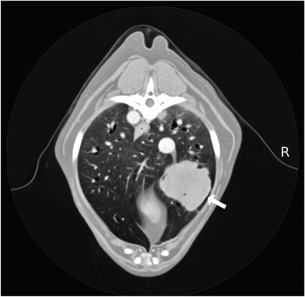
A 9-year-old, 25.3 kg, spayed female Bernese mountain dog was referred for investigation of a right intrathoracic mass that had been diagnosed by thoracic radiography at a primary veterinary hospital. The dog did not present any clinical signs. On general physical examination, the dog was panting, with normal thoracic auscultation. Three-view thoracic radiographs confirmed the presence of an approximately 5.0 × 5.0 cm soft tissue, opaque mass in the right middle- or caudal-lung lobe field. A thoracic ultrasound revealed a hypoechoic solid mass. Cytology of ultrasound-guided fine-needle aspiration of the mass confirmed a malignant epithelial tumor. The abdominal ultrasound examination was normal. Complete hematology and serum biochemistry results were within normal limits. Thoracic computed tomography confirmed a homogeneous contrast-enhanced mass within the right middle lung lobe (Fig. 1). The mass measured 5.2 × 5.2 × 5.7 cm (84.9 cm3, 3.3 cm3/kg) and the intrathoracic lymph nodes were not enlarged. There were one pulmonary artery (diameter of 4.6 mm) and two pulmonary veins (diameter of 4.6 mm, and 2.6 mm). Surgical resection was recommended as a treatment option, and VATS anatomical lobectomy was performed. Informed consent was obtained from the owner.
Fig. 1.
Thoracic computed tomography images showing a mass (arrow) in the right middle lung lobe. Right side of the body (R).
The dog was premedicated with midazolam 0.2 mg/kg intravenously (IV), fentanyl 10 µg/kg IV, and atropine 0.025 mg/kg subcutaneously (SC), and was pre-oxygenated for 5 min prior to anesthetic induction. Anesthesia was induced with propofol 3 mg/kg IV and maintained by total intravenous anesthesia with propofol and fentanyl (propofol up to 0.3–0.7 mg/kg/min and fentanyl 5–20 µg/kg /hr constant rate infusion [CRI]). The rate of infusion was controlled according to the patient monitoring parameters and was recorded. After endotracheal intubation, one-lung ventilation was established using a bronchoscopically placed endobronchial blocker (Arndt endobronchial blocker set 9 Fr, Cook Japan K. K., Tokyo, Japan). The left lung lobes were ventilated mechanically using a volume-controlled ventilator, while the right lobes were not ventilated and remained deflated. Lactated Ringer’s solution was administered throughout the procedure at 5 ml/kg/hr. An intercostal nerve block was performed using 0.5% bupivacaine 2 mg/kg.
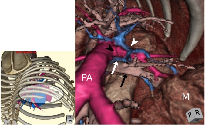
A DICOM workstation (ZioCube, Ziosoft, Inc., Tokyo, Japan) was used to construct 3D images of the hilum (Fig. 2). Using these 3D images, a surgical plan including the sites of ports and small thoracotomy and approaches to the pulmonary vessels and bronchus at the hilum for isolation and ligation were established. The dog was positioned in a left lateral recumbency and the right thorax was clipped and aseptically prepared. A 6-mm port (EZ trocar smart insertion, Hakko Co., Ltd., Osaka, Japan) was placed in the dorsal half of the right ninth intercostal space for the introduction of a 5 mm 30° telescope (1588 AIM, Stryker Japan K. K., Tokyo, Japan). Subsequently, a 6 mm port was placed in the dorsal third of the right ninth intercostal space as an instrument port. A 4 cm, intercostal, small thoracotomy incision was made under thoracoscopic view in the ventral third of the fifth intercostal space, and a 2.5–6 cm wound retractor (Alexis wound protector/retractor, Applied Medical Resources Corp., Rancho Santa Margarita, CA, U.S.A.) was placed. The soft tissue mass was visualized thoracoscopically in the middle lung lobe. The right middle lung lobe was dissected from the right caudal lobe, right pulmonary artery, and right principal bronchus using a vessel sealing device (Liga Sure, Covidien/Medtronic Japan, Tokyo, Japan) and thoracoscopic grasping forceps (T1253/T1269, OLYMPUS, Tokyo, Japan).
Fig. 2.
A computed tomography image of virtual thoracoscopy. Caudal-cranial view from the 9th intercostal camera port. Pulmonary artery of the right middle lung lobe (black arrowhead), right middle bronchus (black arrow), pulmonary vein cranial to the right middle bronchus (white arrowhead), pulmonary vein ventral to the right middle bronchus (white arrow), and right pulmonary artery (PA).
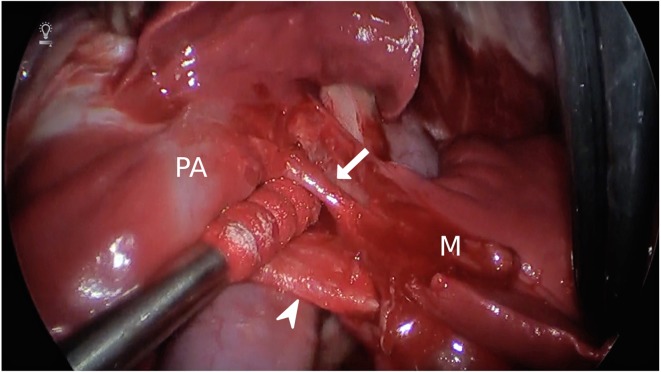
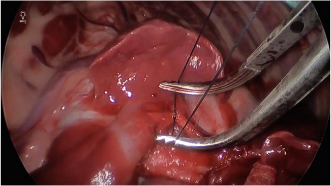
The right middle lung lobe vessels and bronchus were identified. Pulmonary hilum dissection was performed using a combination of thoracoscopic grasping forceps and cotton swabs (Naruke Thoraco Cotton, Kenzmedico Co., Ltd., Saitama, Japan) to provide counter traction (Fig. 3). The pulmonary artery and one of the pulmonary veins (diameter of 4.6 mm) were ligated with simple encircling suture and coagulated using a vessel sealing device. Non-absorbent sutures (2–0 Surgiron, Covidien/Medtronic Japan) were passed around these vessels using conventional right-angle forceps via small thoracotomy. Ligatures of these sutures were tied at proximal end of the vessel using conventional knotting forceps (Naruke D.K. forceps, Kenzmedico Co., Ltd.) via a small thoracotomy (Fig. 4). A vessel sealing device was used to coagulate and transect the distal end of these vessels. In this way, another pulmonary vein (diameter of 2.6 mm) was coagulated and transected using a vessel sealing device. The bronchus of the right middle lung lobe was double ligated with two encircling sutures. Two ligatures of 2–0 non-absorbent sutures were passed around the bronchus using conventional right-angle forceps and were tied at proximal end of the bronchus near the right main bronchus using conventional knotting forceps via small thoracotomy (Fig. 4). The bronchus was transected the distal end of the bronchus using conventional Metzenbaum scissors via small thoracotomy.
Fig. 3.
Dissection and isolation of the pulmonary artery of the right middle lung lobe with a cotton swab. Pulmonary artery of the right middle lobe (arrow), bronchus of the right middle lung lobe (arrowhead), middle lung lobe (M), and the right pulmonary artery (PA).
Fig. 4.
Ligation of the pulmonary artery of the middle lung lobe using knotting forceps.
The right middle lung lobe, including the mass, was removed via a small thoracotomy site, using a specimen retrieval bag (EZ PURES, Hakko Co., Ltd.). Tracheobronchial lymph nodes were directly palpated via a small thoracotomy site. Nodal palpation was performed, and regional tracheobronchial lymph nodes were found to be of normal texture and size. When paired with CT findings, it was elected to not perform resection. After completing the lobectomy, two-lung ventilation was reestablished. A thoracic lavage was performed with warmed sterile saline solution. The edge of the resected pulmonary vessels margin was examined carefully for hemostasis, and the resected bronchial margin was evaluated under positive airway pressure (still 30 cmH2O) to ensure an airtight closure. The pleural cavity was suctioned, and the thorax was closed with absorbent monofilament sutures. An indwelling 12 French thoracostomy tube (NIPRO Trocar Catheter, NIPRO, Osaka, Japan) was placed and secured by means of a finger-trap suture. The duration of the surgery was 101 min. Histopathological analysis of the resected lung lobe confirmed that the mass was a papillary adenocarcinoma, and the margin of surgical resection showed no evidence of neoplasia.
The dog recovered without complications. A few hours after surgery, there was no respiratory distress, and oxygen saturation was maintained above 98% on room air. Pleural effusion was checked every 2–4 hr for 12 hr postoperatively, and thereafter as required. The thoracostomy tube was removed on the day after surgery, because pleural effusion was 0.56 ml/kg/day. Postoperative analgesia consisted of fentanyl 3 µg/kg/hr IV CRI for 1 day postoperatively and then by fentanyl patch for 3 days, and robenacoxib 1 mg/kg SC once a day for 2 days postoperatively. The dog was discharged on the second postoperative day. The incision sites healed without complication, and skin sutures were removed 2 weeks after surgery. At the time of writing (10 months postoperatively), the dog was clinically well.
Lobectomy for lung tumors in veterinary patients has previously been performed via thoracoscopy using stapling devices, without individual isolation of pulmonary vessels and bronchus [1, 4, 5, 8]. To our knowledge, there are no veterinary clinical case report of anatomical lobectomy via thoracoscopy for resection of a primary lung tumor without the use of stapling devices.
The small thoracotomy incision for an instrument port was helpful to accomplish this procedure. Small thoracotomy combined with thoracoscopy for lobectomy is one of the procedures used in human patients [7] and has been termed as hybrid VATS. This is a safe and useful procedure that can obtain multiple viewpoints either from a magnified horizontal view via thoracoscopy or from a direct conventional vertical view via the small thoracotomy site. In addition, it allows manipulation of conventional surgical instruments including forceps and scissors, and direct palpation of intrathoracic organs via the small thoracotomy site. Additionally, an extension of small thoracotomy may be more easily accomplished compared to portal site incisions in cases where emergent conversion is required. In the veterinary literature, two types of techniques are used for lobectomy using thoracoscopy. In the present case, one of the instrument ports was set as a 4 cm small thoracotomy incision that was required to remove the mass from the thoracic cavity. All dissection and transection activities at the hilum were performed in the thoracic cavity. Those complex intrathoracic maneuvers were performed safely from a magnified horizontal view via thoracoscopy or from a direct conventional vertical view, using multiple instruments including conventional surgical instruments and thoracoscopic surgical instruments via the small thoracotomy site.
In the present case, pulmonary vessels and the bronchus were successfully isolated at the hilum and were transected and ligated with sutures and/or using a vessel sealing system, without complications. VATS anatomical lobectomy in dogs had been reported previously in preliminary experimental studies from the same group [2, 9]. Six of 12 dogs in the first study [9], exhibited hemorrhage from the pulmonary vessel avulsion by dissection at the hilum and transaction of pulmonary vessel with an endo stapler. In the second study [2], which used different dissection and ligation techniques, the conventional suture ligation was combined with a vessel sealing device and used for vessel division. Through this combination techniques for vessels, nine of 10 dogs were successfully treated without complications. Although double or triple ligation of pulmonary vessel is recommended for suture ligation technique on total lung lobectomy [6], a combination technique of single ligation and a vessel sealing device was possible without complications. And the thin pulmonary vein (diameter of 2.6 mm) could be coagulated and transected with a vessel sealing device alone, because the vessel sealing device is approved for use on vessels that are up to 7 mm in diameter [6].
In these two studies, the dogs were positioned in a dorsal recumbency position and a transumbilical or transsubxiphoid approach with the single port was used. It was difficult to dissect the hilum of the lung since it could not be easily visualized using these approaches [7]. Using a conventional intracostal thoracotomy approach, it may be necessary to enlarge the thoracotomy wound or transect the anterior and posterior ribs in order to safely visualize, dissect, and transect the pulmonary vasculatures. However, when performing dissection of vessels and bronchus with a thoracoscopic view, the combination of a lateral recumbency position and multiple ports including a small thoracotomy provide sufficient visualization of the surgical field in the present case. In addition, the thoracoscopic view was helpful for the clear visualization of the pulmonary vessels using the magnified horizontal view.
This report describes a success in achieving pulmonary vessels and bronchus isolation at the hilar, without using stapling devices, for anatomical lobectomy via thoracoscopy. Further cases are required to evaluate the effectiveness of hybrid thoracoscopic anatomical lobectomy in small animals.
REFERENCES
- 1.Bleakley S., Duncan C. G., Monnet E.2015. Thoracoscopic lung lobectomy for primary lung tumors in 13 dogs. Vet. Surg. 44: 1029–1035. doi: 10.1111/vsu.12411 [DOI] [PubMed] [Google Scholar]
- 2.Hsieh M. J., Yen-Chu, Wu Y. C., Yeh C. J., Liu C. Y., Liu C. C., Ko P. J., Liu Y. H.2016. Feasibility of subxiphoid anatomic pulmonary lobectomy in a canine model. Surg. Innov. 23: 229–234. doi: 10.1177/1553350615615441 [DOI] [PubMed] [Google Scholar]
- 3.Laksito M. A., Chambers B. A., Yates G. D.2010. Thoracoscopic-assisted lung lobectomy in the dog: report of two cases. Aust. Vet. J. 88: 263–267. doi: 10.1111/j.1751-0813.2010.00587.x [DOI] [PubMed] [Google Scholar]
- 4.Lansdowne J. L., Monnet E., Twedt D. C., Dernell W. S.2005. Thoracoscopic lung lobectomy for treatment of lung tumors in dogs. Vet. Surg. 34: 530–535. doi: 10.1111/j.1532-950X.2005.00080.x [DOI] [PubMed] [Google Scholar]
- 5.Mayhew P. D., Hunt G. B., Steffey M. A., Culp W. T., Mayhew K. N., Fuller M., Johnson L. R., Pascoe P. J.2013. Evaluation of short-term outcome after lung lobectomy for resection of primary lung tumors via video-assisted thoracoscopic surgery or open thoracotomy in medium- to large-breed dogs. J. Am. Vet. Med. Assoc. 243: 681–688. doi: 10.2460/javma.243.5.681 [DOI] [PubMed] [Google Scholar]
- 6.Monnet E.2018. Lungs. pp. 1983–1999. In: Veterinary Surgery Small Animal, 2nd ed. (Johnston, S. A., and Tobias, K. M. eds.), Elsevier Health Sciences, St. Louis. [Google Scholar]
- 7.Okada M., Sakamoto T., Yuki T., Mimura T., Miyoshi K., Tsubota N.2005. Hybrid surgical approach of video-assisted minithoracotomy for lung cancer: significance of direct visualization on quality of surgery. Chest 128: 2696–2701. doi: 10.1378/chest.128.4.2696 [DOI] [PubMed] [Google Scholar]
- 8.Wormser C., Singhal S., Holt D. E., Runge J. J.2014. Thoracoscopic-assisted pulmonary surgery for partial and complete lung lobectomy in dogs and cats: 11 cases (2008–2013). J. Am. Vet. Med. Assoc. 245: 1036–1041. doi: 10.2460/javma.245.9.1036 [DOI] [PubMed] [Google Scholar]
- 9.Yin S. Y., Chu Y., Wu Y. C., Yeh C. J., Liu C. Y., Hsieh M. J., Liu Y. H.2014. Feasibility of transumbilical anatomic pulmonary lobectomy in a canine model. Surg. Endosc. 28: 2980–2987. doi: 10.1007/s00464-014-3561-3 [DOI] [PubMed] [Google Scholar]


