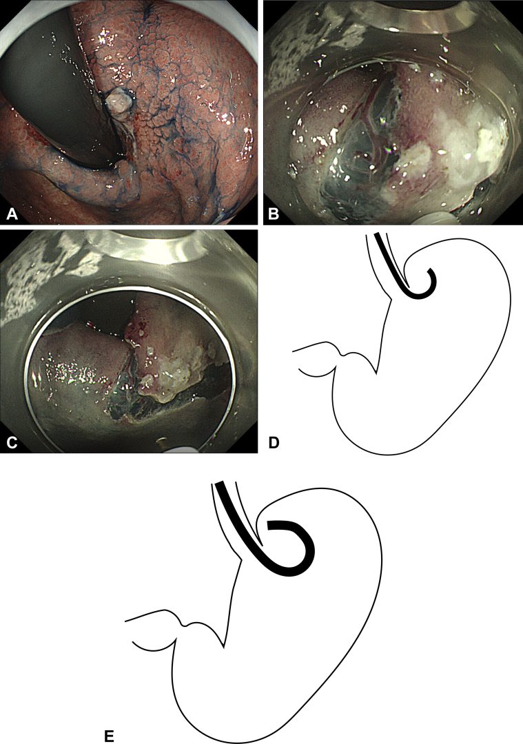
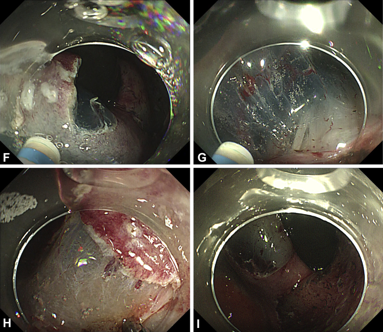
Figure 3.
Patient 2. A, Type 0-IIc lesion at the anterior wall to posterior wall of the upper body of the stomach. B, Incision on the fornix side, the most difficult area to reach with the endoscope. C, Incision on the fornix side. D, Approach to the fornix by use of a conventional endoscope. The endoscope cannot reach the dissection site. E, Approach to the fornix by use of the multibending endoscope. The multibend functionality of the endoscope enables us to efficiently approach and treat the site. F, Extension of the knife from the left channel while incision was made on the posterior wall expanded the right visual field. G, In dissection, when performing a cut toward the right, extending the knife from the left channel secured the visual field in the direction of the cut. H, Close approach to the site enabled us to visually confirm location of the blood vessels, and these vessels can be precisely targeted and grasped with hemostatic forceps. I, Ulcer after endoscopic submucosal dissection.


