Abstract
Traumatic dislocation of the atlanto-axial joint in combination with an odontoid fracture remains a rare entity. Beaucause of its instability, it's alsoo a seriuous injury. A fatal outcome is feared especially in elderly. We report a case of 74-year-old man who presented with neck pain Confusion and spastic tetraparesia after a low energy trauma. Radiographs and computed tomography demonstrated a C1C2 dislocation with odontoid fracture. After an unsuccessful attempt at closed reduction with halo traction, a surgical stabilisation was performed using a posterior approach. Death was occured in early postoperative due to respiratory distress.
Keywords: Atlantoaxial dislocation, elderly patients, neurological deficit, odontoid fracture, surgical management
Introduction
The traumatic atlantoaxial dislocation associated with dens fracture is a rare and serious injury due to its instability. The clinical and radiological diagnosis can be laborious in the elderly in whom brain trauma with low energy can be the cause.
Through an observation of C1-C2 dislocation associated with an odontoid fracture, we study this lesion and discuss its therapeutic management.
Case Report
A 74-year-old male, living alone in a rural area, with no particular history is brought back by his neighbors for headaches associated with impaired general condition and fluctuation of consciousness.
A laborious anamnesis found an episode of head injury without loss of consciousness following a fall of his own height dating back 3 days. The headaches that appeared after the trauma were neglected by the patient. On the other hand, the family was alerted by the installation of intermittent confusion and weakness of the limbs.
On physical examination, the Glasgow coma score was 13 points of 15, and the patient had analgesic attitude of the head with neck pain to the gentle mobilization. The neurological examination showed spastic tetraparesis more pronounced in the lower limbs without achieving sensitivity or reflexes. The examination of the perineum was without abnormalities.
Blood tests were also without anomalies.
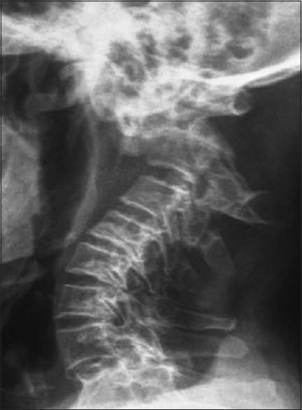
A radiograph of the cervical spine showed an abnormal gap between the vertebral spinous of the atlas and axis associated with a significant anterolisthesis [Figure 1].
Figure 1.

Sagittal plain radiograph showing a high abnormal gap between C1 and C2 spinous process suggesting a dislocation
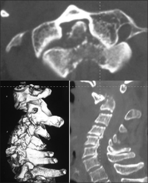
A computed tomography scan eliminated a brain damage and showed a fracture at the junction of the body and the odontoid process corresponding to the type 2 of Anderson and D’Alonzo classification.[1] This fracture was associated with anterior atlantoaxial dislocation of more than 5 mm corresponding to the type III of Fielding classification[2] [Figure 2].
Figure 2.

Sagittal and axial scans showing an anterior C1-C2 dislocation with an odontoïd fracture
A progressive reduction of the dislocation with a cranial traction was initiated. This device was not well tolerated by the patient. So we decided to go for surgery.
Under general anesthesia, reduction and realignment of the odontoid process were obtained by external maneuvers. An instrumented arthrodesis atlanto-occipital posterior approach has been made and completed by the addition of cancellous grafts. The final fluoroscopic control was satisfactory [Figure 3].
Figure 3.

Peroperative fluoroscopy control
Several postoperative ventilator weaning attempts have been initiated. The patient died five days after surgery following respiratory complications.
Discussion
The isolated traumatic atlantoaxial dislocation is a rare injury in adults. The fracture of the odontoid process is not unusual and represents 7%–9% of all fractures of the cervical spine.[3] The combination of these two lesions remains exceptional. Gleizes et al.[4] in an epidemiological study of 14 years have isolated 2 of 784 injuries of the cervical spine with 116 lesions of the upper segment.
Motor vehicle accidents, sports accidents, or high-rise falls are the most common causes of this lesion. However, in the elderly, a low-energy trauma such as a simple fall can be found.
In older patients, the top segment of the cervical spine is most vulnerable, the majority of fractures affecting either C1 or C2 or both.[5] The fracture of the dens is particularly common in this age group and should be systematically raised. The combined presence of osteoporosis of the upper cervical spine and osteoarthritis of the lower cervical spine explains the high incidence of these lesions.
C1-C2 dislocation associated with fracture of the dens is a potentially serious injury because of the vital neurological risk due to the anatomical proximity to the medulla oblongata and the fact that the craniocervical junction is very mobile (axial rotation torque C1-C2) and specifically exposed instability.[6]
Neurological tables are variable, ranging from no abnormalities to complete high tetraplegia to scalable prognosis through simple pyramidal irritation.
When the bulbar neurovegetative disorder begins to settle, confusion or even a pseudodelirium can be noted and it gradually dominates the clinical picture.[7] This particular presentation, occurring in an old person whose initial examination does not include high-energy trauma, may mislead the diagnosis to medical or metabolic etiologies. A C2C2 dislocation can sometimes be a chance discovery on a brain scanner. This could have been the case of our patient with the delirium and neurological deficit dominating the clinical picture. A radiograph of the cervical spine requested for neck pain has objectified the lesion and straightened diagnosis.
For some authors, this neurological status is regressive after orthopedic or surgical stabilization.[7] In the elderly, on the other hand, the existence of neurological signs secondary to a cervical spine injury is considered[8] as a factor of bad prognosis like the case we report. Lefranc et al.[9] in a series of 27 patients older than 70 years treated for a fracture of the odontoid lamented five cases of early death to hospitalization. However, they concluded that it is linked not only to fracture of the odontoid process but also to the presence of cervical lesions. For our patient, the atlantoaxial dislocation would probably influence the morbidity and mortality related to the fracture of the dens.
Radiologically, the dislocation is generally sagittal, but a lateral[10] or longitudinal dislocation[11] has also been reported. In our patient, a hyperflexion associated with an anterior translation seems to be the causal mechanism.
Atlantoaxial traumatic dislocation associated with the fracture of the odontoid is a real therapeutic challenge due to the anatomical complexity of this region. In elderly, the purpose of treatment is a quick recovery to avoid decubitus complications.
An attempt at orthopedic reduction of the dislocation should be made. A halo-corset immobilization can then be proposed in case of realignment and consolidation has been obtained for some authors between 3 and 6 months.[12,13,14]
The reduction under general anesthesia by external maneuvers seems to be dangerous because of the proximity of the medullary bulb. Botelho et al.[15] and Silbergeld et al.[16] reported three cases of death related to these maneuvers.
In the case of failure of the traction, a surgical stabilization is necessary although the installation in the prone position, the manipulation of the spine, and the surgery in an elderly can aggravate the neurological manifestations. The posterior approach is the preferred route of the majority of authors. It allows a fixation C1-C2 or occipitocervical. However, access to joint processes remains difficult and at a high risk of bleeding. The unilateral or bilateral retropharyngeal approach, described by de Andrade and Macnab,[17] circumvents not only this difficulty but also avoids the passage to the prone position.
After reduction of the dislocation, the C1-C2 screwing or more extensive occipitocervical arthrodesis were the two mounting methods advocated by the authors.[18] Only lard[19] conducted a transarticular arthrodesis anterior approach according to Vaccaro technique.
No publication reports an isolated synthesis of the odontoid as a treatment for this lesion.[18]
Despite the neurological deficit, we first attempted reduction by traction hoping regression signs. It was only after the failure of this option a surgical fixation was made. Despite the good reduction of the lesion and the mechanical stability of the assembly, the immediate postoperative course was marked by a fatal outcome. This observation confirms the bad prognosis even dark in the elderly with neurotoxic lesion of the upper cervical spine.
Conclusion
The atlantoaxial dislocation associated with fracture of the odontoid process is a rare entity. Its occurrence in the elderly may follow a simple fall and must fear a fatal evolution despite a correct coverage.
Declaration of patient consent
The authors certify that they have obtained all appropriate patient consent forms. In the form the patient(s) has/have given his/her/their consent for his/her/their images and other clinical information to be reported in the journal. The patients understand that their names and initials will not be published and due efforts will be made to conceal their identity, but anonymity cannot be guaranteed.
Financial support and sponsorship
Nil.
Conflicts of interest
There are no conflicts of interest.
References
- 1.Anderson LD, D’Alonzo RT. Fractures of the odontoid process of the axis. J Bone Joint Surg Am. 1974;56:1663–74. [PubMed] [Google Scholar]
- 2.Fielding JW, Hawkins RJ. Atlanto-axial rotatory fixation.(Fixed rotatory subluxation of the Atlanto-axial joint) J Bone Joint Surg Am. 1977;59:37–44. [PubMed] [Google Scholar]
- 3.Benzel EC, Hart BL, Ball PA, Baldwin NG, Orrison WW, Espinosa M, et al. Fractures of the C-2 vertebral body. J Neurosurg. 1994;81:206–12. doi: 10.3171/jns.1994.81.2.0206. [DOI] [PubMed] [Google Scholar]
- 4.Gleizes V, Jacquot FP, Signoret F, Feron JM. Combined injuries in the upper cervical spine: Clinical and epidemiological data over a 14-year period. Eur Spine J. 2000;9:386–92. doi: 10.1007/s005860000153. [DOI] [PMC free article] [PubMed] [Google Scholar]
- 5.Pepin JW, Bourne RB, Hawkins RJ. Odontoid fractures, with special reference to the elderly patient. Clin Orthop Relat Res. 1985;193:178–83. [PubMed] [Google Scholar]
- 6.Steltzlen C, Lazennec JY, Catonné Y, Rousseau MA. Unstable odontoid fracture: Surgical strategy in a 22-case series, and literature review. Orthop Traumatol Surg Res. 2013;99:615–23. doi: 10.1016/j.otsr.2013.02.007. [DOI] [PubMed] [Google Scholar]
- 7.Argenson C, De Peretti F, Schlatterer B, Hovorka I, Eude P. Traumatisme du rachis cervical. Encycl. med. Chir (Elsevier, paris) Appareil locomoteur. 1998 15-825-A-10:20. [Google Scholar]
- 8.Jubert P, Lonjon G, Garreau de Loubresse C Bone and Joint Trauma Study Group GETRAUM. Complications of upper cervical spine trauma in elderly subjects. A systematic review of the literature. Orthop Traumatol Surg Res. 2013;99:S301–12. doi: 10.1016/j.otsr.2013.07.007. [DOI] [PubMed] [Google Scholar]
- 9.Lefranc M, Peltier J, Fichten A, Desenclos C, Toussaint P, Le Gars D, et al. Odontoid process fracture in elderly patients over 70 years: Morbidity, handicap, and role of surgical treatment in a retrospective series of 27 cases. Neurochirurgie. 2009;55:543–50. doi: 10.1016/j.neuchi.2009.01.021. [DOI] [PubMed] [Google Scholar]
- 10.Lenehan B, Guerin S, Street J, Poynton A. Lateral C1-C2 dislocation complicating a type II odontoid fracture. J Clin Neurosci. 2010;17:947–9. doi: 10.1016/j.jocn.2009.11.025. [DOI] [PubMed] [Google Scholar]
- 11.Przybylski GJ, Welch WC. Longitudinal atlantoaxial dislocation with type III odontoid fracture. Case report and review of the literature. J Neurosurg. 1996;84:666–70. doi: 10.3171/jns.1996.84.4.0666. [DOI] [PubMed] [Google Scholar]
- 12.Autricque A, Lesoin F, Villette L, Franz K, Pruvo JP, Jomin M. Fracture of the odontoid process and C1-C2 lateral luxation 2 cases. Ann Chir. 1986;40:397–400. [PubMed] [Google Scholar]
- 13.Hopf S, Buchalla R, Elhöft H, Rubarth O, Börm W. Atypical dislocated dens fracture type II with rotational atlantoaxial luxation after a riding accident. Unfallchirurg. 2009;112:517–20. doi: 10.1007/s00113-008-1542-5. [DOI] [PubMed] [Google Scholar]
- 14.Spoor AB, Diekerhof CH, Bonnet M, Oner FC. Traumatic complex dislocation of the atlanto-axial joint with odontoid and C2 superior articular facet fracture. Spine (Phila Pa 1976) 2008;33:E708–11. doi: 10.1097/BRS.0b013e31817c140d. [DOI] [PubMed] [Google Scholar]
- 15.Botelho RV, de Souza Palma AM, Abgussen CM, Fontoura EA. Traumatic vertical atlantoaxial instability: The risk associated with skull traction. Case report and literature review. Eur Spine J. 2000;9:430–3. doi: 10.1007/s005860000166. [DOI] [PMC free article] [PubMed] [Google Scholar]
- 16.Silbergeld DL, Laohaprasit V, Grady MS, Anderson PA, Winn HR. Two cases of fatal atlantoaxial distraction injury without fracture or rotation. Surg Neurol. 1991;35:54–6. doi: 10.1016/0090-3019(91)90203-l. [DOI] [PubMed] [Google Scholar]
- 17.de Andrade JR, Macnab I. Anterior occipito-cervical fusion using an extra-pharyngeal exposure. J Bone Joint Surg Am. 1969;51:1621–6. [PubMed] [Google Scholar]
- 18.Moreau PE, Nguyen V, Atallah A, Kassab G, Thiong’o MW, Laporte C. Traumatic atlantoaxial dislocation with odontoid fracture: A case report. Orthop Traumatol Surg Res. 2012;98:613–7. doi: 10.1016/j.otsr.2012.03.012. [DOI] [PubMed] [Google Scholar]
- 19.Riouallon G, Pascal-Moussellard H. Atlanto-axial dislocation complicating a type II odontoid fracture. Reduction and final fixation. Orthop Traumatol Surg Res. 2014;100:341–5. doi: 10.1016/j.otsr.2013.12.026. [DOI] [PubMed] [Google Scholar]


