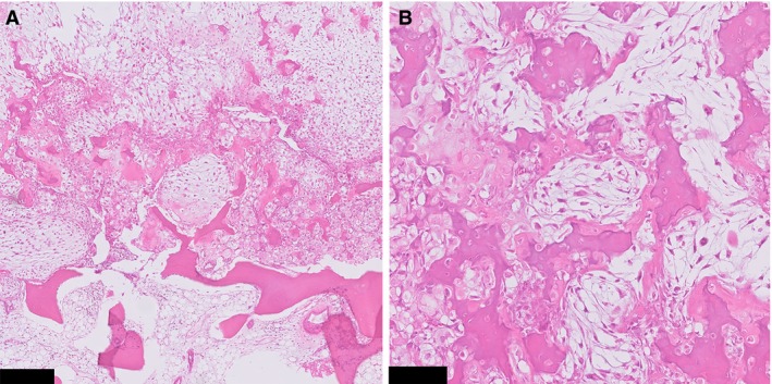
Figure 3.

A, Haphazardly arranged areas with clear cell chondrosarcoma morphology and conventional chondrosarcoma. B, At high power, both components gradually merge together, as opposed to dedifferentiated chondrosarcoma (case 1). Scale bar: 250 μm (A), 100 μm (B).
