Abstract
Objectives
Biologics treatment with antitumour necrosis factor alpha (TNFα) is efficacious in patients with juvenile idiopathic arthritis (JIA). Despite displaying clinical inactivity during treatment, many patients will flare on cessation of therapy. The inability to definitively discriminate patients who will relapse or continue to remain in remission after therapy withdrawal is currently a major unmet medical need. CD4 T cells have been implicated in active disease, yet how they contribute to disease persistence despite treatment is unknown.
Methods
We interrogated the circulatory reservoir of CD4+ immune subsets at the single-cell resolution with mass cytometry (cytometry by time of flight) of patients with JIA (n=20) who displayed continuous clinical inactivity for at least 6 months with anti-TNFα and were subsequently withdrawn from therapy for 8 months, and scored as relapse or remission. These patients were examined prior to therapy withdrawal for putative subsets that could discriminate relapse from remission. We verified on a separate JIA cohort (n=16) the dysregulation of these circulatory subsets 8 months into therapy withdrawal. The immunological transcriptomic signature of CD4 memory in relapse/remission patients was examined with NanoString.
Results
An inflammatory memory subset of CD3+CD4+CD45RA−TNFα+ T cells deficient in immune checkpoints (PD1−CD152−) was present in relapse patients prior to therapy withdrawal. Transcriptomic profiling reveals divergence between relapse and remission patients in disease-centric pathways involving (1) T-cell receptor activation, (2) apoptosis, (3) TNFα, (4) nuclear factor-kappa B and (5) mitogen-activated protein kinase signalling.
Conclusions
A unique discriminatory immunomic and transcriptomic signature is associated with relapse patients and may explain how relapse occurs.
Keywords: Juvenile idiopathic arthritis (JIA), mass cytometry (CyToF), anti-TNFɑ, relapse and therapy withdrawal
Key messages.
What is already known about this subject?
Biologics treatment with tumour necrosis factor alpha inhibitors is efficacious in patients with juvenile idiopathic arthritis, though many patients flare upon cessation of therapy despite achieving clinical inactivity.
There is lack of scientific understanding and definitive biological markers to discriminate patients who will relapse from those who will remain in stable remission after therapy withdrawal.
What does this study add?
This study demonstrates that an inflammatory subset of CD4 memory cells deficient in immune checkpoint receptors is present in patients who will relapse.
T-effector diversification occurs during overt flare in relapse patients.
How might this impact on clinical practice or future developments?
Tracking of these dysregulated CD4 memory subsets may aid in clinical therapeutic management of patients under therapy, and understanding its mechanisms may provide an avenue for future precision therapeutic developments.
Introduction
Targeted therapy of juvenile idiopathic arthritis (JIA) with antitumour necrosis factor alpha (TNFα) biologics is efficacious, with 70%–80% responders and up to 50% achieving clinical inactivity on long-term treatment.1 2 While sustained immune suppression through anti-TNFα is generally well tolerated, clinicians seek to achieve clinical remission off medication to reduce risk of general infection, adverse events and financial burden.2 Drug withdrawal in patients who attain clinical inactivity is complicated by the fact that 50%–80% of patients relapse on therapy discontinuation.3 4 This phenomenon indicates that relapse patients who have attained clinical inactivity on medication, as defined by the Wallace criteria,5 continue to experience subclinical inflammation and persistence of disease without overt presentation of clinical symptoms. Conversely, patients who achieve clinical remission off medication could be spared long-term drug effects. As such, there is a clinical need to address how discontinuing anti-TNFα therapy can be safely implemented, as well as a scientific need to understand the immune mechanisms related to relapse.
The pathogenesis of JIA remains widely debated.6 Unsupervised genome-wide association studies and pathway analysis have highlighted the role of CD4 T-helper cell populations in autoimmune disease progression.7 CD4 T cells were shown to infiltrate the synovium microenvironment,8–11 and corresponding pathogenic CD4 HLA-DR+ T-effector and regulatory subsets possessing strong immune phenotypic and T-cell receptor (TCR) activation correlation with synovial T cells have been found recirculating in the blood during active inflammation.12 13 Furthermore, epigenetic histone modifications associated with enhancer functions have been detected in CD4 T cells of patients with JIA.14 Using network analysis of DNA CpG methylation sites in total CD4 T cells, we have previously demonstrated that T-cell activation pathways are associated with clinical fate on anti-TNFα withdrawal.15 However, the identity of the specific pathogenic CD4 subset that maintains subclinical disease persistence remains elusive.
In this study, we exploited the high-dimensional single-cell resolution capabilities of cytometry by time of flight (CyTOF) and patient samples from a well-defined clinical cohort with the objective of uncovering CD4 T-cell subsets responsible for disease persistence. Patients with JIA who maintained clinical inactivity under anti-TNFα for at least 6 months were subsequently withdrawn from therapy. These patients were rigorously scored for their disease activity across 8 months, and their clinical outcome was defined as relapse or remission. We investigated the circulatory immunome of these patients with JIA to determine the immunological differences at the core of the dichotomic clinical fates. These differences may help provide a framework for understanding the mechanisms of disease relapse on drug withdrawal, thus enabling a knowledge-based guidance for clinical management while proposing some potential new targets for intervention.
Materials and methods
Samples
Peripheral blood mononuclear cells (PBMCs) were obtained from patients with polyarticular JIA recruited through the “Determining Predictors of Safe Discontinuation of Anti-TNF treatment in JIA” trial (ID: NCT00792233).16 Patients treated with anti-TNFα biologics and shown to be in an inactive disease state for 6 months were enrolled into the study. Clinical inactivity was defined by Wallace et al criteria5: (1) absence of active joints; (2) lack of fever, rash and serositis attributable to JIA; (3) no active uveitis; (4) within normal range of erythrocyte sedimentation rate (ESR) unless attributable to JIA; (5) physician global disease activity of ≤0.5 (Likert-like scale); and (6) duration of morning stiffness of ≤15 min. On enrolment, patients are withdrawn from anti-TNFα therapy and accessed through monthly clinical visits for a study period of 8 months. Clinical outcome is designated as relapse or remission depending on six core JIA parameters: (1) number of active joints, (2) number of joints with loss of motion, (3) medical doctor global assessment of current disease activity (Likert-like scale), (4) patient/parent global assessment of overall disease severity in the prior week (Likert-like scale), (5) a validated measure of physical function childhood health assessment questionnaire (CHAQ) and (f) ESR. A patient was considered to be experiencing a relapse if there was ≥30% worsening in more than three of the six JIA core parameters, with no more than one parameter improving by >30%.1 17 For remission individuals, they would have achieved ≥14 months of clinical inactivity from prior recruitment to study end. PBMCs were interrogated by CyTOF from patients (n=20) prior to withdrawal and were designated as (To), and separately from another batch (n=16) at the end of 8 months after withdrawal were designated as (Tend). Patient PBMCs (n=12) were also sorted for CD3+CD4+CD45RO+CD45RA− for NanoString analysis. The demographics/medication history profile of patients with JIA withdrawn from therapy and sample usage breakdown is shown in online supplementary table S1.
annrheumdis-2019-216059supp002.pdf (244.9KB, pdf)
Age-matched healthy controls (n=69) were recruited through the Precision Rheumatology International Platform (PRIP) study conducted at the KK Women’s and Children’s Hospital (KKH). These controls have no indication of inflammation and PBMCs were isolated pre-operatively from patients scheduled for day surgeries. Healthy PBMCs were examined with CyTOF (n=10), NanoString (n=3) or age-matched strata cross validation for receiver operating characteristic (ROC) curve (n=56).
Paired treatment naive/post-treatment patients with JIA (n=4) were also recruited through the study “A Precision Medicine Approach to Understand and Predict Responsiveness to Therapy in Human Arthritis” conducted in KKH for NanoString analysis. These patients with active JIA were initially treatment naive to anti-TNFα and, after a 6-month drug course, exhibited treatment susceptibility determined by complete absence of active joints. The demographics/medication history profile of patients with JIA is shown in online supplementary table S2.
Additional methodological details are available as online supplementary information.
annrheumdis-2019-216059supp003.pdf (103.8KB, pdf)
Results
CD4+CD45RA−TNFα+ T cells were present in patients with JIA prior to relapse
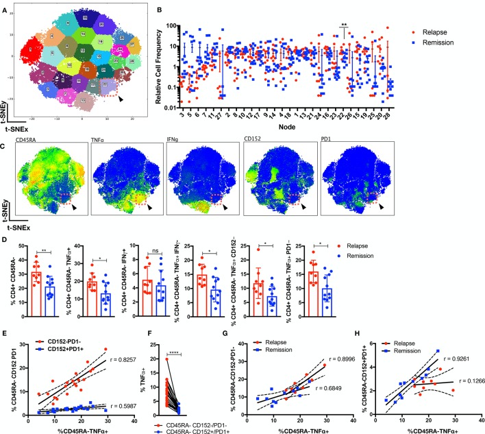
Dsyregulated CD4 T cells are thought to contribute to JIA pathogenesis.8–13 We interrogated the circulatory CD4 landscape of patients with JIA (n=20) prior to therapy withdrawal to understand why certain individuals relapse. At this stage, the patients were clinically scored to be inactive for 6 months; thus, patients who will relapse or remain in remission were clinically indistinguishable prior to withdrawal. We assessed the PBMCs with a CyTOF panel consisting of 31 functional, 6 lineage markers (online supplementary table S3) and CD45 barcoding to facilitate pooling of individuals.18 Batch variability in staining was monitored through an internal biological control (online supplementary figure S1). The debarcoded CD3+CD4+ T cells were exported and normalised for cell events, and the 31 markers were dimensionally reduced with MarVis onto a bivariate X–Y axis through t-distributed stochastic neighbour embedding (t-SNE) (online supplementary figure S2). Clustering with k-means segregated the CD4 cells into distinct nodes (figure 1A), and we detected an enrichment (p<0.01) in node 22 for patients who will relapse that represents CD4+CD45RA−TNFα+IFNγ−CD152−PD1− T cells (figure 1A–C). This was further verified with a separate clustering method, FlowSoM (online supplementary figure S3). To ensure the results were not due to clustering artefacts, we manually gated the preclustering flow cytometry standard (FCS) files (figure 1D, gating strategy in online supplementary figure S4) and determined significant (p<0.05) upregulation in CD4+CD45RA− memory subsets that was restricted within the TNFα+ compartment. In particular, relapse patients were enriched for CD4+CD45RA−TNFα+ T cells, which were absent for IFNγ expression, and were notably deficient in immune checkpoints (PD1/CD152). We investigated the relationship of the dysregulated T effectors and immune checkpoint expression within the memory compartment (figure 1E). There was a stronger positive correlation of CD45RA−TNFα+ with CD45RA−CD152−PD1− (r=0.8257) as opposed to CD45RA−CD152+PD1+ (r=0.5987) cells across the patients. The percentage of TNFα+ cells was significantly higher (p<0.0001) in CD45RA−CD152−/PD1− cells (figure 1F). While relapse patients showed a perceptible increase in T effectors in the absence CD152/PD1, there was a marked drop to upregulate CD152/PD1 as compared with remission patients (figure 1G,H).
Figure 1.
Perturbation in CD4 landscape in patients with JIA who will relapse. Circulatory CD3+CD4+ cells from patients with JIA (n=20; relapse=9, remission=11) prior to therapy withdrawal were stained with 31 functional markers in cytometry by time of flight and were dimensionally reduced onto a bivariate X–Y axis scale with t-SNE. (A) The t-SNE map is segregated into 28 distinct nodes with k-means, and node 22 is highlighted (red-dotted box). (B) The distribution of relative cell frequency in relapse or remission patients across the nodes is shown, with node 22 significantly higher (p<0.01) in relapse individuals. (C) The phenotypic expression of markers is shown for node 22 (red-dotted box). (D) Supervised gating of the preclustering FCS files validates the relevant CD4 memory cellular subsets in relapse versus remission individuals. (E) Correlation analysis of CD45RA−TNFα+ versus CD45RA−CD152−/PD1− or CD45RA−CD152+/PD1+ as percentage of CD3+CD4+ cells across patients with JIA. (F) Percentage of TNFα+ cells in CD45RA−CD152−/PD1− or CD45RA−CD152+/PD1+ compartment. Correlation analysis of CD45RA−TNFα+ versus (G) CD45RA−CD152−/PD1− or (H) CD45RA−CD152+/PD1+ as percentage of CD3+CD4+ cells across relapse or remission patients. Comparison of cellular subsets performed with Mann-Whitney U, unpaired or paired two-tailed test, means±SD. *p<0.05, **p<0.01, ****p<0.0001. Correlation analysis performed with Pearson correlation, two-tailed test. IFN, interferon; TNFα, tumour necrosis factor alpha; t-SNE, t-distributed stochastic neighbour embedding.
annrheumdis-2019-216059supp001.pdf (26.6MB, pdf)
Analysis of the CD4+TNFα+ healthy landscape unveils subclinical T-effector diversification in relapse patients
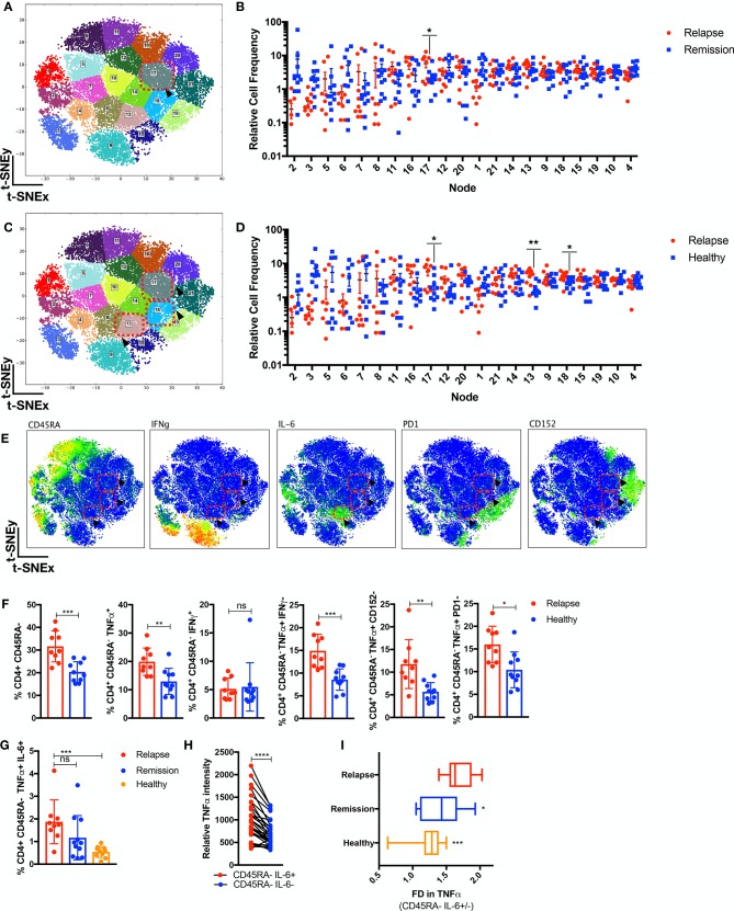
To validate the previously mentioned findings and to investigate the possibility of disease-centric CD4+ cellular subsets that are masked by comparing JIA relapse/remission individuals, we included age-matched paediatric healthy controls. CD4+TNFα+ T cells from JIA relapse/remission (n=20) prior to withdrawal or healthy individuals (n=10) were compared with further delineated key differences within the T-effector compartment. Patients with JIA who will relapse were enriched (p<0.05) for node 17 against remission individuals (figure 2A,B) and additionally for nodes 17, 13 and 18 against healthy controls (p<0.05) (figure 2C,D). Node 17 reaffirms relapse individuals are enriched for CD4+CD45RA−TNFα+IFNγ−CD152−PD1− T cells (figure 2E,F) as compared with remission or healthy individuals. Additionally, node 13 (figure 2E) represents CD4+CD45RA−TNFα+IFNγ−CD152−PD1− T cells that are also interleukin (IL)-6+. Supervised gating (figure 2G gating strategy in online supplementary figure S4) validated that relapse individuals are enriched (p<0.001) with CD4+CD45RA−TNFα+IL-6+ as compared with healthy individuals. The intensity of TNFα was significantly higher (p<0.0001) in CD45RA−IL-6+ as opposed to IL-6− cells (figure 2H), and relapse individuals had a higher fold increase (p<0.05) in TNFα in CD45RA−IL-6+ cells as compared with remission or healthy individuals.
Figure 2.
Subclinical T-effector diversification in relapse individuals. CD4+TNFα+ cells from patients with JIA (n=20; relapse=9, remission=11) prior to therapy withdrawal and healthy paediatric controls (n=10) were analysed through t-SNE. (A) The CD4+TNFα+ t-SNE MAP is segregated into 21 nodes with enrichment in (B) node 17 (p<0.05) for relapse as compared with remission individuals and (C,D) node 17, 18 and 13 (p<0.05) for relapse as compared with healthy individuals. (E) The phenotype expression is shown for node 17, 18 and 13 (red-dotted boxes and arrowheads). (F) Supervised gating of preclustering FCS files validates the relevant CD4 memory cellular subsets in relapse versus healthy individuals. (G) Supervised gating of CD4+CD45RA−TNFα+IL-6+ cells in relapse/remission/healthy individuals. (H) Relative TNFα intensity in CD45RA−IL-6+ or CD45RA−IL-6− cells. (I) Fold difference (FD) in TNFα intensity in CD45RA−IL-6+ over CD45RA−IL-6− cells in relapse/remission/healthy individuals. Comparison of cellular subsets performed with Mann-Whitney U, unpaired or paired two-tailed test, means±SD. *p<0.05, **p<0.01, ***p<0.001, ****p<0.0001. IFN, interferon; IL, interleukin; ns, not significant; TNFα, tumour necrosis factor alpha; t-SNE, t-distributed stochastic neighbour embedding.
Overt T-effector diversification during flare manifestation and quiescence in stable remission
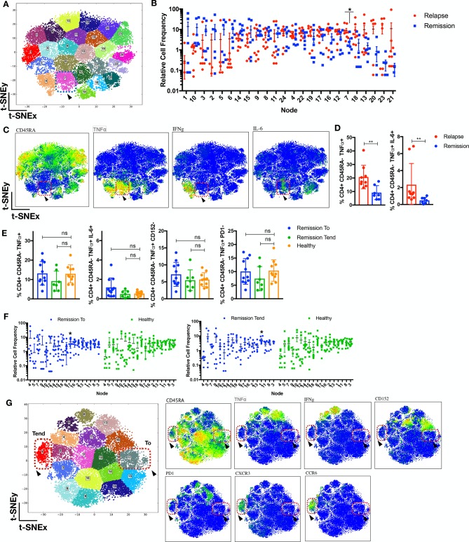
We have observed the presence of CD4 memory T cells in patients prior to relapse. To examine this phenomenon further, we interrogated the CD4 landscape of an independent batch of JIA individuals (n=16) withdrawn from therapy for 8 months that either developed flare or remained in stable remission. Relapse (in flare) patients exhibited the emergence of a previously subclinical CD4+CD45RA−TNFα+IL-6+ subset (figure 3A–D, node 7) as compared with patients who remained in remission after 8 months of withdrawal. To determine the state of immunological quiescence in remission (To: prior withdrawal, Tend: 8 months’ withdrawal) patients, we gated for the relevant dysregulated CD4+CD45RA−TNFα+ subsets and found no difference as compared with healthy individuals (figure 3E). The CD4 landscape of remission patients (To/Tend) as compared with healthy individuals revealed mostly similar profiles (figure 3F) except for nodes 17 and 1 enriched in remission To or Tend patients, respectively. Both nodes were absent for TNFα, with node 17 exhibiting CD45RA+ and node 1 expressing the CD45RA−/+CXCR3+CCR6+ phenotype (figure 3G).
Figure 3.
Emergence of T-effector diversification in flare manifestation and quiescence in stable remission. (A–D) CD4+ T cells from patients with JIA (n=16; relapse=9, remission=7) withdrawn from therapy for 8 months were analysed with t-SNE. (A) The t-SNE MAP is segregated into 24 nodes; node 7 is highlighted (blue-dotted box). (B) The distribution of relative cell frequency across the nodes is shown, with node 7 enriched (p<0.05) in relapse individuals, exhibiting (C) the CD4+CD45RA−TNFα+IL-6+IFNγ− phenotype (node7: red-dotted box and arrowhead). (D) Supervised gating of preclustering FCS files for CD4+CD45RA−TNFα+IL-6+ in relapse and remission patients. (E) Supervised gating of relevant CD4+CD45RA−TNFα+ subsets in remission patients (to: prior to withdrawal or tend: 8 months after withdrawal) or healthy individuals. (F,G) CD4+ T cells from JIA remission patients (n=18; To=11, Tend=7) and healthy controls (n=10) were analysed with t-SNE. (F) The distribution of relative cell frequency across the nodes is shown, with node 17 enriched (p<0.05) in remission to and node 1 enriched (p<0.05) in remission tend as compared with healthy individuals. (G) Nodes 1 and 17 phenotypes are depicted. Comparison of cellular subsets performed with Mann-Whitney U, two-tailed test, means±SD. *p<0.05, **p<0.01. IFN, interferon; IL, interleukin; ns, not significant; TNFα, tumour necrosis factor alpha; t-SNE, t-distributed stochastic neighbour embedding.
Corresponding increase of memory Tregs and CD45RA−TNFα+ prior to relapse
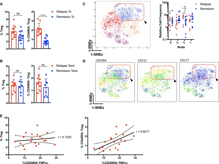
As Tregs have been previously implicated in JIA pathogenesis,11 13 19–21 we examined their role in patients with JIA prior/after therapy withdrawal (figure 4A,B; gating strategy in online supplementary figure S4). While no differential total Treg frequencies was detected, we observed higher levels of CD45RA−Treg (p<0.0001) in patients with JIA prior to relapse. We further verified this with t-SNE analysis of Tregs from relapse/remission patients prior to withdrawal and determined an enrichment for CD45RA−CD152+CD127−Tregs for relapse individuals (figure 4C,D). While no correlation (r=0.1532) with total Tregs was observed for CD45RA−TNFα+ cells (figure 4E), there was a positive correlation (r=0.6017) with CD45RA−Treg.
Figure 4.
Corresponding increase of memory Tregs and CD45RA−TNFα+ prior to relapse. Supervised gating of Tregs and CD45RA−Tregs in patients with JIA (A) prior (n=20) and (B) after withdrawal (n=16) of therapy. (C–D) Tregs cells from patients with JIA (n=20; relapse=9, remission=11) prior to withdrawal were analysed with t-SNE and (C) segregated into four nodes, with node 3 (p<0.05) enriched in relapse individuals. (D) Phenotype of node 3 is depicted (red-dotted box and arrowhead). (E) Correlation analysis of frequency of Tregs or CD45RA−Tregs with CD45RA−TNFα+. Comparison of cellular subsets performed with Mann-Whitney U, two-tailed test, means±SD. *p<0.05, ****p<0.0001. Correlation analysis performed with Pearson correlation, two-tailed test. ns, not significant; TNFα, tumour necrosis factor alpha; t-SNE, t-distributed stochastic neighbour embedding.
Transcriptomic divergence in disease-centric pathways that persists despite therapy
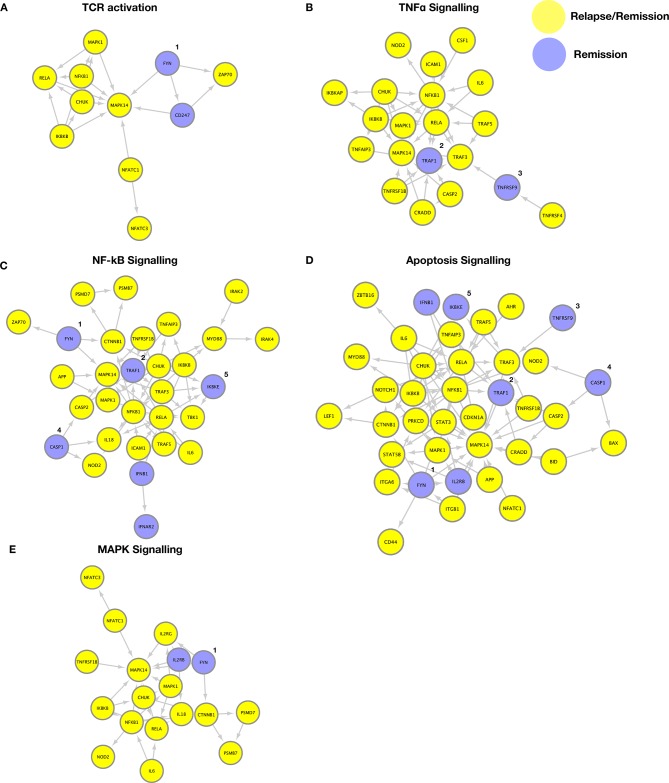
We have previously shown that patients with JIA who developed active disease on therapy withdrawal have stable epigenetic DNA CpG modifications in CD4 T cells that predisposed towards T-cell activation and TCR signalling.15 We wanted to investigate if CD4 memory T cells from JIA relapse/remission patients were differential in transcriptomic profile when their TCR is activated. We sorted for CD3+CD4+CD45RA−CD45RO+ T cells (online supplementary figure S5), stimulated 24 hours with anti-CD3/CD28, and profiled 579 immunological genes through NanoString. Functional gene enrichment analysis (DAVID) of JIA relapse (n=6) or remission (n=6) patients versus healthy individuals (n=3) identified five common disease-centric pathways that persisted from prior to after withdrawal of therapy (online supplementary figure S6 and tables S4 and S5). Dysregulation in UBE2L3, IL-6, STAT4, TYK2, TNFAIP3 and PTPN2 were found in both relapse and remission individuals, have been previously associated with JIA.22 23 We examined through Cytoscape and Reactome database for the gene associations involved in the five pathways (figure 5A–E): (1) TCR activation, (2) apoptosis, (3) TNFα, (4) nuclear factor-kappa B (NF-κB) and (5) mitogen-activated protein kinase (MAPK) signalling, and found a considerable overlap between relapse and remission individuals. This overlap of pathways in patients with relapse/remission JIA may arise from their shared susceptibility to clinical control with continued anti-TNFα therapy. Indeed, we observe a similar disease-centric pathway persistence in a separate cohort of patients with JIA (n=4) that are responsive to anti-TNFα therapy, from the point of pretreatment (active) until post-treatment (recent clinical inactivity) (online supplementary figures S7and S8 and table S3 and S6). Despite this overlap, we detected selective divergence within these pathways (figure 5A–E), with remission individuals expressing higher levels of FYN, TNFRSF9, CASP1, TRAF1 and IKBKE, which are involved in the termination or resolution of these pathways.24–33
Figure 5.
Transcriptomic divergence in disease-centric pathways that persist despite therapy. Genes enriched (p<0.05, fold difference >1.5) in patients with JIA (n=6 relapse or n=6 remission) that were persistent from prior to after therapy withdrawal, as compared with healthy individuals, were exported to David for functional gene-set enrichment, and gene associations were constructed with Cytoscape using the Reactome database. Five major pathways were dysregulated in relapse and remission patients with JIA compared with healthy controls: (A) TCR activation, (B) TNFα signalling, (C) NF-κB signalling, (D) apoptosis, (E) MAPK signalling (yellow=relapse/remission, blue=remission only). Genes enriched in remission individuals include 1, Fyn; 2, TRAF1; 3, TNFRSF9; 4, CASP1; and 5, IKBKE. MAPK, mitogen-activated protein kinase; NF-κB, nuclear factor-kappa B; TCR, T-cell receptor; TNFα, tumour necrosis factor alpha.
CD4+CD45RA−TNFα+ discriminates clinical fate prior to withdrawal of therapy
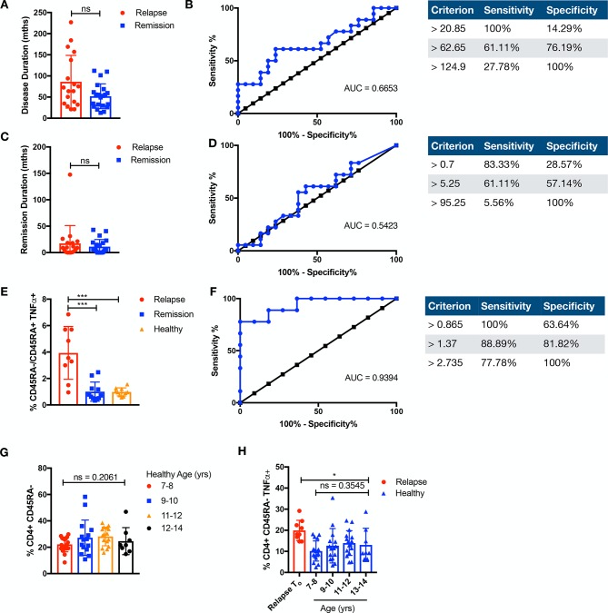
We explored whether disease or remission duration of patients with JIA (n=39) prior to study enrolment could differentiate clinical fate, and found no significant difference between patients with relapse JIA or patients with remission JIA (figure 6A–D). As we have shown that an inflammatory memory subset of CD4+CD45RA−TNFα+ is present in relapse individuals prior to therapy withdrawal, we tested whether this could afford for discrimination in clinical fate. There was a significant difference (p<0.001) in the ratio of CD45RA−TNFα+/CD45RA+TNFα+ cells in relapse as compared with remission individuals prior to therapy withdrawal, allowing for an ROC curve of area under the curve (AUC)=0.9394 (figure 6E,F). As age could be a serious potential cofounder for immunological memory, we determined that, in the relevant age groups (7–14 years), there was no significant difference in CD45RA− or CD45RA−TNFα+ cells among healthy individuals (n=56) (figure 6G,H). Relapse individuals had significantly higher CD45RA−TNFα+ cells as compared across all the relevant age groups in healthy individuals (figure 6H).
Figure 6.
CD4+CD45RA−TNFα+ discriminates clinical fate. Duration and ROC of (A,B) disease (months) or (C,D) remission (months) prior to study enrolment is compared among relapse (+=18) or remission (n=21) individuals. (E) The ratio of memory CD4+CD45RA−TNFα+ over naive CD4+CD45RA+TNFα+ of relapse or remission prior to withdrawal and healthy individuals is compared. Comparison of cellular subsets performed with Mann-Whitney U, two-tailed test, means±SD. *p<0.05. (F) ROC of ratio of CD4+CD45RA−TNFα+ over CD4+CD45RA+TNFα+ of relapse or remission prior to withdrawal. (G) CD3+CD4+CD45RA−CD45RO+ T cells were compared in healthy paediatric controls (n=56) across the relevant age groups of 7–8 years (n=17), 9–10 years (n=15), 11–12 years (n=16) and 12–14 years (n=8) or (H) against relapse (n=9) prior to therapy withdrawal for CD3+CD4+CD45RA−TNFα+. Comparison of cellular subsets performed with Kruskal-Wallis test, means±SD. AUC, area under the curve; ns, not significant; ROC, receiver operating characteristic; TNFα, tumour necrosis factor alpha.
Discussion
With a large proportion of patients with JIA achieving clinical inactivity as a result of efficacious treatment with anti-TNFα biologics,2 it becomes increasing pertinent to address the lack of definitive withdrawal guidelines. Here, we investigated with CyTOF the heterogenous CD4 landscape of patients who achieved clinical inactivity prior to therapy withdrawal. We have identified for the first time an inflammatory CD4 memory subset (CD3+CD4+CD45RA−TNFα+) that remains elevated in patients with JIA prior to relapse, and could notably discriminate clinical fate prior to therapy withdrawal (AUC=0.939).
Remarkably, the presence of this inflammatory subset, despite therapy, was associated with a deficit in immune checkpoint (PD1−CD152−). This is consistent with the phenomenon of rheumatic immune-related adverse events, where the application of immune checkpoint therapy (anti-PD1/anti-CD152) in cancer results in rheumatic diseases.34 The presence of this inflammatory subset was further verified when we separately compared relapse against healthy non-JIA individuals. The CD4+TNFα+ healthy landscape helped reveal the subclinical diversification of T-effector mechanisms in relapse individuals, with the emergence of CD3+CD4+CD45RA−TNFα+PD1−CD152− T cells that are IL-6+. Particularly, CD4+CD45RA− T cells that are IL-6+ express higher levels of TNFα. Recently, a case series of three patients with cancer and developed severe polyarthritis following immune blockade therapy, reported successful treatment with tocilizumab (anti-IL-6).35 This reflects a level of commonality between inflammatory and disease resolution mechanisms operating in autoimmune disorders and cancer.
We further examined a separate cohort of patients with JIA who developed flare or remained in remission 8 months into withdrawal of therapy, and verified the overt presence of T-effector diversification (CD45RA−TNFα+IL-6+) during flare. Whereas in patients who continued to remain in remission 8 months into withdrawal, there was no differential display of any CD45RA−TNFα+ subsets as compared with the healthy CD4 landscape, though a CD4+CXCR3+CCR6+ subset was detected. It remains speculative if the CD4+CXCR3+CCR6+ subset seen in remission patients is an early disease-driven subset or side effect of therapy as it extends beyond the scope of the study and will require a longer follow-up duration. In rheumatoid arthritis, the CD4+CXCR3+CCR6+ subset is known to express high levels of IFNγ with poor secretion of IL-17A,36 though we did not observe any associated cytokine profile in our remission patients.
In patients prior to relapse, the prevalence of the inflammatory CD4+CD45RA−TNFα+ T cells within the T-effector compartment is parallel by a corresponding increase in CD4+CD45RA−CD152+ Tregs in the regulatory arm. This suggests there is a compensatory regulatory response towards subclinical inflammation prior to relapse, though it remains to be seen whether the suppression suffices or is defective. Others have shown that synovium T effectors are resistant to Treg suppression,20 which can be alleviated in patients with JIA undergoing anti-TNFα therapy.37 Though it is noted that in a subset of patients, in vitro blockade of IL-6+ additionally alleviated T-effector resistance to Treg suppression.37
Transcriptomic profiling of immunological genes in response to TCR signalling reveals five common disease-centric pathways (TCR activation, apoptosis, TNFα, NF-κB and MAPK signalling) in relapse/remission patients as compared with healthy controls. Separately, the same dysregulated pathways were also detected in patients with JIA that are treatment naive and later responsive to anti-TNFα therapy. This suggests that the disease pathways affected are likely an indication of susceptibility to anti-TNFɑ treatment rather than an artefact of prolonged medication. As the afflicted pathways remain dysregulated prior and after therapy withdrawal in both relapse and remission patients, exogenous therapy by sequestration of TNFα may accomplish little to re-establish healthy physiological TCR response. Despite this, we detected selective divergence in certain genes (eg, TRAF1) with these pathways that may aid in termination or resolution of TCR-induced signalling for remission individuals. Notably, disease association within the TRAF1-C5 locus38–40 and epigenetic dysregulation within the TRAF1 locus of CD4 T cells have been detected in patients with JIA.41 Studies using TRAF1−/ − mice revealed a TRAF1 negative regulatory role in T-cell response to TCR and TNFα signalling.25 TRAF1 −/ − T cells show enhanced proliferation in response to TCR and TNFα stimulation resulting in NF-κB and AP-1 activation.25
In summary, by applying a combination of high-dimensionality technologies, we have identified functional perturbations of the immunome in patients with arthritis who will relapse on withdrawal for anti-TNFα therapy. These aberrations show that relapse of clinical disease relies on a foundation of complex and diverse interlacing immune mechanisms, which affect both the effector and regulatory arms of adaptive T-cell immunity. Our findings have an immediate translational valency. Indeed, there is a potential diagnostic advantage42 in tracking these dysregulated CD4 subsets to affect clinical management of therapy withdrawal. Also, the occurrence of T-effector diversification in relapse patients and insights into key divergence in disease-centric pathways for remission patients are mechanistically relevant as, altogether, they define a cluster of deranged mechanisms that can be target of focused intervention.
Footnotes
Handling editor: Josef S Smolen
Collaborators: PRCSGPediatric Rheumatology Collaborative Study Group (PRCSG): Daniel J. Lovell, Anne L. Johnson, Steven J. Spalding, Beth S. Gottlieb, Paula W. Morris, Yukiko Kimura, Karen Onel, Suzanne C. Li, Alexei A. Grom, Janalee Taylor, Hermine I. Brunner, Jennifer L. Huggins, James J. Nocton, Kathleen A. Haines, Barbara S. Edelheit, Michael Shishov, Lawrence K. Jung, Calvin B. Williams, Melissa S. Tesher, Denise M. Costanzo, Lawrence S. Zemel, Jason A. Dare, Murray H. Passo, Kaleo C. Ede, Judyann C. Olson, Elaine A. Cassidy, Thomas A. Griffin, Linda Wagner-Weiner, Jennifer E. Weiss, Larry B. Vogler, Kelly A. Rouster-Stevens, Timothy Beukelman, Randy Q. Cron, Daniel Kietz, Kenneth Schikler, Jay Mehta, Tracy V. Ting, James W. Verbsky, B. Anne Eberhard, Bin Huang, Chen Chen, Edward H. Giannini. Authors involved in the clinical trial are listed in the Acknowledgement and affiliations in the online supplementary. (The position of this study group in the author list is shown in the main document)
Contributors: JYL performed the mass cytometry (cytometry by time of flight
(CyTOF)), sorting and NanoString experiments and analysis. PC and SLP helped with the CyTOF and NanoString runs. FA, CC and SNH helped with mRNA processing and NanoString. LP helped write the R scripts for the CyTOF analysis. LL helped with the CyToF. LTB helped with sample processing. DL, JGY, ESCL, TA and PRSCG helped with patient recruitment. SA was the lead principal investigator.
Funding: Grant support from the National Medical Research Council (NMRC) (NMRC/STaR/020/2013, NMRC/MOHIAFCAT2/2/08, MOHIAFCAT2/0001/2014, NMRC MOHIAFCAT1-6003, centre grants, TCR15Jun006, NMRC/CIRG/1460/2016, MH 095:003\016-0002), Duke-NUS, A*STAR-BMRC (IAF311020), BMRC (SPF2014/005) is gratefully acknowledged.
Competing interests: None declared.
Patient and public involvement statement: This research was done without patient involvement. Patients were not invited to comment on the study design and were not consulted to develop patient relevant outcomes or interpret the results. Patients were not invited to contribute to the writing or editing of this document for readability or accuracy.
Patient consent for publication: Not required.
Ethics approval: All samples were collected upon informed consent. All experiments were conducted according to the principles expressed in the Declaration of Helsinki, and institutional review board approved by the Sanford-Burnham or KK Women’s and Children’s Hospital IRB Committee.
Provenance and peer review: Not commissioned; externally peer reviewed.
Data availability statement: Data are available upon reasonable request.
Contributor Information
Pediatric Rheumatology Collaborative Study Group:
Daniel J. Lovell, Anne L. Johnson, Steven J. Spalding, Beth S. Gottlieb, Paula W. Morris, Yukiko Kimura, Karen Onel, Suzanne C. Li, Alexei A. Grom, Janalee Taylor, Hermine I. Brunner, Jennifer L. Huggins, James J. Nocton, Kathleen A. Haines, Barbara S. Edelheit, Michael Shishov, Lawrence K. Jung, Calvin B. Williams, Melissa S. Tesher, Denise M. Costanzo, Lawrence S. Zemel, Jason A. Dare, Murray H. Passo, Kaleo C. Ede, Judyann C. Olson, Elaine A. Cassidy, Thomas A. Griffin, Linda Wagner-Weiner, Jennifer E. Weiss, Larry B. Vogler, Kelly A. Rouster-Stevens, Timothy Beukelman, Randy Q. Cron, Daniel Kietz, Kenneth Schikler, Jay Mehta, Tracy V. Ting, James W. Verbsky, B. Anne Eberhard, Bin Huang, Chen Chen, and Edward H. Giannini
References
- 1. Lovell DJ, Giannini EH, Reiff A, et al. Etanercept in children with polyarticular juvenile rheumatoid arthritis. pediatric rheumatology collaborative Study Group. N Engl J Med 2000;342:763–9. 10.1056/NEJM200003163421103 [DOI] [PubMed] [Google Scholar]
- 2. Taddio A, Cattalini M, Simonini G, et al. Recent advances in the use of Anti-TNFα therapy for the treatment of juvenile idiopathic arthritis. Expert Rev Clin Immunol 2016;12:641–9. 10.1586/1744666X.2016.1146132 [DOI] [PubMed] [Google Scholar]
- 3. Baszis K, Garbutt J, Toib D, et al. Clinical outcomes after withdrawal of anti-tumor necrosis factor α therapy in patients with juvenile idiopathic arthritis: a twelve-year experience. Arthritis Rheum 2011;63:3163–8. 10.1002/art.30502 [DOI] [PubMed] [Google Scholar]
- 4. Iglesias E, Torrente-Segarra V, Bou R, et al. Non-Systemic juvenile idiopathic arthritis outcome after reaching clinical remission with anti-TNF-α therapy: a clinical practice observational study of patients who discontinued treatment. Rheumatol Int 2014;34:1053–7. 10.1007/s00296-013-2884-z [DOI] [PubMed] [Google Scholar]
- 5. Wallace CA, Giannini EH, Huang B, et al. American College of rheumatology provisional criteria for defining clinical inactive disease in select categories of juvenile idiopathic arthritis. Arthritis Care Res 2011;63:929–36. 10.1002/acr.20497 [DOI] [PubMed] [Google Scholar]
- 6. Leong JY, Guan YJ, Albani S, et al. Recent advances in our understanding of the pathogenesis of juvenile idiopathic arthritis and their potential clinical implications. Expert Rev Clin Immunol 2018;14:933–44. 10.1080/1744666X.2018.1529757 [DOI] [PubMed] [Google Scholar]
- 7. Eyre S, Orozco G, Worthington J. The genetics revolution in rheumatology: large scale genomic arrays and genetic mapping. Nat Rev Rheumatol 2017;13:421–32. 10.1038/nrrheum.2017.80 [DOI] [PubMed] [Google Scholar]
- 8. Hunter PJ, Nistala K, Jina N, et al. Biologic predictors of extension of oligoarticular juvenile idiopathic arthritis as determined from synovial fluid cellular composition and gene expression. Arthritis Rheum 2010;62:896–907. 10.1002/art.27284 [DOI] [PMC free article] [PubMed] [Google Scholar]
- 9. Cosmi L, Cimaz R, Maggi L, et al. Evidence of the transient nature of the Th17 phenotype of CD4+CD161+ T cells in the synovial fluid of patients with juvenile idiopathic arthritis. Arthritis Rheum 2011;63:2504–15. 10.1002/art.30332 [DOI] [PubMed] [Google Scholar]
- 10. Issekutz AC, Quinn PJ, Lang B, et al. Coexpression of chemokine receptors CCR5, CXCR3, and CCR4 and ligands for P- and E-selectin on T lymphocytes of patients with juvenile idiopathic arthritis. Arthritis Rheum 2011;63:3467–76. 10.1002/art.30521 [DOI] [PubMed] [Google Scholar]
- 11. Prakken B, Albani S, Martini A. Juvenile idiopathic arthritis. Lancet 2011;377:2138–49. 10.1016/S0140-6736(11)60244-4 [DOI] [PubMed] [Google Scholar]
- 12. Spreafico R, Rossetti M, van Loosdregt J, et al. A circulating reservoir of pathogenic-like CD4+ T cells shares a genetic and phenotypic signature with the inflamed synovial micro-environment. Ann Rheum Dis 2016;75:459–65. 10.1136/annrheumdis-2014-206226 [DOI] [PMC free article] [PubMed] [Google Scholar]
- 13. Rossetti M, Spreafico R, Consolaro A, et al. Tcr repertoire sequencing identifies synovial Treg cell clonotypes in the bloodstream during active inflammation in human arthritis. Ann Rheum Dis 2017;76:435–41. 10.1136/annrheumdis-2015-208992 [DOI] [PMC free article] [PubMed] [Google Scholar]
- 14. Jiang K, Zhu L, Buck MJ, et al. Disease-Associated single-nucleotide polymorphisms from noncoding regions in juvenile idiopathic arthritis are located within or adjacent to functional genomic elements of human neutrophils and CD4+ T cells. Arthritis Rheumatol 2015;67:1966–77. 10.1002/art.39135 [DOI] [PMC free article] [PubMed] [Google Scholar]
- 15. Spreafico R, Rossetti M, Whitaker JW, et al. Epipolymorphisms associated with the clinical outcome of autoimmune arthritis affect CD4+ T cell activation pathways. Proc Natl Acad Sci U S A 2016;113:13845–50. 10.1073/pnas.1524056113 [DOI] [PMC free article] [PubMed] [Google Scholar]
- 16. Determining predictors of safe discontinuation of anti-TNF treatment in JIA. Available: https://clinicaltrials.gov/show/NCT00792233
- 17. Brunner HI, Lovell DJ, Finck BK, et al. Preliminary definition of disease flare in juvenile rheumatoid arthritis. J Rheumatol 2002;29:1058–64. [PubMed] [Google Scholar]
- 18. Lai L, Ong R, Li J, et al. A CD45-based barcoding approach to multiplex mass-cytometry (CyTOF). Cytometry A 2015;87:369–74. 10.1002/cyto.a.22640 [DOI] [PMC free article] [PubMed] [Google Scholar]
- 19. Collison J. Paediatric rheumatology: novel Treg cell subset discovered in JIA. Nat Rev Rheumatol 2016;12:436–7. 10.1038/nrrheum.2016.115 [DOI] [PubMed] [Google Scholar]
- 20. Haufe S, Haug M, Schepp C, et al. Impaired suppression of synovial fluid CD4+CD25- T cells from patients with juvenile idiopathic arthritis by CD4+CD25+ Treg cells. Arthritis Rheum 2011;63:3153–62. 10.1002/art.30503 [DOI] [PubMed] [Google Scholar]
- 21. Henderson LA, Volpi S, Frugoni F, et al. Next-Generation sequencing reveals restriction and clonotypic expansion of Treg cells in juvenile idiopathic arthritis. Arthritis Rheumatol 2016;68:1758–68. 10.1002/art.39606 [DOI] [PMC free article] [PubMed] [Google Scholar]
- 22. Hinks A, Cobb J, Marion MC, et al. Dense genotyping of immune-related disease regions identifies 14 new susceptibility loci for juvenile idiopathic arthritis. Nat Genet 2013;45:664–9. 10.1038/ng.2614 [DOI] [PMC free article] [PubMed] [Google Scholar]
- 23. Hersh AO, Prahalad S. Immunogenetics of juvenile idiopathic arthritis: a comprehensive review. J Autoimmun 2015;64:113–24. 10.1016/j.jaut.2015.08.002 [DOI] [PMC free article] [PubMed] [Google Scholar]
- 24. Ebata T, Mogi S, Hata Y, et al. Rapid induction of CD95 ligand and CD4+ T cell-mediated apoptosis by CD137 (4-1BB) costimulation. Eur J Immunol 2001;31:1410–6. [DOI] [PubMed] [Google Scholar]
- 25. Tsitsikov EN, Laouini D, Dunn IF, et al. Traf1 is a negative regulator of TNF signaling. enhanced TNF signaling in TRAF1-deficient mice. Immunity 2001;15:647–57. [DOI] [PubMed] [Google Scholar]
- 26. Filby A, Seddon B, Kleczkowska J, et al. Fyn regulates the duration of TCR engagement needed for commitment to effector function. J Immunol 2007;179:4635–44. 10.4049/jimmunol.179.7.4635 [DOI] [PubMed] [Google Scholar]
- 27. Salmond RJ, Filby A, Qureshi I, et al. T-Cell receptor proximal signaling via the Src-family kinases, Lck and Fyn, influences T-cell activation, differentiation, and tolerance. Immunol Rev 2009;228:9–22. 10.1111/j.1600-065X.2008.00745.x [DOI] [PubMed] [Google Scholar]
- 28. Harr MW, McColl KS, Zhong F, et al. Glucocorticoids downregulate Fyn and inhibit IP(3)-mediated calcium signaling to promote autophagy in T lymphocytes. Autophagy 2010;6:912–21. 10.4161/auto.6.7.13290 [DOI] [PMC free article] [PubMed] [Google Scholar]
- 29. Nagila A, Netsawang J, Suttitheptumrong A, et al. Inhibition of p38MAPK and CD137 signaling reduce dengue virus-induced TNF-α secretion and apoptosis. Virol J 2013;10:105 10.1186/1743-422X-10-105 [DOI] [PMC free article] [PubMed] [Google Scholar]
- 30. Doitsh G, Galloway NLK, Geng X, et al. Cell death by pyroptosis drives CD4 T-cell depletion in HIV-1 infection. Nature 2014;505:509–14. 10.1038/nature12940 [DOI] [PMC free article] [PubMed] [Google Scholar]
- 31. Nishimoto T, Seta N, Anan R, et al. A single nucleotide polymorphism of TRAF1 predicts the clinical response to anti-TNF treatment in Japanese patients with rheumatoid arthritis. Clin Exp Rheumatol 2014;32:211–7. [PubMed] [Google Scholar]
- 32. Lee D-J, Du F, Chen S-W, et al. Regulation and function of the caspase-1 in an inflammatory microenvironment. J Invest Dermatol 2015;135:2012–20. 10.1038/jid.2015.119 [DOI] [PMC free article] [PubMed] [Google Scholar]
- 33. Zhang J, Feng H, Zhao J, et al. Iκb kinase ε is an NFATc1 kinase that inhibits T cell immune response. Cell Rep 2016;16:405–18. 10.1016/j.celrep.2016.05.083 [DOI] [PMC free article] [PubMed] [Google Scholar]
- 34. Calabrese L, Mariette X. The evolving role of the rheumatologist in the management of immune-related adverse events (irAEs) caused by cancer immunotherapy. Ann Rheum Dis 2018;77:162–4. 10.1136/annrheumdis-2017-212061 [DOI] [PubMed] [Google Scholar]
- 35. Kim ST, Tayar J, Trinh VA, et al. Successful treatment of arthritis induced by checkpoint inhibitors with tocilizumab: a case series. Ann Rheum Dis 2017;76:2061–4. 10.1136/annrheumdis-2017-211560 [DOI] [PubMed] [Google Scholar]
- 36. Mellado M, Martínez-Muñoz L, Cascio G, et al. T cell migration in rheumatoid arthritis. Front Immunol 2015;6:384 10.3389/fimmu.2015.00384 [DOI] [PMC free article] [PubMed] [Google Scholar]
- 37. Wehrens EJ, Vastert SJ, Mijnheer G, et al. Anti-Tumor necrosis factor α targets protein kinase B/c-Akt-induced resistance of effector cells to suppression in juvenile idiopathic arthritis. Arthritis Rheum 2013;65:3279–84. 10.1002/art.38132 [DOI] [PubMed] [Google Scholar]
- 38. Albers HM, Kurreeman FAS, Houwing-Duistermaat JJ, et al. The TRAF1/C5 region is a risk factor for polyarthritis in juvenile idiopathic arthritis. Ann Rheum Dis 2008;67:1578–80. 10.1136/ard.2008.089060 [DOI] [PubMed] [Google Scholar]
- 39. Behrens EM, Finkel TH, Bradfield JP, et al. Association of the TRAF1-C5 locus on chromosome 9 with juvenile idiopathic arthritis. Arthritis Rheum 2008;58:2206–7. 10.1002/art.23603 [DOI] [PubMed] [Google Scholar]
- 40. Pers Y-M, Le Blay P, Ludwig C, et al. Association of TRAF1-C5 with risk of uveitis in juvenile idiopathic arthritis. Joint Bone Spine 2017;84:305–8. 10.1016/j.jbspin.2016.04.014 [DOI] [PubMed] [Google Scholar]
- 41. Zhu L, Jiang K, Webber K, et al. Chromatin landscapes and genetic risk for juvenile idiopathic arthritis. Arthritis Res Ther 2017;19 10.1186/s13075-017-1260-x [DOI] [PMC free article] [PubMed] [Google Scholar]
- 42. Leong JY, Albani S. Using a theragnostic approach in juvenile idiopathic arthritis. Expert Opin Orphan Drugs 2016;4:269–80. 10.1517/21678707.2016.1129944 [DOI] [Google Scholar]
Associated Data
This section collects any data citations, data availability statements, or supplementary materials included in this article.
Supplementary Materials
annrheumdis-2019-216059supp002.pdf (244.9KB, pdf)
annrheumdis-2019-216059supp003.pdf (103.8KB, pdf)
annrheumdis-2019-216059supp001.pdf (26.6MB, pdf)


