Abstract
Background: Closed reduction and percutaneous pinning (CRPP) is traditionally performed following full surgical prep and draping. The semisterile technique utilizes minimal prep and draping, which was proven to be a viable alternative when treating pediatric supracondylar humerus fractures. The purpose of this study was to investigate the safety and benefits of the semisterile technique for CRPP of pediatric upper extremity fractures. Methods: A retrospective cohort study was conducted of pediatric patients who underwent CRPP of an upper extremity fracture over a 4-year period. Demographic data, fracture type/location, and the type of prep technique (full-prep vs semisterile) were recorded. Qualities of intraoperative care were assessed, and postoperative care parameters were compared. Patient outcomes for the 2 techniques were compared using bivariate analyses. Results: In total, 219 patient records were reviewed including 160 in the semisterile group and 59 in the full-prep group. When comparing intraoperative parameters between the full-prep and semisterile techniques, the average room setup time was similar (20.6 vs 18.8 minutes, P = .52). However, the procedure times (32.1 vs 26.9 minutes, P = .04) were significantly shorter in the semisterile group. Nearly a 10-minute decrease in total time in the operating room was present while utilizing the semisterile technique (62.8 vs 53.6 minutes, P < .01). There were no statistical differences in complication rates between prep groups (P = .31), and there were no infections while utilizing the semisterile technique. Conclusions: The semisterile technique is a safe and efficient alternative that may be used when performing CRPP of pediatric upper extremity fractures.
Keywords: pediatric, upper extremity, fracture, closed reduction and percutaneous pinning (CRPP), semisterile technique
Introduction
Upper extremity fractures are commonly treated injuries in the pediatric and adolescent population. Closed reduction and percutaneous pinning (CRPP) is the most commonly performed operative treatment for displaced pediatric upper extremity fractures. The procedure is traditionally performed following a full sterile prep and draping of the upper extremity. Furthermore, the surgeon(s) and scrub nurse/technician maintain sterility by wearing a sterile gown and gloves while performing the procedure. However, the utilization of these practices is often unnecessary, as it increases medical waste and health care costs without providing a clear advantage in lowering patient morbidity. The benefits of the semisterile technique are alike to wide-awake hand surgery, which has been proven to decrease operative-related health care costs.7,13
The concept of using a semisterile technique when performing a CRPP procedure was first introduced by Iobst and colleagues for the treatment of pediatric supracondylar humerus fractures.6 The authors only prepped the extremity at the site of pin placement using Betadine solution and created a sterile operative field using only sterile towels. The surgeon(s) wore sterile gloves, but no sterile gowns or drapes were used during the procedure. In their cohort of 304 patients, there were no pin site or deep site infections using this semisterile technique.6 Bashyal et al retrospectively compared the complication profile of treating pediatric supracondylar humerus fractures using a full-prep or various semisterile prep techniques and determined there was no statistically significant correlation between the type of skin preparation and infection rate.1
Through observation and discussion with colleagues, we believe the semisterile technique has been adopted by many surgeons and institutions when performing CRPP procedures, but it remains uncertain how widespread this is and whether or not it is utilized for all upper extremity CRPP procedures. In addition, to our knowledge there are no comparative studies assessing the complications and treatment outcomes of CRPP using the semisterile and full-prep techniques for these fractures.
The purpose of the current study was to investigate the safety and benefits of the semisterile technique for CRPP of all pediatric and adolescent upper extremity fractures.
Materials and Methods
A retrospective cohort study was conducted of all pediatric patients who underwent CRPP of an upper extremity fracture over a 4-year period (2012-2015) at a single academic institution after obtaining institutional review board approval. During this time period, there was a transition by the senior author (J.M.A.) from utilizing a traditional full-prep and drape for all pediatric upper extremity CRPP procedures to the use of the semisterile technique for all CRPP procedures being performed to treat pediatric upper extremity fractures.
Patient demographics including age, sex, and race were recorded. Injury characteristics, including fracture type and location, mechanism of injury, associated injuries, preoperative neurovascular status, and time to procedure, were documented. Operative reports were assessed using the electronic medical record to determine intraoperative parameters including the type of prep technique utilized (full-prep vs semisterile), the time required to prep the operative site, the room setup time, the anesthesia time, the procedure time, and the operating room turnover/clean time. The times entered were documented into the operative record by the circulating nurse. Complications of the injury and/or procedure were recorded including infection rate, neurovascular injury, pin migration, loss of reduction, decreased range of motion, and need for reoperation. Postoperative care measures were compared including length of follow-up and time to pin removal.
CRPP Utilizing the Semisterile Technique
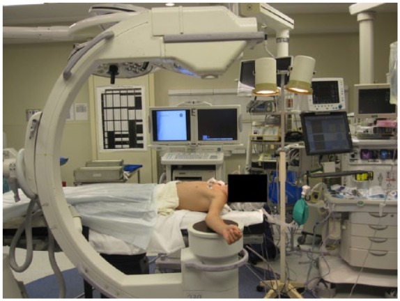
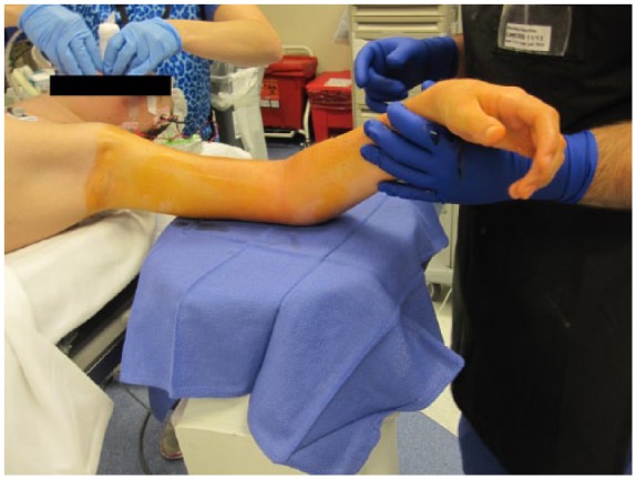
The semisterile technique performed during the study period included a prep utilizing a single chlorhexidine paint brush to locally sterilize the operative site and placement of the “prepped” area onto a sterile towel (see online Supplemental Video). Typically, an inverted fluoroscopy unit or mini C-arm was utilized as the operating surface (Figure 1), and therefore the sterile towel was placed on these locations. The surgeon(s) and scrub nurse/technician only donned sterile gloves (Figure 2). The fracture was then reduced under direct visualization and fluoroscopic guidance, and subsequently percutaneous pinning was performed using Kirschner wire (K-wire) fixation (see online Supplemental Figure). Sterile dressings were then applied followed by splint or cast immobilization.
Figure 1.

Intraoperative photograph of the operating room setup for a closed reduction and percutaneous pinning procedure in which an inverted fluoroscopy unit is utilized as the operating surface.
Figure 2.

Intraoperative photograph of an upper extremity prepped using the semisterile technique. Note that the operating field created uses only a sterile towel and only the surgeon’s gloves are sterile.
Statistical Analysis
Baseline patient characteristics were described using means and standard deviations for continuous variables and frequencies and proportions for categorical variables. Bivariate analyses were conducted to compare the full-prep versus the semisterile technique for all variables of interest. Pearson chi-square tests were used to compare patient gender and race by treatment group. Fisher exact tests were used to compare clinical outcomes by treatment group. Student t tests or Wilcoxon rank sum tests were used to compare for continuous data, depending on the distribution of the data. All tests were 2-tailed and a P value of <.05 was considered significant. All analyses were performed using JMP Pro Version 12 (Cary, North Carolina).
Results
In total, 219 patients were identified who underwent CRPP of an upper extremity fracture, with a total of 222 upper extremity fractures treated. One hundred sixty patients were treated using the semisterile technique and 59 using the full-prep technique. During the study period, there was a gradual transition from utilizing the full-prep and drape technique for all CRPP procedures of the upper extremity to the semisterile technique for all CRPP procedures of the upper extremity.
Patient demographics are summarized in Table 1. The average age of the cohort treated with the full-prep technique was 8.6 years (SD = 4.3) versus 7.9 years (SD = 4.3) treated with the semisterile technique (P = .26). About 61% of the patients were male, and 46% were Caucasian. Patients treated using the full-prep technique were on average 8.5 days from injury compared with 6.2 days with the semisterile technique (P = .04).
Table 1.
Baseline Characteristics of Included Patients (n = 219).
| Variable | Full-prep (n = 59) |
Semisterile (n = 160) |
P value |
|---|---|---|---|
| Age, mean (SD) | 8.6 (4.3) | 7.9 (4.3) | .26 |
| Male, n (%) | 37 (62.7) | 96 (60.0) | .71 |
| Race, n (%) | |||
| White | 22 (37.3) | 78 (48.8) | .51 |
| Black | 23 (39.0) | 50 (31.3) | |
| Other | 12 (20.3) | 27 (17.1) | |
| Unknown | 2 (3.4) | 5 (3.1) | |
| Time to procedure (days), mean (SD) | 8.5 (6.9) | 6.2 (7.5) | .04 |
The types and locations of fractures treated with CRPP prepped with either technique are summarized in Table 2. The most common fractures involved the humerus (45.9%), distal radius (24.3%), and phalanges (22.1%). The preoperative neurovascular status of the cohort is summarized in Table 3. Overall, 3 patients had a pulseless extremity and 14 (6.4%) of the patients had a nerve palsy, most commonly affecting the radial nerve (n = 7). There was no statistical difference in the preoperative neurovascular status between the full-prep and semisterile groups (Table 3).
Table 2.
Fracture Type and Location.
| Fracture type and location | Total | Full-prep | Semisterile |
|---|---|---|---|
| Total | 222 | 59 | 163 |
| Humerus | 102 | 23 | 79 |
| Proximal | 2 | 1 | 1 |
| Distal (transphyseal/SH II) | 3 | 2 | 1 |
| Supracondylar–Type II | 56 | 11 | 45 |
| Supracondylar–Type III | 34 | 6 | 28 |
| Lateral condyle | 7 | 3 | 4 |
| Radius | 54 | 12 | 42 |
| Distal | 54 | 12 | 42 |
| Ulna | 1 | — | 1 |
| Distal | 1 | — | 1 |
| Metacarpal | 16 | 3 | 13 |
| Neck | 11 | 2 | 9 |
| Shaft | 5 | 1 | 4 |
| Phalanges | 49 | 21 | 28 |
| P1 | 41 | 18 | 23 |
| Intra-articular | 4 | 2 | 2 |
| Neck | 11 | 6 | 5 |
| Shaft | 5 | 3 | 2 |
| SH II | 20 | 7 | 13 |
| SH III | 1 | − | 1 |
| P2 | 5 | 2 | 3 |
| Neck | 4 | 1 | 3 |
| SH I | 1 | 1 | − |
| P3 | 3 | 1 | 2 |
| Bony Mallet | 2 | — | 2 |
| SH II | 1 | 1 | — |
Note. SH = Salter-Harris.
Table 3.
Preoperative Neurovascular Status.
| Nerve | Total | Full-prep | Semisterile | P-value |
|---|---|---|---|---|
| Nerve palsy | 14 | 2 (3.4) | 12 (7.5) | .36 |
| Radial nerve | 7 | 1 (1.7) | 6 (3.8) | .68 |
| Anterior interosseous nerve | 4 | — | 4 (2.5) | .58 |
| Median nerve | 2 | 1 (1.7) | 1 (0.6) | .47 |
| Ulnar nerve | 1 | — | 1 (0.6) | 1.00 |
The average time in the operating room utilizing the full-prep technique was 62.8 minutes (SD = 22.0) while the average time utilizing the semisterile technique was 53.6 minutes (SD = 18.4) (P < .01). The average room setup time was similar between the two treatment groups (full-prep: 20.6 vs semisterile: 18.8 minutes, P = .52) (Table 4). There was no statistical difference in the average surgical site preparation time between the full-prep (5.5 minutes) and semisterile prep group (5.1 minutes) (P = .80). However, the semisterile technique led to a 14% reduction in anesthesia time, 16% reduction in operating time, and 16% reduction in overall patient in room time compared with the full-prep technique (Table 4).
Table 4.
Duration of Setup, Operation, and Clean Times (Minutes).
| Full-prep |
Semisterile |
P value | |||
|---|---|---|---|---|---|
| n | Mean (SD) | n | Mean (SD) | ||
| Room setup time | 24 | 20.6 (11.6) | 78 | 18.8 (8.6) | .52 |
| Preparation time | 15 | 5.5 (4.6) | 52 | 5.1 (4.6) | .80 |
| Anesthesia time | 55 | 61.9 (23.0) | 151 | 53.4 (17.7) | .02 |
| Operating time | 54 | 32.1 (16.5) | 150 | 26.9 (12.2) | .04 |
| Patient time in room | 55 | 62.8 (22.0) | 150 | 53.6 (18.4) | <.01 |
| Clean time | 21 | 19.0 (12.4) | 56 | 17.0 (13.6) | .56 |
Note. The values in bold are statistically significant.
There was no statistical difference in the total complication rate between the full-prep group (8.5%) and semisterile group (4.4%) (P = .31) (Table 5). There was one pin site infection in the full-prep group and zero in the semisterile group. The average days to pin removal in the full-prep and semisterile prep groups was 27.5 days (SD = 5.2) and 27.5 days (SD = 8.3), respectively (P = .95). Patients were followed up for a median of 48 days (interquartile range [IQR]: 31-70) post injury. This included a median of 21 days (IQR: 0-42) follow-up after the pins were removed. There was no significant difference in follow-up time between the treatment groups.
Table 5.
Postoperative Data.
| Postoperative outcome | Full-prep | Semisterile | P value |
|---|---|---|---|
| Complications, n (%) | 5 (8.5) | 7 (4.4) | .31 |
| Infection | 1 (1.7) | 0 | .27 |
| Physeal arrest | 1 (1.7) | 0 | .27 |
| Drainage/granulation | 2 (3.4) | 3 (1.9) | .61 |
| Loss of flexion | 1 (1.7) | 2 (1.3) | 1.00 |
| Pin migration | 0 | 2 (1.3) | 1.00 |
| Days to pin removal, mean (SD) | 27.5 (5.2) | 27.5 (8.3) | .95 |
Discussion
In this retrospective study, the authors sought to assess the safety and benefits of using the semisterile technique in the treatment of pediatric upper extremity fractures as compared with a full-prep and drape. A total of 222 fractures, in 219 patients, underwent closed reduced and percutaneous pinning using either a full-prep and drape or the semisterile prep technique. Overall, there was a 16% decrease in total perioperative time when the semisterile technique was utilized. There were no significant differences in complication rates between treatment groups, and the infection rate was 0% when using the semisterile technique.
K-wire fixation theoretically presents an increased risk of infection, as an external source is percutaneously introduced into a sterile environment.14 The infection rate after CRPP of pediatric extremity fractures has been variably reported. Tosti and colleagues retrospectively reviewed patients who underwent CRPP over a 16-year period and reported an overall infection rate of 1.6% (12/884).14 The most implicated pathogens included Staphylococcus, Pseudomonas, and Streptococcus species leading to cellulitis, abscess formation, and osteomyelitis.14 Battle and Carmichael reported an overall infection rate of 7.9% with smooth K-wire fixation in pediatric patients, but the overall major infection rate was 2% with these cases necessitating irrigation and debridement in the operating room.2
Infection rates following CRPP have also been investigated in the setting of surgical delays8 and the use of postoperative antibiotics11 when treating supracondylar humerus fractures. Larson et al retrospectively reviewed Gartland type II supracondylar humerus fractures treated within 24 hours or >24 hours from the time of injury.8 There was no difference in the complication rate in patients treated with or without surgical delay, and the overall infection rate was 1.5%. Schroeder and colleagues’ retrospective study evaluated the effect of postoperative antibiotics in preventing surgical site infections when treating supracondylar humerus fractures with CRPP.11 The authors reported an overall infection rate of 1.8%, and patients who received postoperative antibiotics did not have a lower rate of surgical site infections (P = .883).11
Previous studies have provided outcome and complication data for CRPP procedures of upper extremity fractures when preparing the limb using the full-prep technique.2,4,5,8,10-12,14,15 The semisterile technique, as first described by Iobst et al in the treatment of supracondylar humerus fractures, is a cost-effective substitute to the full-prep and drape technique that does not increase the rate of infection and reduces medical waste.6 In our study, there was a 0% infection rate (0/163) using the semisterile technique to sterilize the upper extremity. Furthermore, the current study demonstrates the safety beyond that of just utilizing the technique for CRPP of supracondylar humerus fractures as previously reported.1,6 Through discussion with colleagues, we believe the semisterile technique has been utilized by many surgeons to sterilize the upper extremity for CRPP procedures, but there is limited data assessing its benefits. Furthermore, to our knowledge there is no data assessing its use in fractures other than pediatric supracondylar humerus fractures. The current study documented a 0% infection rate using the semisterile technique for all pediatric upper extremity fractures. Therefore, we recommend that surgeons consider performing CRPP of upper extremity fractures in the pediatric and adolescent population utilizing the semisterile technique.
Continued utilization of the full-prep technique increases the economic burden on the health care system, as the technique requires a scrub prep tray, surgical drapes, and sterile gowns. The monetary costs to prepare and dispose of this equipment increases the overall cost of health care delivery and substantially increases the need to dispose of additional medical waste. It is difficult to estimate the exact positive financial impact the semisterile technique can have on the treatment of pediatric upper extremity fractures, as this study was limited to a single-center investigation. However, the obvious benefit to the “Earth” is present as there was a decreased need to dispose of additional medical waste. The benefits of the semisterile technique are alike to wide-awake hand surgery.7 Theil and colleagues created a “minimal” custom pack of disposable surgical supplies and prospectively compared 178 patients undergoing wide-awake hand surgery.13 The study determined there was an overall 13% decrease in medical waste, and 55% decrease ($125) in costs when compared with standard pack designs.13 The proposed benefits of the semisterile technique are just as profound, as there is no formal draping required, and using a multicenter prospective investigation, the decrease in health related costs can be readily identified in a prospective setting.
Furthermore, the current study noted an approximately 10-minute decrease in the total time in the operating room utilizing the semisterile technique as compared with the full-prep and drape technique. Although the authors cannot put an exact monetary figure on this decreased time, the potential for substantial benefits to the health care system is apparent given how common CRPP type procedures are. An additional operating room case may be able to be placed in a particular operating room due to the time saved utilizing this technique.
The clinical sequelae of K-wire infections can range from local cellulitis and purulent infections to more devastating consequences such as septic arthritis and osteomyelitis,3,9,14 which might prevent surgeons from uniformly adopting the semisterile technique. However, the current study demonstrates the safety and benefits of the semisterile technique without increasing patient morbidity.
Several limitations exist in this study. Given the nature of the retrospective review, reported outcomes were dependent on the quality of documentation. Intraoperative documentation by the operating room staff was not always consistent when recording all of the intraoperative parameters assessed in this study as there were no prior definitions noted to differentiate between the variables assessed such as cleaning time, preparation time, and operative time. The circulating nurse was responsible for inputting the data into the electronic medical record; therefore, it is unknown how much variation exists between nurses when documenting when the prep or procedure time begins and ends. For example, does the procedure begin at the start of prepping of the limb, at the time of the first closed reduction maneuver, or at the time of K-wire placement? The average surgical site preparation time was 5.5 minutes in the full-prep group and 5.1 minutes in the semisterile group (P = .80). The prep time was expected to be much longer in the full-prep group as it requires a thorough scrubbing of the extremity followed by placement of the drapes once the prep has dried. However, the data only demonstrated a small difference in the prep time, which can most likely be attributed to the inconsistent documentation. Therefore, the average total time in the operating room might be the most representative for both treatment groups. Nearly a 10-minute decrease was present utilizing the semisterile technique compared with the full-prep technique. However, it is possible that surgeon technique improved or other environmental factors changed over the study period, accounting for the difference in total perioperative time. Furthermore, a decrease of 10 minutes may not permit an institution to add an additional case to the schedule; therefore, the 10-minute decrease would only be beneficial if enough semisterile procedures were performed on a given day to permit an additional case to be added to the operating room schedule.
The authors are unable to comment on the reproducibility to other centers as this study was performed at a single institution. Last, it is important to recognize that there may have been a selection bias for the full-prep technique in some cases later in the cohort if there was concern preoperatively that closed reduction techniques would fail thus necessitating an open reduction and internal fixation. Therefore, even though all of these fractures were treated using CRPP, the intraoperative times might have been longer due to increased difficulty reducing the fracture and achieving fracture stability.
Future studies should aim to prospectively evaluate the semisterile technique when treating upper and lower extremity fractures in the pediatric and adolescent population. Furthermore, these studies should try to gauge the economic and opportunity costs of performing these procedures utilizing either sterilization technique.
The semisterile technique is an alternative to a full-prep and drape that can be used for CRPP procedures of all pediatric and adolescent upper extremity fractures. The use of the semisterile technique for pediatric upper extremity CRPP procedures may lower health care costs and medical waste without increasing patient morbidity, but future studies are needed to determine this.
Supplemental Material
Supplemental material, DS_10.1177_1558944718787310 for The Safety and Benefits of the Semisterile Technique for Closed Reduction and Percutaneous Pinning of Pediatric Upper Extremity Fractures by Karan Dua, Charles J. Blevins, Nathan N. O’Hara and Joshua M. Abzug in HAND
Footnotes
Supplemental material is available in the online version of the article.
Authors’ Note: JMA is the primary investigator.
Ethical Approval: This study was approved by our institutional review board.
Statement of Human and Animal Rights: This article does not contain any studies with human or animal subjects.
Statement of Informed Consent: Informed consent was obtained when necessary.
Declaration of Conflicting Interests: The author(s) declared no potential conflicts of interest with respect to the research, authorship, and/or publication of this article.
Funding: The author(s) received no financial support for the research, authorship, and/or publication of this article.
References
- 1. Bashyal RK, Chu JY, Schoenecker PL, et al. Complications after pinning of supracondylar distal humerus fractures. J Pediatr Orthop. 2009;29:704-708. [DOI] [PubMed] [Google Scholar]
- 2. Battle J, Carmichael KD. Incidence of pin track infections in children’s fractures treated with Kirschner wire fixation. J Pediatr Orthop. 2007;27:154-157. [DOI] [PubMed] [Google Scholar]
- 3. Botte MJ, Davis JL, Rose BA, et al. Complications of smooth pin fixation of fractures and dislocations in the hand and wrist. Clin Orthop Relat Res. 1992(276):194-201. [PubMed] [Google Scholar]
- 4. Boyer JS, London DA, Stepan JG, et al. Pediatric proximal phalanx fractures: outcomes and complications after the surgical treatment of displaced fractures. J Pediatr Orthop. 2015;35:219-223. [DOI] [PMC free article] [PubMed] [Google Scholar]
- 5. de Haseth KB, Neuhaus V, Mudgal CS. Dorsal fracture-dislocations of the proximal interphalangeal joint: evaluation of closed reduction and percutaneous Kirschner wire pinning. Hand (N Y). 2015;10:88-93. [DOI] [PMC free article] [PubMed] [Google Scholar]
- 6. Iobst CA, Spurdle C, King WF, et al. Percutaneous pinning of pediatric supracondylar humerus fractures with the semisterile technique: the Miami experience. J Pediatr Orthop. 2007;27:17-22. [DOI] [PubMed] [Google Scholar]
- 7. Lalonde DH. Conceptual origins, current practice, and views of wide awake hand surgery. J Hand Surg Eur Vol. 2017;42(9):885-895. [DOI] [PubMed] [Google Scholar]
- 8. Larson AN, Garg S, Weller A, et al. Operative treatment of type II supracondylar humerus fractures: does time to surgery affect complications? J Pediatr Orthop. 2014;34:382-387. [DOI] [PubMed] [Google Scholar]
- 9. Maraqa NF, Gomez MM, Rathore MH. Outpatient parenteral antimicrobial therapy in osteoarticular infections in children. J Pediatr Orthop. 2002;22:506-510. [PubMed] [Google Scholar]
- 10. Ponce BA, Hedequist DJ, Zurakowski D, et al. Complications and timing of follow-up after closed reduction and percutaneous pinning of supracondylar humerus fractures: follow-up after percutaneous pinning of supracondylar humerus fractures. J Pediatr Orthop. 2004;24:610-614. [DOI] [PubMed] [Google Scholar]
- 11. Schroeder NO, Seeley MA, Hariharan A, et al. Utility of postoperative antibiotics after percutaneous pinning of pediatric supracondylar humerus fractures. J Pediatr Orthop. 2017;37:363-367. [DOI] [PubMed] [Google Scholar]
- 12. Skaggs DL, Sankar WN, Albrektson J, et al. How safe is the operative treatment of Gartland type 2 supracondylar humerus fractures in children? J Pediatr Orthop. 2008;28:139-141. [DOI] [PubMed] [Google Scholar]
- 13. Theil CL, Fiorin Carvalho R, Hess L, et al. Minimal custom pack design and wide-awake hand surgery: reducing waste and spending in the orthopedic operating room [published online ahead of print, 2017]. Hand (N Y). doi: 10.1177/1558944717743595. [DOI] [PMC free article] [PubMed] [Google Scholar]
- 14. Tosti R, Foroohar A, Pizzutillo PD, et al. Kirschner wire infections in pediatric orthopaedic surgery. J Pediatr Orthop. 2015;35:69-73. [DOI] [PubMed] [Google Scholar]
- 15. Yung PS, Lam CY, Ng BK, et al. Percutaneous transphyseal intramedullary Kirschner wire pinning: a safe and effective procedure for treatment of displaced diaphyseal forearm fracture in children. J Pediatr Orthop. 2004;24:7-12. [DOI] [PubMed] [Google Scholar]
Associated Data
This section collects any data citations, data availability statements, or supplementary materials included in this article.
Supplementary Materials
Supplemental material, DS_10.1177_1558944718787310 for The Safety and Benefits of the Semisterile Technique for Closed Reduction and Percutaneous Pinning of Pediatric Upper Extremity Fractures by Karan Dua, Charles J. Blevins, Nathan N. O’Hara and Joshua M. Abzug in HAND


