Abstract
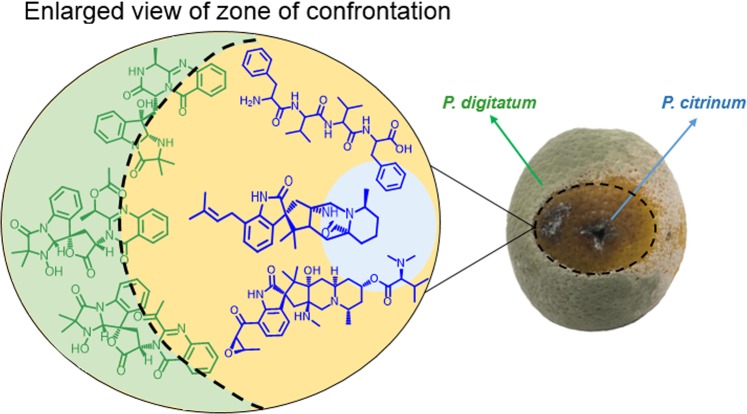
Numerous postharvest diseases have been reported that cause substantial losses of citrus fruits worldwide. Penicillium digitatum is responsible for up to 90% of production losses, and represent a problem for worldwide economy. In order to control phytopathogens, chemical fungicides have been extensively used. Yet, the use of some artificial fungicides cause concerns about environmental risks and fungal resistance. Therefore, studies focusing on new approaches, such as the use of natural products, are getting attention. Co-culture strategy can be applied to discover new bioactive compounds and to understand microbial ecology. Mass Spectrometry Imaging (MSI) was used to screen for potential antifungal metabolites involved in the interaction between Penicillium digitatum and Penicillium citrinum. MSI revealed a chemical warfare between the fungi: two tetrapeptides, deoxycitrinadin A, citrinadin A, chrysogenamide A and tryptoquialanines are produced in the fungi confrontation zone. Antimicrobial assays confirmed the antifungal activity of the investigated metabolites. Also, tryptoquialanines inhibited sporulation of P. citrinum. The fungal metabolites reported here were never described as antimicrobials until this date, demonstrating that co-cultures involving phytopathogens that compete for the same host is a positive strategy to discover new antifungal agents. However, the use of these natural products on the environment, as a safer strategy, needs further investigation. This paper aimed to contribute to the protection of agriculture, considering health and ecological risks.
Subject terms: Antifungal agents, Fungi
Introduction
Citrus fruits have an important impact on world’s economy since they have the largest production compared with other fruits1; for example, the global orange production for 2018/19 is forecast to reach 54.3 million tons2. The major factor affecting the quality of citrus is the postharvest fungal diseases, in particular the green mold caused by Penicillium digitatum that is responsible for 90% of citrus losses during postharvest period1,3.
To control post-harvest diseases, synthetic fungicides are widely used4, causing health and environmental issues5. Furthermore, some fungi strains have developed resistance to commonly used fungicides6. As antifungal resistance is becoming a significant concern, the search for new bioactive compounds has an important role to bypass the extensive use of fungicides7.
Microorganisms are one of the main sources for natural products with useful biological activities, i.e., potential antifungals, antibiotics, anticancer agents, surfactants8,9. However, besides the genetic potential and diversity of microbes, many microbial biosynthetic genes are not activated in the unnatural cultivation conditions used in laboratory and only a few part of the metabolites are accessible10. Several strategies are implemented to overcome the limitations in natural products discovery from microbial sources, i.e., OSMAC approach10,11, the use of epigenetic modifications12,13 and co-cultivation14.
Co-cultures that mimic natural environments, exhibiting microbial competition for limited space and nutrients, have revealed to be a major ecological force that could activate silent gene clusters and defense mechanisms that can lead to the production of bioactive secondary metabolites14–17. Co-culture experiments are highly relevant for allowing not only the identification of new compounds, but also to investigate chemical events that govern interactions between microorganisms in nature18–21.
To date, there is no information about how citrus pathogen fungi interact with other microorganisms in the same environment and what are the mechanisms of attack and defense against endophytic microorganisms or other phytopathogens. This information may lead to the discovery of new secondary metabolites involved in microbial interactions and provide better knowledge about the importance of microbial ecology during infection process.
Here we show an interaction between Penicillium digitatum and Penicillium citrinum, aiming to search for new antifungal compounds that could be used to control postharvest diseases. Considering this purpose, co-culture strategy and MSI were applied to induce the production of secondary metabolites and provide initial insights concerning their biological role based on their spatial distribution in the interaction. The metabolites of interest were isolated and all the structures were elucidated based on Mass spectrometry and NMR experiments. Yet, antifungal assays and confocal microscopy analyses were performed in order to investigate the potential of the fungal metabolites as new antifungal agents.
Materials and Methods
Fungi culture
The P. digitatum (PD) strain used in the studies is deposited with the Spanish Type Culture Collection (CECT) under the accession code CECT20796. P. citrinum (PC) strain was provided by Sylvio Moreira Citrus Center (Cordeirópolis, SP, Brazil). P. digitatum and P. citrinum were cultivated on commercial potato dextrose agar (PDA) (Acumedia). PDA was autoclaved at 103 KPa (121 °C) for 15 min. PDA plates were stored at 25 °C for 7 days in darkness. Spores were harvested by washing the agar surface with sterile distilled water and diluted to a final concentration of 106 or 105 spore mL−1.
Co-culture growth conditions
In vitro co-culture was prepared in 20 mL of PDA plates and 5 µL of each fungal spore solution (106 spore mL−1) was inoculated, on opposite sides. The plates were incubated in darkness at 25 °C for 7 days.
For MSI analyses and confocal microscopy, the in vitro co-culture was prepared by placing a sterile microscope slide in the Petri dish, followed by the pouring of 11 ml of PDA22 and the inoculums were made above the microscope slide. The plates were incubated in darkness at 25 °C for 72 h.
For in vivo co-culture, mature oranges (Citrus sinensis) were surface sterilized and wounded23. A small piece of PDA containing P. citrinum was inoculated in the wound site. Infected and control oranges were stored in sterile 500 mL beakers, in darkness at 25 °C. After 10 days, the fruits were wounded on the opposite equatorial region of P. citrinum inoculum and infected with P. digitatum 106 spore mL−1 solution. The fruits were stored for more 5 days in darkness at 25 °C.
Mass Spectrometry Imaging (MSI) analysis and MS image generation
After the incubation period, the microscope slides were removed from the Petri dishes and put in a vacuum desiccator, for 1 hour, for complete agar dehydration (Angolini et al., 2015). MSI analyses were performed directly on the microscope containing the co-culture, in positive mode, using a desorption electrospray ionization (DESI) source Prosolia Model Omni Spray 2D®-3201) coupled to a Thermo Scientific QExactive® Hybrid Quadrupole-Orbitrap Mass Spectrometer. MSI data was acquired with a mass resolving power of 70.000 at m/z 200. The DESI configuration used was the same set by previous work22. Images were generated with a bin width of Δm/z = ± 0.07 using Firefly data conversion software (version 2.1.05) and processed using BioMap software (version 3.8.0.4) developed by Novartis Institutes for BioMedical Research. In BioMap, color scaling was adjusted to a fixed value during the processing of each image. MS spectra were processed with Xcalibur software (version 3.0.63) developed by Thermo Fisher Scientific.
Extraction of metabolites from the co-culture experiments
The whole contents of the co-culture in vitro were cut into small pieces and transferred to an Erlenmeyer flask. The extraction was performed using methanol. The flasks were sonicated during 1 h in ultrasonic bath and vacuum filtered. Solvent was removed under reduced pressure and the final extract stored at −20 °C.
For in vivo co-culture, the orange peels were cut (2 cm × 2 cm) in the interface zone between the microorganisms and extraction was performed with 5 mL of methanol during 1 h in ultrasonic bath. Extracts were filtered, dried under N2 and stored at −20 °C.
Mass Spectrometry analysis (MS)
Extracts were diluted in methanol and analyzed on a Thermo Scientific QExactive® Hybrid Quadrupole-Orbitrap Mass Spectrometer. Analyses were performed in the positive mode with m/z range of 115–1500, capillary voltage of 3.4 kV, inlet capillary temperature of 280 °C, S-lens 100 V. 5 µL of sample were injected. Stationary phase: Thermo Scientific column Accucore C18 2.6 µm (2.1 mm × 100 mm). Mobile phase: 0.1% formic acid (A) and acetonitrile (B). Eluent profile (A/B): 95/5 up to 2/98 within 15 min, hold for 5 min, up to 95/5 within 1.2 min and hold for 7.8 min. The total run time was 29 min for each run and the flow rate, 0.2 mL min−1. Injection volume: 5 µL.
MS/MS was performed by the collision induced dissociation (CID) with m/z range of 100–800 and the collision energy ranged from 10 to 50 V. The samples were directly infused by electrospray with 5.0 µL min−1 flow rate. MS and MS/MS data was processed with Xcalibur software (version 3.0.63) developed by Thermo Fisher Scientific.
Metabolite separation (HPLC analysis)
Secondary metabolites separations were achieved using a Phenomenex column Luna 5 µm Phenyl-Hexyl (250 × 4.6 mm) and a SHIMADZU prominence HPLC LC-20AT, equipped with CBM-20A communication bus module, SPD-M20A photodiode array detector and SIL-20A auto sampler. Mobile phase: water (0.1% formic acid) (A) and acetonitrile (B). Eluent profile (A/B): 65/35 up to 50/50 within 50 min, up to 40/60 within 20 min. The total run time was 70 min and the flow rate of 1.0 mL min−1. Injection volume was 5 µL. Preparative HPLC purifications were performed on a Phenomenex column Luna 5 µm Phenyl-Hexyl (250 × 10 mm) using a Waters 1525 Binary HPLC Pump equipped with Waters 2998 Photodiode Array Detector and Waters Fraction Collector III using the same optimized gradient conditions. The flow rate was set at 4.7 mL min−1 and the injection volume was 200 µL.
NMR Spectroscopy
1H NMR, 13C NMR and 2D experiments were performed on a Bruker Avance III 500 (1H 500.13 MHz and 13C 125.7 MHz) and Bruker Avance III 600 (1H 600.17 MHz). Deuterated chloroform (CDCl3; 7.23 ppm), dimethyl sulfoxide (DMSO; 2.50 ppm and 39.51 ppm) and tetramethylsilane (TMS; 0.0 ppm) were used as a solvent and internal reference. Chemical shifts (δ) were expressed in (ppm) and the coupling constants (J) in Hertz (Hz).
Molecular Networking analysis
A molecular network for P. citrinum metabolites was created using the online workflow at Global Natural Products Social Molecular Networking (GNPS) (http://gnps.ucsd.edu). The data was filtered by removing all MS/MS peaks within + /−17 Da of the precursor m/z. MS/MS spectra were window filtered by choosing only the top 6 peaks in the + /−50 Da window throughout the spectrum. The data was then clustered with MS-Cluster with a parent mass tolerance of 0.2 Da and a MS/MS fragment ion tolerance of 0.2 Da to create consensus spectra. Further, consensus spectra that contained less than 2 spectra were discarded. A network was then created where edges were filtered to have a cosine score above 0.65 and more than 2 matched peaks. Further edges between two nodes were kept in the network only if each of the nodes appeared in each other’s respective top 10 most similar nodes. The spectra in the network were then searched against GNPS’ spectral libraries. The library spectra were filtered in the same manner as the input data. All matches kept between network spectra and library spectra were required to have a score above 0.5 and at least 5 matched peaks. The resulting molecular networking is available at https://gnps.ucsd.edu/ProteoSAFe/status.jsp?task=c6f716c6fd044ba985eacd96935ee0c3.
Antifungal assays
A stock solution of the co-culture extract was prepared in methanol and further diluted in PDA to the concentration of 0.5 mg mL−1. 15 mL of the resultant solution was poured in a Petri dish followed by the inoculation of 15 μl of a 105 spore mL−1 P. digitatum solution on the center of the agar plate. A control assay was also performed. The plates were incubated in darkness at 25 °C for 96 h.
For antifungal assays, 2.5 mL of PDA was supplemented with 6, 9 and 10 (400 µg mL−1) and each solution were poured in a 6-well microplate. 5 μl of a 105 spore mL−1 P. digitatum solution was inoculated on the center of each agar plate. Negative controls were performed in triplicate. The microplate was incubated in darkness at 25 °C for 96 h.
For determination of minimum inhibitory concentration (MIC) of compounds 1 and 4, microbroth dilution assay was performed as recommended by Clinical and Laboratory Standards Institute (2008)24 with few modifications. Stock solutions of 1 and 4, were prepared in water (5% methanol) and further diluted in YES media in a range of concentrations to 600 µg mL−1 to 1 µg mL−1. 195 μl of each solution were transferred to a 96-well microplate followed by the inoculation of 5 μl of a 105 spore mL−1 P. digitatum solution. Assays were made in duplicate and controls in triplicate. Itraconazole (100 µg mL−1) were used as positive control. Negative controls were performed with methanol in YES. The microplates were incubated in darkness at 25 °C for 96 h.
Confocal laser scanning microscopy
After the incubation period, the microscopes slides were removed from the Petri dish. The in vitro co-culture samples were stained with Congo Red (0.25% w/v in water) for 20 minutes and briefly washed in distilled water. Samples were analyzed with Leica TCS SP5 microscope. Excitation was by the 543 nm emission line of the He-Ne laser, and light emitted between 570 and 680 nm was collected25.
Results and Discussion
MSI reveals potential antifungals in the interaction between P. digitatum and P. citrinum
To screen for new antifungal compounds with potential to protect citrus fruits and control postharvest diseases, we applied a co-culture strategy involving P. digitatum and another citrus pathogen, P. citrinum. Co-culture is a strategy inspired by nature in which the competition between the microorganisms can induce the production of new metabolites8. In previous work, the co-cultivation between Trichophyton rubrum and Bionectria ochroleuca induced the production of a new sulfated analogue of PS-990, suggesting that this compound is further sulfated during the fungal interaction26. Also, another example of a compound derived from fungi interaction is the tetrapeptide cyclo-(L-leucyl-trans-4-hydroxy-L-prolyl-D-leucyl-trans-4-hydroxy-L-proline) isolated from the co-culture broth of Phomopsis sp. K38 and Alternaria sp. E33; the cyclic tetrapeptide exhibited moderate to high inhibitory activity against phytopathogenic fungi when compared to the commercial fungicide triadimefon27. Thus, co-cultivation experiments are a viable approach to find compounds that can inhibit the main citrus phytopathogens, specially, the green mold caused by P. digitatum.
In co-cultivation performed in both orange (in vivo) and synthetic media (in vitro) we visually observed a long-distance growth inhibition between P. citrinum and P. digitatum. In a fungi interaction, silent genes can be activated and harmful metabolites can be diffused from one partner to the other16,28. These induced metabolites are usually localized at the zone of confrontation in solid media of co-cultures16. However, regular approaches used to detect and elucidate metabolites such as mass spectrometry coupled to liquid (LC-MS) or gas (GC-MS) chromatography do not provide information about the spatial distribution of the molecules29.
The information about molecular spatial distribution can be obtained by mass spectrometry imaging (MSI), a powerful tool that generates images for each ion detected in the mass spectrum16. The use of MSI to understand microbial systems and their secondary metabolites is not new and studies where this technique was successfully applied can be found in the literature30. By example, MSI was applied to investigate the interaction between Bacillus subtilis 3610 and Streptomyces coelicolor A3. DESI imaging of the bacterial co-culture revealed 57 signals spatially localized to bacterial colonies, leading to the identification of some secondary metabolites such as surfactin and plipastatin31. Furthermore, MSI analysis also showed that S. coelicolor has the production of certain secondary metabolites inhibited in the presence of B. subtilis, revealing an interaction between bacteria31.
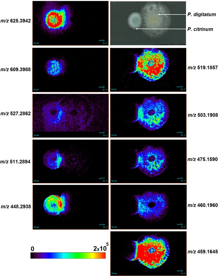
Therefore, to detect the secondary metabolites involved in the interaction between the citrus pathogens, we applied DESI-MSI directly on the surface of the co-culture agar, to visualize the diffusion of compounds to the zone of confrontation. MSI signals were obtained for ions [M + H]+ at m/z 519.1857, m/z 503.1908, m/z 475.1590, m/z 460.1960, m/z 459.1645, m/z 625.3942, m/z 609.3988, m/z 527.2862, m/z 511.2894 and m/z 448.2938 (Figs. S1–S10). We observed that all the ions mentioned were detected and concentrated in the zone of confrontation between the fungi (Fig. 1). These compounds may be related to the fungus-fungus interaction and could be potentially new antimicrobial agents. Ions at m/z 625, 609, 527, 511 and 448 were produced by P. citrinum, while ions at m/z 519, 503, 475, 460 and 459, seemed to be a counter-attack of P. digitatum, revealing a chemical warfare between these two citrus pathogens. The ions detected in vitro through MSI analyses were also detected in the extracts of the co-cultures in vivo (Figs. S11–S20) using oranges as substrate (Fig. 2).
Figure 1.
Imaging analysis by (+) DESI-MS of citrus pathogens co-culture. Each MS image shows the spatial distribution of a m/z ratio over the co-culture surface. All images are plotted on the same color scale from 0 (black) to 2 × 10⁵ (red) yet ion concentration cannot be compared across images since differences in chemical structures can lead to variation in ionization efficiency30.
Figure 2.
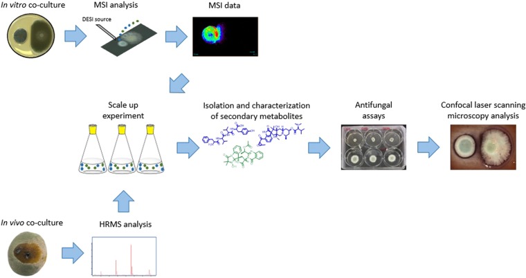
Overview of the experimental setup and strategies applied to analyze the secondary metabolites involved in citrus pathogens interaction. P. digitatum and P. citrinum were co-cultured in orange (in vivo) and in PDA media (in vitro) to induce the production of secondary metabolites. MSI was applied, in vitro, to detect the secondary metabolites produced in the zone of confrontation. HRMS analysis was performed to confirm the fungus-fungus interaction in vivo. The metabolites of interest were isolated from a scale up experiment. Subsequently, antifungal assays and confocal laser scanning microscopy analysis were performed to investigate antimicrobial activity and fungal cell morphology, respectively.
To characterize the metabolites involved in the interaction, a co-culture extract was obtained from a scale up cultivation experiment and the compounds of interest, detected initially through MSI analyses, were isolated by preparative HPLC and characterized through tandem mass spectrometry and NMR analyses.
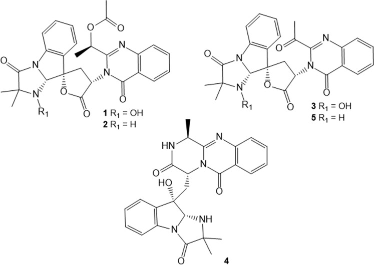
Through the exact masses obtained by DESI-MSI analyses (Table 1) it was possible to confirm the presence of indole alkaloids produced by P. digitatum. Ions [M + H]+ at m/z 519.1857, m/z 503.1908, m/z 475.1590, m/z 460.1960 and m/z 459.1645 which correspond respectively to tryptoquialanine A (1), tryptoquialanine C (2), tryptoquialanone (3), 15-dimethyl-2-epi-fumiquinazoline A (4) and deoxytryptoquialanone (5) (chemical structures represented in Fig. 3). These compounds are part of the tryptoquialanines biosynthetic pathway32 and were previously detected during DESI-MSI analysis of oranges infected with the green mold disease23.
Table 1.
DESI-MSI data obtained for the secondary metabolites involved in P. digitatum and P. citrinum interaction.
| Compound | Ion formula ([M + H]+) | Calculated m/z | Experimental m/z | Error (ppm) |
|---|---|---|---|---|
| Tryptoquialanine A | C27H27N4O7 | 519.1874 | 519.1857 | −3.4 |
| Tryptoquialanine C | C27H27N4O6 | 503.1925 | 503.1908 | −3.5 |
| Tryptoquialanone | C25H23N4O6 | 475.1612 | 475.1590 | −4.6 |
| 15-dimethyl-2-epi-fumiquinazoline A | C25H26N5O4 | 460.1979 | 460.1960 | −4.2 |
| deoxytryptoquialanone | C25H23N4O5 | 459.1663 | 459.1645 | −4.0 |
| Citrinadin A | C35H53N4O6 | 625.3960 | 625.3942 | −2.8 |
| Deoxycitrinadin A | C35H53N4O5 | 609.4010 | 609.3988 | −3.7 |
| Phe-Val-Val-Tyr | C28H39N4O6 | 527.2864 | 527.2862 | −0.4 |
| Phe-Val-Val-Phe | C28H39N4O5 | 511.2915 | 511.2894 | −4.1 |
| Chrysogenamide A | C28H38N3O2 | 448.2959 | 448.2938 | −4.6 |
Figure 3.
Chemical structures of indole alkaloids produced by P. digitatum: tryptoquialanine A (1), tryptoquialanine C (2), tryptoquialanone (3), 15-dimethyl-2-epi-fumiquinazoline A (4) and deoxytryptoquialanone (5). These compounds were detected through DESI-MSI in the fungal confrontation zone.
Compound 1 was reported as the major secondary metabolite produced by P. digitatum33. Yet, the deletion of tqaA gene, responsible to regulate 1 biosynthesis, showed that 1 is not involved in the pathogenicity of P. digitatum against citrus since the infection ability of the mutants was not altered34. The exact biological role of the tryptoquialanines is still unknown34 and this study provides a new biological activity of the tryptoquialanines with the involvement of these alkaloids in the fungal-fungal interaction.
The MS/MS of the ion [M + H]+ at m/z 625.3951 yielded to fragments at m/z 594.3533, 576.3427, 449.2430 and 431.2325 (Fig. S21), the same fragmentation pattern obtained for citrinadin A (6) in a study involving co-culture between P. citrinum and Pseudoalteromonas sp. OT5919. Also GNPS database suggested that the ion at m/z 625 could be citrinadin A. The tandem mass spectrum obtained for this compound shared five fragments in common with the tandem mass spectrum of 6 deposited in the database (Fig. S22). In addition, it shares similar MS/MS fragmentation pattern with citrinadin A reported by Moree et al. (2014). Citrinadins were reported being higher produced in co-culture situations and concentrated in co-culture interfaces, suggesting a defensive response of P. citrinum against other microorganisms19,35.
Molecular networking analysis of the isolated compounds revealed that another citrinadin is involved in the co-culture interaction. We observed that ion at m/z 609, detected initially by MSI analysis, is grouped in the same cluster of compound 6 (m/z 625) (Fig. S23), suggesting that this compound is a citrinadin-like metabolite. In a molecular networking analysis, related metabolites are grouped in same clusters since they have similar MS/MS spectrum36.
The exact mass obtained for ion [M + H]+ at m/z 609.4010 corresponds to a compound with molecular formula C35H53N4O5. Fragmentation pattern yielded to fragments at m/z 578.3583, 464.2905, 451.2586 and 433.2482 (Supplementary Fig. S24). 1H NMR and 1H-1H COSY spectra obtained for the purified metabolite (Figs. S25–S26 and Table S1) exhibited similar signals of a synthesized deoxycitrinadin A that was reported by Bian et al. (2013)37. The epoxide characteristic signal at δH 4.0 was absent in this derivative and a signal for vynilic hydrogen could be observed at δH 6.9, indicating the lack of the epoxide group and the presence of a double bond in comparison with citrinadin A37. It is the first time that deoxycitrinadin A (7) is reported in the literature as a secondary metabolite produced by a microorganism.
Exact masses obtained by DESI-MSI for ions [M + H]+ at m/z 527.2862 and 511.2894 indicated compounds with elemental composition of C28H38N4O6 and C28H38N4O5, respectively. MS/MS revealed that the ion at m/z 511 has similar structure compared to m/z 527, except by an absence of an oxygen atom. Fragmentation of the ion at m/z 527 yielded to fragments m/z 281, 247, 219, 182 and 120 (Fig. S27), while ion at m/z 511 yielded to m/z 265, 247, 219, 166 and 120 (Fig. S30). Same fragmentation pattern was reported for the sequence of tetrapeptides Phe-Val-Val-Tyr (8) of Penicillium canescens38. Yet, the tetrapeptides 8 and Phe-Val-Val-Phe (9) were recently reported as secondary metabolites produced by Penicillium roqueforti39. 1H and 13C NMR analyses of the isolated compounds (Figs. S28–S32 and Tables S2-S3) confirmed the results obtained through MS/MS, concluding that ions at m/z 527 and m/z 511 correspond to 8 and 9, respectively. The production of these tetrapeptides in fungal chemical warfare is not surprising because small peptides are known for their antimicrobial activity40. This is the first report of 8 and 9 as secondary metabolites of P. citrinum.
For ion [M + H]+ at m/z 448.2938, the exact mass suggested a compound with molecular formula C28H38N3O2, the same composition of the secondary metabolite chrysogenamide A (10) (error = −4.6 ppm). Compound 10 was isolated and 1H and 13C NMR analyses (Figs. S33–S34 and Table S4) confirmed its structure; it is the first report that chrysogenamide A is involved in a fungal-fungal interaction. Compound 10 was first reported as a secondary metabolite of Penicillium chrysogenum No. 005, an endophytic fungus associated with the plant Cistanche deserticola41. Also, 10 was reported as a secondary metabolite of a P. citrinum strain42. The structures of the metabolites produced by P. citrinum are represented in Fig. 4.
Figure 4.
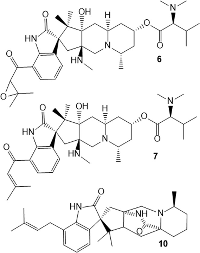
Chemical structures of secondary metabolites produced by P. citrinum during chemical warfare against P. digitatum: citrinadin A (6), deoxycitrinadin A (7) and chrysogenamide A (10).
Antifungal assays
We investigated the antifungal activity of the metabolites produced in the co-culture and P. digitatum was inoculated in agar media containing co-culture extract. Compared to the control, we observed a reduction of 67% in P. digitatum radial growth (Fig. S35), indicating that the metabolites involved in the fungal warfare have potential as antifungal agents. To confirm this hypothesis, we tested the isolated compounds and observed that compounds 6, 9 and 10 (400 µg mL−1) reduced 48%, 41% and 61% of P. digitatum radial growth, respectively, when compared to control (Fig. 5), confirming the antifungal activity.
Figure 5.
P. digitatum growing on PDA with 400 µg ml−1 of (A) chrysogenamide A (B) citrinadin A and (C) tetrapeptide Phe-Val-Val-Phe. In 96 h, an inhibition in radial growth is observed in the presence of the isolated metabolites when compared to (D) control.
Citrinadins were found to be involved in the response of P. citrinum against other microorganisms in co-cultures, but the biological role of them in biological environments are still unknown to this date19. Compound 6 was tested for Anti-buruli ulcer activity on Mycobacterium ulcerans MN209, but no interesting MIC was observed43; also, cytotoxicity activity of 6 against leukemia and carcinoma cells was reported44. Our results are the first report of an antimicrobial activity of citrinadins in literature and can provide first insights about the biological role of these compounds in fungal-fungal interactions.
In addition, chrysogenamide A was never reported as an antimicrobial agent until now. Compound 10 exhibited a protective effect on neurocytes against oxidative stress-induced cell death41, however no other biological activity for 10 was reported in the literature.
Antifungal assays applying D-Phe-L-Val-D-Val-L-Tyr revealed that this tetrapeptide has inhibitory activity against B. subitllis and the soybean phytopathogen Fusarium virguliforme38. In contrast, no inhibition of E. coli, B. subtilis and S. cerevisiae in presence of D-Phe-L-Val-D-Val-L-Tyr or D-Phe-L-Val-D-Val-L-Phe was observed39.
The antifungal activity of the tryptoquialanines was also evaluated, since these compounds seemed to be a chemical response of P. digitatum against P. citrinum in the chemical warfare. Tryptoquialanines 1 and 4 were tested and revealed an antifungal activity against P. citrinum. Compounds 1 and 4 had MIC of 300 µg mL−1, inhibiting P. citrinum spore production (Fig. S36). To the best of our knowledge, it is the first report of an antimicrobial activity of the tryptoquialanines. Recently, 1 was demonstrated as an insecticidal compound against Ae. aegypti larvae23; the antifungal activity can provide more understanding about the role of the tryptoquialanines in the citrus-pathogen environment once these compounds are not required for P. digitatum virulence34.
Chemical warfare alters P. digitatum cell wall in co-culture
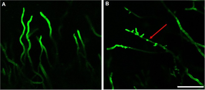
To investigate the action of the secondary metabolites during the fungal interaction, co-culture samples were stained with Congo Red and observed through confocal laser scanning microscopy. Congo Red is commonly used to stain polysaccharides containing β 1,4 linkages as, by example, the fungal cell wall component chitin45,46. P. digitatum hyphae were observed in the confrontation zone (sample) and compared with hyphae distant to the interface region (control) (Fig. S37). Control hyphae were homogeneous stained with Congo Red while hyphae in the interface region exhibited an altered staining pattern (Fig. 6), with irregular patches. Similar staining patterns were obtained for knockout mutant fungi which the deleted genes had a role in cell wall organization; as result, mutants exhibited defective cell walls and irregular staining25,46,47. Yet, abnormal staining with Calcofluor White was observed for P. ostreatus P89 treated with 36 °C; high temperature altered chitin distribution and cell wall integrity48.
Figure 6.
Confocal laser scanning microscopy of Congo Red-stained P. digitatum hyphae (A) distant from P. citrinum and (B) in the zone of confrontation. Patches of Congo Red indicates a defective fungal cell wall. Bars = 5.0 µm.
This data shows that P. digitatum hyphae, in contact with the metabolites diffused during the co-culture, have a defective cell wall since Congo Red bounds to fungal cell wall structures. The fungal cell wall is an attractive target of antimicrobials because they are not present in mammalian cells49,50. In conclusion, the microscopy analysis and the antifungal assays reinforce that the metabolites involved in the fungal interaction have potential as antifungal agents and may be the mechanism in nature that these phytopathogens developed to compete against other microorganisms for the host (Fig. 7).
Figure 7.
Chemical warfare between P. citrinum and P. digitatum in citrus fruit. Tryptoquialanines, citrinadins, chrysogenamide A, tetrapeptides and other metabolites are involved in the long-distance inhibition observed.
Conclusions
The search for new natural antimicrobials is a promising field in natural products research concerning the economic impact of postharvest diseases to worldwide agriculture. Furthermore, the appearance of fungi strains resistant to fungicides makes the discovery of new antifungal agents to replace synthetic compounds extremely important. Using co-cultivation between phytopathogens that compete for the same host, P. digitatum and P. citrinum, we observed a fungal interaction. Through MSI technique, we detected secondary metabolites diffused to the interface zone between the microorganisms. Tryptoquialanines, citrinadins, chyrsogenamide A and tetrapeptides exhibited great antifungal activity, confirming that co-cultures and MSI technique are a good combination in the search of new natural antimicrobials.
Until this date, there has been no information about the interaction between citrus pathogenic fungi. Our data revealed compounds that play a role in the citrus microbial ecology. In addition, we demonstrated that the metabolites studied have great potential as antifungal agents since fungal cell walls are one of the main targets of antifungal compounds. The use of the identified compounds as natural antifungals instead of synthetic fungicides should be further investigated. This paper opens new research possibilities and contributes to the environmental and human health, helping in the search of safer strategies for agriculture through the use of compounds obtained from natural sources.
Supplementary information
Acknowledgements
This study was financed in part by the Coordenação de Aperfeiçoamento de Pessoal de Nível Superior - Brasil (CAPES) - Finance Code 001, Fundação de Amparo a Pesquisa no Estado de São Paulo [grant number 2017/24462-4 and 2019/06359-7], Deutsche Forschungsgemeinschaft (SFB 1127 ChemBioSys) and L’Oréal Brazil, together with ABC and UNESCO in Brazil; CFFA was recipient of a postdoctoral Fellowship from CNPq (grant number 400577/2015-1). TPF was recipient of a postdoctoral Fellowship from Capes/Humboldt. We would like to thank Dr. Katia Kupper for the P. citrinum strain, Dr. Marcos Nogueira Eberlin for the orbitrap mass spectrometer and Guerline François.
Author contributions
J.H.C. and C.I.W. wrote the manuscript. C.F.F.A and C.I.W. performed the MSI analysis. J.H.C., T.P.F, K.S. and C.I.W analyzed MSI data, isolated and characterized the secondary metabolites, performed the antimicrobial assays and conducted microscopy analysis. T.P.F. oversaw the project. T.P.F., C.F.F.A., K.S. and C.H. reviewed the manuscript. T.P.F. and J.H.C. submitted the manuscript.
Data availability
All data generated or analyzed during this study are included in this published article and its Supplementary Information file.
Competing interests
The authors declare no competing interests.
Footnotes
Publisher’s note Springer Nature remains neutral with regard to jurisdictional claims in published maps and institutional affiliations.
These authors contributed equally: Jonas Henrique Costa and Cristiane Izumi Wassano.
Supplementary information
is available for this paper at 10.1038/s41598-019-55204-9.
References
- 1.Ghooshkhaneh NG, Golzarian MR, Mamarabadi M. Detection and classification of citrus green mold caused by Penicillium digitatum using multispectral imaging. J Sci Food Agric. 2018;98(9):3542–3550. doi: 10.1002/jsfa.8865. [DOI] [PubMed] [Google Scholar]
- 2.United States Department of Agriculture - Foreign Agricultural Service, 2019. Citrus: World Markets and Trade, https://apps.fas.usda.gov/psdonline/circulars/citrus.pdf (accessed 06 August 2019).
- 3.Macarisin D, et al. Penicillium digitatum suppresses production of hydrogen peroxide in host tissue during infection of citrus fruit. Phytopathology. 2007;97:1491–1500. doi: 10.1094/PHYTO-97-11-1491. [DOI] [PubMed] [Google Scholar]
- 4.Hao W, Li H, Hu M, Yang L, Rizwan-ul-Haq M. Integrated control of citrus green and blue mold and sour rot by Bacillus amyloliquefaciens in combination with tea saponin. Postharvest Biol Technol. 2011;59:316–323. doi: 10.1016/j.postharvbio.2010.10.002. [DOI] [Google Scholar]
- 5.Frisvad JC, Samson RA. Polyphasic taxonomy of Penicillium subgenus Penicillium A guide to identification of food and air-borne terverticillate Penicillia and their mycotoxins. Stud Mycol. 2004;49:1–174. [Google Scholar]
- 6.Kanetis L, Förster H, Adaskaveg JE. Determination of natural resistance frequencies in Penicillium digitatum using a new air-sampling method and characterization of Fludioxonil- and Pyrimethanil-Resistant isolates. Phytopathology. 2010;100:738–743. doi: 10.1094/PHYTO-100-8-0738. [DOI] [PubMed] [Google Scholar]
- 7.Strano Maria C., Altieri Giuseppe, Admane Naouel, Genovese Francesco, Di Renzo Giovanni C. Citrus Pathology. 2017. Advance in Citrus Postharvest Management: Diseases, Cold Storage and Quality Evaluation. [Google Scholar]
- 8.Azzollini A, et al. Dynamics of Metabolite Induction in Fungal Co-cultures by Metabolomics at Both Volatile and Non-volatile Levels. Front Microbiol. 2018;9:72. doi: 10.3389/fmicb.2018.00072. [DOI] [PMC free article] [PubMed] [Google Scholar]
- 9.Demain AL. Importance of microbial natural products and the need to revitalize their discovery. J Ind Microbiol Biotechnol. 2014;41:185–201. doi: 10.1007/s10295-013-1325-z. [DOI] [PubMed] [Google Scholar]
- 10.Bode, H. B., Bethe, B., Höfs, R. & Zeeck, A. Big effects from small changes: Possible ways to explore nature’s chemical diversity. ChemBioChem, 3, 619–627, 10.1002/1439-7633(20020703)3:7<619::AID-CBIC619>3.0.CO;2-9 (2002). [DOI] [PubMed]
- 11.Pan R, Bai X, Chen J, Zhang H, Wang H. Exploring Structural Diversity of Microbe Secondary Metabolites Using OSMAC Strategy: A Literature Review. Front Microbiol. 2019;10:294. doi: 10.3389/fmicb.2019.00294. [DOI] [PMC free article] [PubMed] [Google Scholar]
- 12.Keller NP. Fungal secondary metabolites: regulation, function and drug discovery. Nat Rev Microbiol. 2019;17:167–180. doi: 10.1038/s41579-018-0121-1. [DOI] [PMC free article] [PubMed] [Google Scholar]
- 13.Aghcheh RK, Kubicek CP. Epigenetics as an emerging tool for improvement of fungal strains used in biotechnology. Appl Microbiol Biotechnol. 2015;99(15):6167–6181. doi: 10.1007/s00253-015-6763-2. [DOI] [PubMed] [Google Scholar]
- 14.Marmann A, Aly AH, Lin W, Wang B, Proksch P. Co-Cultivation – A Powerful Emerging Tool for Enhancing the Chemical Diversity of Microorganisms. Mar Drugs. 2014;12:1043–1065. doi: 10.3390/md12021043. [DOI] [PMC free article] [PubMed] [Google Scholar]
- 15.Netzker T, et al. Microbial communication leading to the activation of silent fungal secondary metabolite gene clusters. Front Microbiol. 2015;6:299. doi: 10.3389/fmicb.2015.00299. [DOI] [PMC free article] [PubMed] [Google Scholar]
- 16.Bertrand S, et al. Metabolite induction via microorganism co-culture: A potential way to enhance chemical diversity for drug discovery. Biotechnol Adv. 2014;32(6):1180–1204. doi: 10.1016/j.biotechadv.2014.03.001. [DOI] [PubMed] [Google Scholar]
- 17.Vinale F, et al. Co-Culture of Plant Beneficial Microbes as Source of Bioactive Metabolites. Sci. Rep. 2017;7:14330. doi: 10.1038/s41598-017-14569-5. [DOI] [PMC free article] [PubMed] [Google Scholar]
- 18.Chagas FO, Dias LG, Pupo MT. A mixed culture of endophytic fungi increases production of antifungal polyketides. J Chem Ecol. 2013;39(10):1335–1342. doi: 10.1007/s10886-013-0351-7. [DOI] [PubMed] [Google Scholar]
- 19.Moree WJ, et al. Microbiota of Healthy Corals Are Active against Fungi in a Light-Dependent Manner. ACS Chem Biol. 2014;9(10):2300–2308. doi: 10.1021/cb500432j. [DOI] [PMC free article] [PubMed] [Google Scholar]
- 20.Partida-Martinez LP, Hertweck C. Pathogenic fungus harbous endosymbiotic bacteria for toxin production. Nature. 2005;437:884–888. doi: 10.1038/nature03997. [DOI] [PubMed] [Google Scholar]
- 21.Heine D, et al. Chemical warfare between leafcutter ant symbionts and a co-evolved pathogen. Nat. Commun. 2018;9:2208. doi: 10.1038/s41467-018-04520-1. [DOI] [PMC free article] [PubMed] [Google Scholar]
- 22.Angolini CFF, et al. Direct Protocol for Ambient Mass Spectrometry Imaging on Agar Culture. Anal Chem. 2015;87:6925–6930. doi: 10.1021/acs.analchem.5b01538. [DOI] [PubMed] [Google Scholar]
- 23.Costa JH, et al. Monitoring indole alkaloid production by Penicillium digitatum during infection process in citrus by Mass Spectrometry Imaging and molecular networking. Fungal Biol. 2019;123(8):594–600. doi: 10.1016/j.funbio.2019.03.002. [DOI] [PubMed] [Google Scholar]
- 24.CLSI. Reference Method for Broth Dilution Antifungal Susceptibility Testing of Filamentous Fungi; Approved Standard – Second Edition. CLSI document M38-A2. Wayne, PA: Clinical and Laboratory Standards Institute; 2008.
- 25.Siriputthaiwan P, Jauneau A, Herbert C, Garcin D, Dumas B. Functional analysis of CLPT1, a Rab/GTPase required for protein secretion and pathogenesis in the plant fungal pathogen Colletotrichum lindemuthianum. J Cell Sci. 2005;118:323–329. doi: 10.1242/jcs.01616. [DOI] [PubMed] [Google Scholar]
- 26.Bertrand S, et al. De Novo Production of Metabolites by Fungal Co-culture of Trichophyton rubrum and Bionectria ochroleuca. J Nat Prod. 2013;76:1157–1165. doi: 10.1021/np400258f. [DOI] [PubMed] [Google Scholar]
- 27.Li C, Wang J, Luo C, Ding W, Cox DG. A new cyclopeptide with antifungal activity from the co-culture broth of two marine mangrove fungi. Nat. Prod. Res. 2014;28:616–621. doi: 10.1080/14786419.2014.887074. [DOI] [PubMed] [Google Scholar]
- 28.Xu XY, et al. Metabolomics Investigation of an Association of Induced Features and Corresponding Fungus during the Co-culture of Trametes versicolor and Ganoderma applanatum. Front Microbiol. 2018;8:2647. doi: 10.3389/fmicb.2017.02647. [DOI] [PMC free article] [PubMed] [Google Scholar]
- 29.Parrot D, Papazian S, Foil D, Tasdemir D. Imaging the Unimaginable: Desorption Electrospray Ionization - Imaging Mass Spectrometry (DESI-IMS) in Natural Product Research. Planta Med. 2018;84:584–593. doi: 10.1055/s-0044-100188. [DOI] [PMC free article] [PubMed] [Google Scholar]
- 30.Dunham SJB, Ellis JF, Li B, Sweedler JV. Mass Spectrometry Imaging of Complex Microbial Communities. Acc Chem Res. 2017;50:96–104. doi: 10.1021/acs.accounts.6b00503. [DOI] [PMC free article] [PubMed] [Google Scholar]
- 31.Watrous J, Hendricks N, Meehan M, Dorrestein PC. Capturing Bacterial Metabolic Exchange Using Thin Film Desorption Electrospray Ionization-Imaging Mass Spectrometry. Anal Chem. 2010;82:1598–1600. doi: 10.1021/ac9027388. [DOI] [PMC free article] [PubMed] [Google Scholar]
- 32.Gao X, et al. Fungal indole alkaloid biosynthesis: genetic and biochemical investigation of the tryptoquialanine pathway in Penicillium aethiopicum. J Am Chem Soc. 2011;133(8):2729–2741. doi: 10.1021/ja1101085. [DOI] [PMC free article] [PubMed] [Google Scholar]
- 33.Ariza MR, Larsen TO, Petersen BO, Duus JØ, Barrero AF. Penicillium digitatum metabolites on synthetic media and citrus fruits. J Agric Food Chem. 2002;50:6361–6365. doi: 10.1021/jf020398d. [DOI] [PubMed] [Google Scholar]
- 34.Zhu, C. et al. Identification of secondary metabolite biosynthetic gene clusters associated with the infection of citrus fruit by Penicillium digitatum. Postharvest Biol Technol, 134, 17–21, https://doi.org/0.1016/j.postharvbio.2017.07.011 (2017).
- 35.Raimundo I, Silva SG, Costa R, Keller-Costa T. Bioactive Secondary Metabolites from Octocoral-Associated Microbes – New Chances for Blue Growth. Mar Drugs. 2018;16(12):485. doi: 10.3390/md16120485. [DOI] [PMC free article] [PubMed] [Google Scholar]
- 36.Nguyen DD, et al. MS/MS networking guided analysis of molecule and gene cluster families. Proc Natl Acad Sci USA. 2013;110:E2611–E2620. doi: 10.1073/pnas.1303471110. [DOI] [PMC free article] [PubMed] [Google Scholar]
- 37.Bian Z, Marvin CC, Martin SF. Enantioselective Total Synthesis of (-)-Citrinadin A and Revision of Its Stereochemical Structure. J Am Chem Soc. 2013;135:10886–10889. doi: 10.1021/ja405547f. [DOI] [PMC free article] [PubMed] [Google Scholar]
- 38.Bertinetti BV, Peña NI, Cabrera GM. An Antifungal Tetrapeptide from the Culture of Penicillium canescens. Chem Biodivers. 2009;6:1178–1184. doi: 10.1002/cbdv.200800336. [DOI] [PubMed] [Google Scholar]
- 39.Hammerl R, Frank O, Schmittnägel T, Ehrmann MA, Hofmann T. Functional Metabolome Analysis of Penicillium roqueforti by Means of Differential Off-Line LC-NMR. J Agric Food Chem. 2019;67(18):5135–5146. doi: 10.1021/acs.jafc.9b00388. [DOI] [PubMed] [Google Scholar]
- 40.Sharma, K., Aaghaz, S., Shenmar, K. & Jain, R. Short Antimicrobials Peptides. Recent Pat Antiinfect Drug Discov, 13(1), 12–52, https://doi.org/0.2174/1574891X13666180628105928 (2018). [DOI] [PubMed]
- 41.Lin Z, et al. Chrysogenamide A from an Endophytic Fungus Associated with Cistanche deserticola and Its Neuroprotective Effect on SH-SY5Y Cells. J Antibiot. 2008;61(2):81–85. doi: 10.1038/ja.2008.114. [DOI] [PubMed] [Google Scholar]
- 42.Andrade JAS, et al. Citrinadin A derivates from Penicillium citrinum, an endophyte from the marine alga Dichotomaria marginata. Planta Med. 2014;80(10):776. doi: 10.1055/s-0034-1382419. [DOI] [Google Scholar]
- 43.Kyeremeh K, Owusu KB, Ofosuhene M, Ohashi M, Agyapong J. Anti-Proliferative and Anti-Plasmodia Activity of Quinolactacin A2, Citrinadin A and Butrecitrinadin co-isolated from a Ghanaian Mangrove Endophytic Fungus Cladosporium oxysporum strain BRS2A-AR2F. J Chem Applications. 2017;3(1):12. [Google Scholar]
- 44.Tsuda M, et al. Citrinadin A, a Novel Pentacyclic Alkaloid from Marine-Derived Fungus Penicillium citrinum. Org Lett. 2004;6(18):3087–3089. doi: 10.1021/ol048900y. [DOI] [PubMed] [Google Scholar]
- 45.Hawkins PJ, Geddes BA, Oresnik IJ. Common dyes used to determine bacterial polysaccharides on agar are affected by medium acidification. Can J Microbiol. 2017;63(6):559–562. doi: 10.1139/cjm-2016-0743. [DOI] [PubMed] [Google Scholar]
- 46.Gaulin E, et al. The CBEL glycoprotein of Phytophthora parasitica var-nicotianae is involved in cell wall deposition and adhesion to cellulosic substrates. J Cell Sci. 2002;115:4565–4575. doi: 10.1242/jcs.00138. [DOI] [PubMed] [Google Scholar]
- 47.Mitic M, et al. Disruption of calcineurin catalytic subunit (cnaA) in Epichloë festucae induces symbiotic defects and intrahyphal hyphae formation. Mol Plant Pathol. 2017;16(6):1414–1426. doi: 10.1111/mpp.12624. [DOI] [PMC free article] [PubMed] [Google Scholar]
- 48.Qiu Z, Wu X, Gao W, Zhang J, Huang C. High temperature induced disruption of the cell wall integrity and structure in Pleurotus ostreatus mycelia. Appl Microbiol Biotechnol. 2018;102(15):6627–6636. doi: 10.1007/s00253-018-9090-6. [DOI] [PubMed] [Google Scholar]
- 49.Mazu TK, Bricker BA, Flores-Rozas H, Ablordeppey SY. The Mechanistic Targets of Antifungal Agents: An Overview. Mini Rev Med Chem. 2016;16(7):555–578. doi: 10.2174/1389557516666160118112103. [DOI] [PMC free article] [PubMed] [Google Scholar]
- 50.Georgopapadakou NH, Tkacz JS. The Fungal cell wall as a drug target. Trends Microbiol. 1995;3(3):98–104. doi: 10.1016/S0966-842X(00)88890-3. [DOI] [PubMed] [Google Scholar]
Associated Data
This section collects any data citations, data availability statements, or supplementary materials included in this article.
Supplementary Materials
Data Availability Statement
All data generated or analyzed during this study are included in this published article and its Supplementary Information file.


