Abstract
Objective
Erythropoietin (EPO) is a clinically available hematopoietic cytokine. EPO has shown beneficial effects in the context of spinal cord injury and other neurological conditions. The aim of this study was to evaluate the effect of EPO on a rat model of spinal cord compression-induced cervical myelopathy and to explore the possibility of its use as a pharmacological treatment.
Methods
To develop the compression-induced cervical myelopathy model, an expandable polymer was implanted under the C5-C6 laminae of rats. EPO administration was started 8 weeks after implantation of a polymer. Motor function of rotarod performance and grip strength was measured after surgery, and motor neurons were evaluated with H-E, NeuN and choline acetyltransferase staining. Apoptotic cell death was assessed with TUNEL and Caspase-3 staining. The 5HT, GAP-43 and synaptophysin were evaluated to investigate the protection and plasticity of axons. Amyloid beta precursor protein (APP) was assessed to evaluate axonal injury.
To assess transfer of EPO into spinal cord tissue, the EPO levels in spinal cord tissue were measured with an ELISA for each group after subcutaneous injection of EPO.
Results
High-dose EPO maintained motor function in the compression groups. EPO significantly prevented the loss of motor neurons and significantly decreased neuronal apoptotic cells. Expression of 5HT and synaptophysin was significantly preserved in the EPO group. APP expression was partly reduced in the EPO group. The EPO levels in spinal cord tissue were significantly higher in the high-dose EPO group than other groups.
Conclusion
EPO improved motor function in rats with compression-induced cervical myelopathy. EPO suppressed neuronal cell apoptosis, protected motor neurons, and induced axonal protection and plasticity. The neuroprotective effects were produced following transfer of EPO into the spinal cord tissue. These findings suggest that EPO has high potential as a treatment for degenerative cervical myelopathy.
Introduction
As the population ages, degenerative changes in the cervical spine progress. The spinal canal gradually narrows due to cervical spondylosis, disc hernia, and ossification of the posterior longitudinal ligament [1] [2]. This chronic compression of the cervical spinal cord causes degenerative cervical myelopathy. The symptoms of degenerative cervical myelopathy such as motor weakness, sensory disturbances, decreased fine motor coordination, and spastic gait gradually progress over time. Mechanical stress as a consequence of focal compression, which induces spinal cord ischemia at the compressed segment, is an important component in the pathogenesis of degenerative cervical myelopathy [3]. At this time, surgical decompression is often performed to treat degenerative cervical myelopathy [4] [5] [6]. At present, however, there is no accredited medical treatment which improve the neurological status in patients with worsening degenerative cervical myelopathy.
To elucidate the biological mechanism of degenerative cervical myelopathy and develop a treatment strategy for it, a co-author, Kim, established a novel experimental model of spinal cord compression-induced cervical myelopathy [7]. This model is created by inserting a sheet of water-absorbing urethane-compound polymer under the laminae of rats. This model induces delayed motor dysfunction and reproduces the characteristic course of clinical delayed degenerative cervical myelopathy. Using this model, we have previously demonstrated that pharmacological agents, such as Limaprost alfadex, prostaglandin E1 derivative, and Cilostazol, a selective type III phosphodiesterase inhibitor, ameliorate compression-induced cervical myelopathy [8] [9]. However, functional recovery from developing compression myelopathy has not been elucidated in those studies.
We recently confirmed that granulocyte colony-stimulating factor (G-CSF) improves motor function in the progressive phase of compression myelopathy and preserves anterior horn motor neurons in the rat spinal cord compression-induced cervical myelopathy model [10]. However, in healthy people, G-CSF causes marked leukocytosis, which commonly results in fever, arthralgia, and rarely, thromboembolism and splenomegaly [11].
Erythropoietin (EPO) is a physiological hematopoietic cytokine similar to G-CSF. EPO is a 30.4-kDa glycoprotein secreted from the kidney that stimulates red blood cell (RBC) production (erythropoiesis) after binding to the EPO receptor in the bone marrow [12]. EPO is commonly used in anemic patients undergoing chronic hemodialysis or suffering from cancer and undergoing chemotherapy [13] [14]. EPO is also used for preoperative autologous blood donation in hematologically healthy individuals [15]. Therefore, EPO can often be used safely, even in elderly patients or those with critical disease.
In addition, EPO has multifunctional tissue-protective effects, including anti-apoptotic, anti-inflammatory, anti-oxidative, and angiogenic effects [16] [17] [18]. During the last two decades, a number of studies have described its neuroprotective effects in cerebral infarction, brain contusion, and acute spinal cord injury (SCI) in laboratory investigations [19] [20] [21] [22] [23]. Those papers reported beneficial effects of EPO on neuroprotection, angiogenesis, and anti-apoptosis in the brain and spinal cord [18] [24]. Recently, recombinant human EPO (rhEPO) was preliminarily used in a randomized clinical trial of acute SCI, and results indicated the possibility of treating acute SCI with EPO [25].
However, no reports have shown the neuroprotective effect of EPO for spinal cord compression-induced cervical myelopathy in experimental or clinical studies.
Here, we investigated the neuroprotective effects of EPO for degenerative cervical myelopathy using our established rat model of spinal cord compression [7].
Materials and methods
Animal maintenance
This study was approved by the Institutional Animal Care and Use Committee of Yokohama City University School of Medicine (IRB: F-A-15-022). Male Wistar rats (12 weeks old, weight 250–300 g; Japan SLC Inc., Hamamatsu, Japan) were housed in cages for 3 weeks before surgery for adaptation to the environment. All rats were trained to exercise on the rotarod device and to undergo forepaw grip strength measurement for 2 weeks before surgery. Throughout this experimental period, the rats had free access to water and food. Body weight was recorded every week during this study.
Surgical procedure to create the spinal cord compression-induced cervical myelopathy model
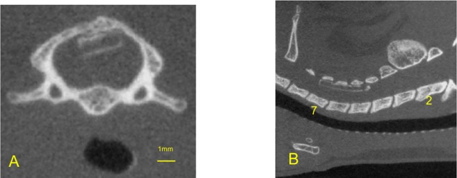
The detailed surgical procedure to create the spinal cord compression-induced cervical myelopathy model has been described [7]. Under general anesthesia with 2% isoflurane, a midline incision was made in the nuchal area, and the C3-Th1 laminae were exposed. A sheet of expandable urethane compound polymer (size 2 × 6 × 0.7 mm; Aquaprene C®, Sanyo Chemical Industries, Ltd., Tokyo, Japan) was inserted into the sublaminar space of C5-C6 (Fig 1A and 1B). This sheet gradually expands to 230% of the original volume over 48–72 hours by absorbing water in the tissue. In this model, the decline in motor function is delayed, with a latency period after compression introduction, and then gradual progression, whereas no acute damage suggestive of SCI is observed. This model reproduces the characteristic course and features of clinical degenerative cervical myelopathy [7].
Fig 1. The spinal cord compression-induced cervical myelopathy model.
A: Computed tomography (CT) axial view at the C5 levels, 0.5 mm above the intervertebral foramen. B: CT sagittal view of the cervical spine. Aquaprene® (expandable urethane compound sheet, size 2 × 6 × 0.7 mm) was inserted under the C5-C6 laminae.
Experimental design
Preliminary experiment
In a preliminary experiment, we confirmed the course of motor function decline in this model to determine when to administer EPO in the treatment experiment.
Briefly, 40 rats were allocated to two groups; sham operation group (n = 15) and compression group (n = 25). In the sham group, rats underwent a sham operation; the polymer sheet was placed under the laminae and removed immediately. In the compression group, this polymer sheet was left in place and continued to compress the spinal cord chronically (Fig 1A and 1B). The motor functions were evaluated once a week from 1 week before surgery to 26 weeks after surgery.
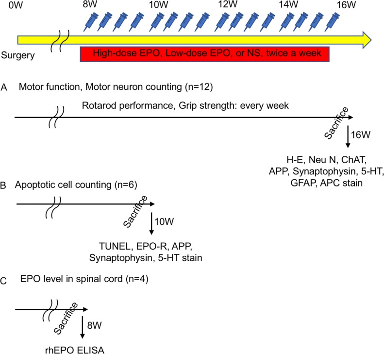
Treatment experiment (Fig 2)
Fig 2. Treatment experiment: Experimental design.
Administration of low-dose and high-dose EPO and normal saline was started twice a week at 8 weeks postoperatively. A: Forty-eight rats were divided into four groups (sham, vehicle, high-dose EPO, and low-dose EPO). The motor functions of rotarod performance and grip strength were evaluated once a week before surgery to 16 weeks after surgery. Every rat was sacrificed, and histological analysis was performed (H-E staining and NeuN staining). B: Another 18 rats were divided into three groups (sham, vehicle, and high-dose EPO). Treatment was done from 8 weeks to 10 weeks after surgery, and all rats were sacrificed at 10 weeks after surgery. Apoptotic cells were evaluated with TUNEL staining at 10 weeks after surgery. C: Another 12 rats were divided into three groups (vehicle, high-dose EPO, and low-dose EPO). Each single treatment was done 8 weeks after surgery. All rats were sacrificed 12 hours after injection, and the EPO levels in the spinal cord was measured using a rhEPO enzyme-linked immunosorbent assay (ELISA).
In the treatment experiment, 48 rats were allocated to four groups; sham group (sham operation + normal saline [NS]; n = 12), vehicle group (compression + NS; n = 12), low-dose EPO group (compression + EPO low dose; n = 12), and high-dose EPO group (compression + EPO high dose; n = 12). From the results of the preliminary experiments, the motor function was significantly decreased 8 weeks after surgery. Therefore, administration of rhEPO or NS was started from 8 weeks after surgery and lasted until 16 weeks; the frequency of administration was twice a week. In the sham group, rats underwent the sham operation and received administration of NS subcutaneously. In the vehicle group, rats underwent polymer sheet implantation and received administration of NS subcutaneously. In the low-dose EPO group, spinal cord compression-induced cervical myelopathy model rats received rhEPO 500 IU/kg/day (rhEPO; kindly provided by Chugai Pharmaceutical Co., Ltd., Tokyo, Japan) subcutaneously. In the high-dose EPO group, spinal cord compression-induced cervical myelopathy rats received administration of rhEPO 5000 IU/kg/day subcutaneously. The motor functions were also evaluated once a week from 1 week before surgery to 16 weeks after surgery. Histological assessment of the anterior horn (H-E, Neu N, ChAT) was evaluated at 16 weeks after surgery (Fig 2A).
All rats were weighed weekly.
Motor function analysis
Rotarod performance
Rotarod performance was assessed by using the rotarod device (ENV-557, Med Associates Inc., St. Albans, VT, USA). Based on our previous research, a moderate rotation speed of 10 rpm was set [7] [8] [9] [10]. All rats could walk on the rotarod for more than 300 seconds before surgery. Therefore, 300 seconds was set as the cut-off. Three trials in each session were performed for all rats. We recorded the longest duration time of the three trials.
Forelimb grip strength
Forelimb grip strength was assessed by using a digital force meter (MK-380CM/F, Muromachi Kikai, Tokyo, Japan). We assessed grip strength according to the methods of Meyer et al [26]. The animals were evaluated before surgery and once a week after surgery. All rats also performed three trials in each session, and the maximum score (in newtons: N) was used for data analysis.
Histological analysis
Hematoxylin and eosin (H-E) staining at C5-6 levels
At 16 weeks after surgery, transcardial perfusion was performed with 4% paraformaldehyde in phosphate-buffered saline (PBS) in all rats. The spinal cord segment at C5-6 was removed en bloc and placed in 4% paraformaldehyde solution for 3 days. After this process, these C5-6 segments were embedded in paraffin and sectioned at a slice thickness of 5 μm and a gap interval of 5 μm over 1000 μm length, according to stereological considerations of motor neurons [7] [8] [9] [10]. One hundred specimens of all rats were stained with H-E. Motor neurons have large nuclei and well-developed, densely stained Nissl bodies in the cytoplasm. The characteristic large nucleolus has a uniform diameter of approximately 5 μm [27] [28]. In H-E-stained sections, we regarded such cells as motor neurons. Motor neurons on both sides of the anterior horn gray matter were counted.
H-E staining at C2, Th4, Th12 levels
At 16 weeks after surgery, transcardial perfusion was performed with 4% paraformaldehyde in phosphate-buffered saline (PBS) in another 9 rats (sham group; n = 3, vehicle group; n = 3, and high-dose EPO group; n = 3). Each segment was cut into 10 sections in the same manner as described above. Ten specimens of all rats were stained with H-E. Motor neurons on both sides of the anterior horn gray matter were counted.
NeuN staining
NeuN protein appears in neuron-specific nuclei. The nucleus of the motor neuron is more clearly detected with NeuN staining compared with H-E staining.
In the treatment experiment, 10 specimens (thickness = 5 μm, gap interval = 50 μm) of five rats in all groups were stained with immunohistochemistry. Both sides of the anterior horn were also evaluated with NeuN staining following application of the chromogen diaminobenzidine (Dako North America, Santa Clala, CA, USA, 1:100) using the labeled streptavidin biotin technique [29]. Rabbit anti-NeuN (EMD Millipore Corporation, Burlington, MA, USA, 1:100) was used as the primary antibody. NeuN-positive cells on both sides of the anterior horn gray matter were counted.
Choline Acetyltransferase (ChAT) staining
In spinal cord, ChAT is expressed in motor neurons and pre-ganglionic autonomic neurons. Here, ChAT staining was used as specification of motor neurons.
In the treatment experiment, 3 specimens (thickness = 5 μm, gap interval = 50 μm) of one rat in vehicle and high-dose EPO groups at 16 weeks after surgery were stained with immunohistochemistry. Both sides of the anterior horn were also evaluated with ChAT staining following application of the chromogen diaminobenzidine (Dako North America, Santa Clala, CA, USA, 1:100) using the labeled streptavidin biotin technique [29]. Rabbit anti-ChAT (GeneTex, Alton Pkwy Irvine, CA, USA, 1:100) was used as the primary antibody. ChAT-positive cells on both sides of the anterior horn gray matter were counted.
Glial fibrillary acid protein (GFAP) staining
GFAP is an intermediate filament protein that is specifically expressed in cells of the astroglia lineage and is widely used as an astrocytic marker in the brain and spinal cord. In the treatment experiment, 3 specimens (thickness = 5 μm, gap interval = 50 μm) of one rat in vehicle and high-dose EPO groups at 16 weeks after surgery were stained with immunohistochemistry. Both sides of the anterior horn were also evaluated with GFAP staining following application of the chromogen diaminobenzidine (Dako North America, Santa Clala, CA, USA, 1:100) using the labeled streptavidin biotin technique [29]. Rabbit anti-GFAP (NOVUS, Centennial, CO, USA, 1:100) was used as the primary antibody. GFAP positive astrocytes on both sides of the anterior horn gray matter were counted.
Allophycocyanin (APC) staining
APC is expressed in the oligodendrocyte in the brain and spinal cord. Here, APC staining was used as specification of oligodendrocytes.
In the treatment experiment, 3 specimens (thickness = 5 μm, gap interval = 50 μm) of one rat in Vehicle and high-dose EPO groups at 16 weeks after surgery were stained with immunohistochemistry. Both sides of the anterior horn were also evaluated with APC staining following application of the chromogen diaminobenzidine (Dako North America, Santa Clala, CA, USA, 1:100) using the labeled streptavidin biotin technique [29]. Rabbit anti-APC (abcam, Cambridge, Cambridgeshire, UK, 1:100) was used as the primary antibody. APC positive oligodendrocytes on both sides of the anterior horn gray matter were evaluated.
Erythropoietin receptor (EPO-R) staining
In the treatment experiment, 3 specimens (thickness = 5 μm, gap interval = 50 μm) of one rat in vehicle and high-dose EPO groups at 10 weeks after surgery were stained with immunohistochemistry. Both sides of the anterior horn were also evaluated with EPO-R staining following application of the chromogen diaminobenzidine (Dako North America, Santa Clala, CA, USA, 1:100) using the labeled streptavidin biotin technique [29]. Rabbit anti-EPO-R (NOVUS, Centennial, CO, USA, 1:100) was used as the primary antibody. EPO-R-positive cells on both sides of the anterior horn gray matter were counted.
5-hydroxytryptamine (5-HT) staining
5-hydroxytryptamine (5-HT) (Serotonin) is a monoamine neurotransmitter in raphespinal axons. Here, 5-HT staining was used as an axonal marker of raphespinal tract.
In the treatment experiment, 3 specimens (thickness = 5 μm, gap interval = 50 μm) of one rat in vehicle and high-dose EPO groups at 10 and 16 weeks after surgery were stained with immunohistochemistry. Both sides of the anterior horn were also evaluated with 5-HT staining following application of the chromogen diaminobenzidine (Dako North America, Santa Clala, CA, USA, 1:100) using the labeled streptavidin biotin technique [29]. Rabbit anti-5-HT (NOVUS, Centennial, CO, USA, 1:100) was used as the primary antibody. 5-HT-positive axons on both sides of the anterior horn gray matter were evaluated.
Growth Associated Protein 43 (GAP-43) staining
GAP-43 is a membrane bound protein expressed in extending axons and its expression likely represents a high-growth state [30].
In the treatment experiment, 3 specimens (thickness = 5 μm, gap interval = 50 μm) of one rat in vehicle and high-dose EPO groups at 10 and 16 weeks after surgery were stained with immunohistochemistry. Both sides of the anterior horn were also evaluated with GAP-43 staining following application of the chromogen diaminobenzidine (Dako North America, Santa Clala, CA, USA, 1:100) using the labeled streptavidin biotin technique [29]. Rabbit anti-GAP-43 (Proteintech Group, Chicago, IL, USA, 1:100) was used as the primary antibody. GAP-43-positive cells on both sides of the anterior horn gray matter were evaluated.
Synaptophysin staining
Synaptophysin is a synaptic vesicle glycoprotein in neurons in the brain and spinal cord. Here, synaptophysin was used as an axonal synaptic marker in spinal cord.
In the treatment experiment, 3 specimens (thickness = 5 μm, gap interval = 50 μm) of one rat in the vehicle and high-dose EPO groups at 10 and 16 weeks after surgery were stained with immunohistochemistry. Both sides of the anterior horn were also evaluated with synaptophysin staining following application of the chromogen diaminobenzidine (Dako North America, Santa Clala, CA, USA, 1:100) using the labeled streptavidin biotin technique [29]. Rabbit anti-Synaptophysin (NOVUS, Centennial, CO, USA, 1:100) was used as the primary antibody. Synaptophysin positive axons on both sides of the anterior horn white matter were counted.
Amyloid Precursor Protein (APP) staining
APP is an integral membrane protein expressed in the synapses of neurons. APP is well known as the precursor molecule whose proteolysis generates beta amyloid. Increased APP in axons is thought to represent protein accumulation due to disruption of axon flow [31].
Here, APP staining was used as a marker of axonal and neuronal damage [32].
In the treatment experiment, 3 specimens (thickness = 5 μm, gap interval = 50 μm) of one rat in vehicle and high-dose EPO groups at 10 and 16 weeks after surgery were stained with immunohistochemistry. Both sides of the anterior horn were also evaluated with APP staining following application of the chromogen diaminobenzidine (Dako North America, Santa Clala, CA, USA, 1:100) using the labeled streptavidin biotin technique [29]. Rabbit anti-APP (GeneTex, Alton Pkwy Irvine, CA, USA, 1:100) was used as the primary antibody. APP-positive cells on both sides of the anterior horn gray matter and white matter were counted.
Terminal deoxynucleotidyl transferase-mediated deoxyuridine triphosphate-biotin nick end labeling (TUNEL) staining
Apoptotic cell death was investigated 10 weeks after surgery. Another 18 rats (Sham group; n = 6, Vehicle group; n = 6, high-dose EPO group; n = 6) were perfused transcardially with 4% paraformaldehyde in PBS (Fig 2B). The C5-6 segment of the spinal cord was embedded in optimal cutting temperature compound and frozen in liquid nitrogen. Three sections from C5-6 segments (thickness = 20 μm, gap interval = 50 μm) were cut in a cryostat and stained with the In Situ Cell Death Detection Kit, POD (Roche, Basel, Switzerland) according to the manufacturer’s recommendations. Nuclei were counterstained with 4’,6-diamidino-2-phenylindole (DAPI, 1:5000 in PBS) (Molecular Probes, Eugene, OR, USA). The TUNEL stain signal was observed under an FV300 confocal microscope (Olympus Optical Company, Ltd., Tokyo, Japan). TUNEL- and DAPI-positive cells were counted, and the ratios of apoptotic cells to total nuclei were evaluated in each group.
Caspase-3 staining
Caspase-3 staining was evaluated as an indicator of the course of apoptotic cell death.
In the treatment experiment, 3 specimens (thickness = 5 μm, gap interval = 50 μm) of one rat in the vehicle and high-dose EPO groups at 10 weeks after surgery were stained with immunohistochemistry. Both sides of the anterior horn were also evaluated with Caspase-3 staining following application of the chromogen diaminobenzidine (Dako North America, Santa Clala, CA, USA, 1:100) using the labeled streptavidin biotin technique [29]. Rabbit anti-Caspase-3 (NOVUS, Centennial, CO, USA, 1:100) was used as the primary antibody. Caspase-3-positive cells on both sides of the anterior horn gray matter were evaluated.
Hematological assessment
Another 12 rats were divided into three groups. All these rats underwent the operation to place the polymer sheet under the C5-6 laminae and were treated with NS or rhEPO twice a week from 8 weeks after surgery. The vehicle group, low-dose EPO group, and high-dose EPO group were examined. All rats were subjected to inhalation anesthesia with 2% isoflurane. Blood samples (0.5 ml/body) were collected by venipuncture from the tail vein at 2, 4, and 6 weeks after the first EPO administration. Blood samples were collected into blood collection tubes with EDTA 2K (BD Microtainer, Japan Becton, Dickinson and Company, Tokyo, Japan) immediately. RBC, hemoglobin (Hb), and hematocrit (Ht) values were assessed with an automated hematology analyzer (XE 2100, Sysmex, Hyogo, Japan).
Assessing rhEPO levels in spinal cord tissue
To assess whether subcutaneously injected EPO was transferred to the spinal cord, we measured EPO levels in the spinal cord with an enzyme-linked immunosorbent assay. Another 12 rats were divided into three groups; vehicle group, low-dose EPO group, and high-dose EPO group. All these rats underwent the operation in which the polymer sheet remained under the C5-6 laminae. They received NS or rhEPO 8 weeks after surgery. Twelve hours after subcutaneous injection of NS or rhEPO, all rats were sacrificed under anesthesia, and blood was completely removed by transcardial perfusion with PBS to exclude rhEPO from blood (Fig 2C). The spinal cord segment at the C5-6 levels was removed en bloc. These tissues were homogenized in IP buffer with an ultra Turrax homogenizer and centrifuged at 12000 rpm at 4°C for 5 min. Supernatants were removed and analyzed to determine the levels of rhEPO in the spinal cord tissue. The total protein of the spinal cord tissue was determined using bovine serum albumin as a standard. The rhEPO concentration in spinal cord tissue was measured with a rhEPO enzyme-linked immunosorbent assay kit (R&D Systems Europe, Abingdon, UK) according to the manufacturer’s instructions. The concentration was described as the rhEPO levels per 1 g tissue (mIU/g) and tissue dose % of injected dose (%ID).
Statistical analysis
GraphPad Prism 6 software for Windows (GraphPad Software Inc., La Jolla, CA, USA) was used for statistical analysis. Data are expressed as the mean ± standard error of the mean. The duration of walking on the rotarod, forelimb grip strength, body weight and hematological data were analyzed using two-way repeated-measures analysis of variance (ANOVA), which takes into account the passage of time. The number of anterior horn motor neurons with H-E, NeuN, and TUNEL staining and the rhEPO levels in spinal cord were tested using one-way ANOVA, which does not take into account the passage of time. The number of ChAT, APP, EPO-R-positive cells and Synaptophysin positive axons were tested using unpaired t-test. P values <0.05 were regarded as significant.
Results
Motor function
Preliminary experiment
Rotarod performance declined gradually with a latency period of 4 weeks in the compression group. At 7 weeks after surgery, the walking duration significantly decreased in the compression group compared to the sham group (P < 0.001: two-way ANOVA). In the compression group, the duration declined gradually and reached a plateau after 16 weeks (Fig 3A).
Fig 3. Preliminary experiment.
A: Time course of rotarod performance measured by walking time on a rotarod (cut-off 300 seconds). In the compression group, the walking time gradually started to decline from 4 weeks and showed a significant decrease at 7 weeks after surgery (P < 0.001: two-way ANOVA). The performance reached a plateau with a low duration of about 50 seconds after 15 weeks. B: Time course of forelimb grip strength. In the compression group, grip strength decreased at 1 week after surgery due to surgery, but gradually increased as body weight increased. However, grip strength gradually declined from 6 weeks, and showed a significant difference at 7 weeks after surgery (P < 0.0001: two-way ANOVA) After that, the strength continued to decrease gradually, reaching approximately 10.5 N at 26 weeks postoperatively.
Forelimb grip strength increased with weight gain in all groups until 5 weeks after surgery. In the sham group, it reached a plateau after 5 weeks from surgery. On the other hand, it started to decrease from 6 weeks in the compression group. The strength gradually declined and significantly decreased after 7 weeks compared with the sham group (P < 0.001: two-way ANOVA) (Fig 3B).
Based on these results, we decided to administer EPO beginning 8 weeks after surgery as a treatment experiment.
Treatment experiment
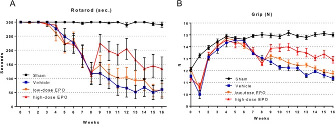
The rotarod performance of the compression groups (vehicle, low-dose EPO, and high-dose EPO groups) declined gradually from 5 weeks after surgery, and a significant decrease was seen from 7 weeks compared with the sham group (P < 0.005: two-way ANOVA) as in the preliminary experiments (Fig 4A).
Fig 4. Treatment experiment: Motor function.
A: Time course of rotarod performance measured by walking time on a rotarod (cut-off 300 seconds). In the compression models (vehicle, low-dose EPO, and high-dose EPO groups), rotarod performance gradually declined from 3 weeks after surgery, and showed a significant difference at 7 weeks after surgery. After administration of EPO from 8 weeks, rotarod performance improved in the EPO groups. Especially in the high-dose EPO group, performance markedly improved. This effect was maintained with a significant difference by week 13 after surgery (P < 0.01: two-way ANOVA). In the low-dose EPO group, slight improvement in rotarod performance was observed, but it did not reach statistical significance compared with the vehicle group. B: Time course of forelimb grip strength. In the compression groups, the strength started to decline from 6 weeks after surgery, and a significant decline was observed at 7 weeks. EPO was administered at 8 weeks, and grip strength improved, especially in the high-dose EPO group. Significant improvement was seen from 9 weeks in the high-dose EPO group (P < 0.0001: two-way ANOVA) and continued up to 16 weeks after surgery. In the low-dose EPO group, grip strength slightly improved, but no significant difference was found compared with the vehicle group.
After EPO administration beginning 8 weeks after surgery, rotarod performance started to improve in the treatment groups (low-dose and high-dose EPO groups).
Especially in the high-dose EPO group, rotarod performance significantly improved compared with the other compression groups (vehicle and low-dose EPO groups), although the performance gradually declined from 13 weeks. The effects of EPO continued for 5 weeks after EPO administration (P < 0.01: two-way ANOVA). Furthermore, the high-dose EPO group improved to the levels at which no significant difference in motor function was seen between the sham and high-dose EPO groups at 9 weeks after surgery (N.S: one-way ANOVA). The low-dose EPO group showed slightly improved rotarod performance, but did not show significant improvement compared with the vehicle group (Fig 4A).
The forelimb grip strength of the compression groups decreased at 1 week after surgery but started to recover gradually from 2 weeks after surgery. The strength of the compression group showed an improvement course equal to that of the sham group from 2 weeks after surgery and then started to decrease gradually from 7 weeks; at this time, the strength was significantly decreased compared with the sham group (P < 0.001: two-way ANOVA) (Fig 4B).
After EPO administration at 8 weeks after surgery, grip strength started to improve in the treatment groups (low-dose EPO, high-dose EPO groups).
In the high-dose EPO group, the strength significantly improved compared with the other compression groups (vehicle and low-dose EPO groups) (P < 0.0001: two-way ANOVA). Its effects continued throughout the period of EPO administration (9 to 16 weeks after surgery), although the strength gradually decreased from 4 weeks after EPO administration.
In contrast, the low-dose EPO group showed a slight improvement in strength, but it did not show significant improvement compared with the vehicle group (Fig 4B).
Histopathological analysis
H-E staining in C5-6 levels
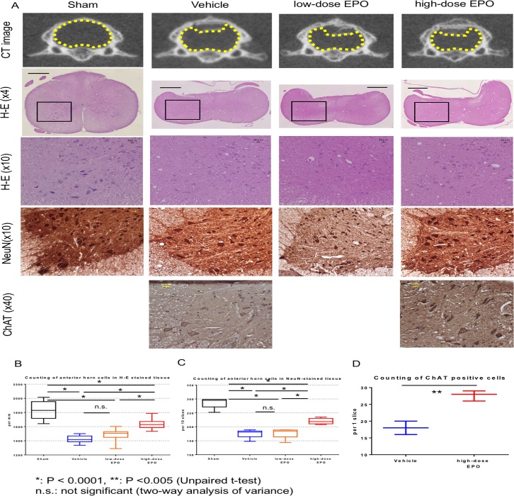
At 16 weeks after surgery, the loss of anterior horn motor neurons and vacuolar degeneration in the spinal cord were observed in H-E-stained sections from the compression groups (vehicle, low-dose EPO, and high-dose EPO group) (Fig 5A). The numbers of motor neurons were 1834.7 ± 115.4 (sham group), 1421.6 ± 50.1 (vehicle group), 1484.7 ± 74.2 (low-dose EPO group), and 1640.0 ± 66.9 (high-dose EPO group). The number of motor neurons on both sides of the anterior horn was significantly decreased in every compression group compared to the non-compression sham group (P < 0.0001: one-way ANOVA). In the high-dose EPO group, however, the motor neurons were significantly preserved compared with the other compression groups (vehicle and low-dose EPO groups; P < 0.0001: one-way ANOVA) (Fig 5B).
Fig 5. Treatment experiment: Anterior motor neurons.
A: Top panels: CT axial view in C5. In the compression groups (vehicle, low-dose EPO, and high-dose EPO), the spinal cord was compressed by Aquaprene® (expandable urethane compound sheet, size 2 × 6 × 0.7 mm). The yellow dotted figure shows the outline of the spinal cord. Second panels: The spinal cord at the C5 levels was sliced into 5-μm thick sections at 16 weeks after surgery. Hematoxylin and eosin staining of cross sections of spinal cord is shown (original magnification ×4, scale bar = 100 μm). In the compression groups, the spinal cord was flattened. Black box shows the region of the anterior horn. Third panels: The black box in the second panel was magnified (×10, scale bar 20 μm). Cells with large nuclei and well-developed, densely stained Nissl bodies in the cytoplasm indicate motor neurons. In the vehicle and low-dose EPO groups, motor neurons decreased, and vacuolar degeneration was obvious. In the high-dose EPO group, motor neurons were preserved, although vacuolar degeneration was present. Fourth panels: NeuN staining of the anterior horn (×10, scale bar 20 μm). The nuclei of motor neurons are clearly detected with NeuN staining compared with H-E staining. Motor neurons decreased in the vehicle and low-dose EPO groups, but they were preserved in the high-dose EPO group. Bottom panels: ChAT staining of the anterior horn (×40, scale bar 20 μm). Motor neurons were stained with ChAT staining. Motor neurons were preserved in the high-dose EPO groups compared to Vehicle groups. B: Counting of anterior horn cells in H-E-stained tissue. The number of cells with large nuclei in the anterior horn was counted in every group. The number was significantly decreased in the compression groups compared with the sham group (*P < 0.0001: one-way ANOVA). However, in the high-dose EPO group, the number was significantly preserved compared with the other two compression groups (vehicle and low-dose EPO groups). C: Counting of anterior horn cells in NeuN-stained tissue. NeuN-positive cells were significantly decreased in the compression groups compared with the sham group (*P < 0.0001: one-way ANOVA). However, in the high-dose EPO group, the number was significantly preserved compared with the other two compression groups (*P < 0.0001: one-way ANOVA). The tendency in the cell count was similar to that with H-E staining. D: Counting of anterior horn cells in ChAT-stained tissue. ChAT-positive cells were significantly preserved in the high-dose EPO groups compared with the vehicle group (**P < 0.005: one-way ANOVA).
Cell counting of NeuN-positive cells
The number of NeuN-positive cells in 10 slices of each group was 286.8 ± 17.6 (sham group), 176.0 ± 14.3 (Vehicle group), 178.0 ± 17.1 (low-dose EPO group), and 220.4 ± 9.4 (high-dose EPO group). NeuN-positive cells in each compression group decreased compared with the sham group (P < 0.0001: one-way ANOVA), but the number in the high-dose EPO group was significantly preserved compared with the vehicle and low-dose EPO groups (P < 0.0001: one-way ANOVA). This tendency was similar to that of the number of motor neurons in H-E-stained sections (Fig 5B and 5C).
Cell counting of ChAT-positive cells
The number of ChAT-positive cells in 1 slice of each group was 18.0 ± 1.6 (Vehicle group) and 27.7 ± 1.2 (High-dose EPO group). ChAT-positive cells in the high-dose EPO group were significantly preserved compared with the vehicle group (P < 0.005: one-way ANOVA) (Fig 5A and 5D).
H-E staining in C2, Th4 and Th12 levels
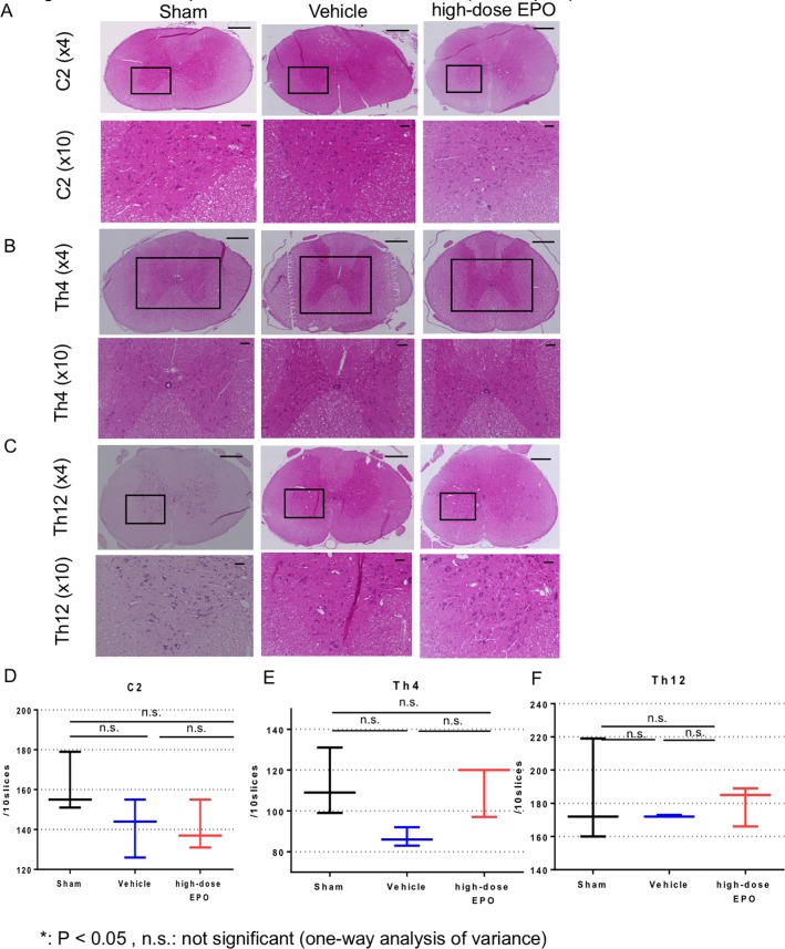
At 16 weeks after surgery, H-E staining was also performed at C2, Th4 and Th12 levels in the sham, vehicle and high-dose EPO groups (Fig 6A, 6B and 6C). In C2 levels, the numbers of motor neurons were 161.6 ± 12.4 (sham group), 141.6 ± 12.0 (vehicle group) and 141.0 ± 10.2 (high-dose EPO group). There was no significant difference in the number of motor neurons in all groups (Fig 6D). In Th4 levels, the numbers of motor neurons were 113.0 ± 13.4 (Sham group), 87.0 ± 3.7 (Vehicle group) and 112.3 ± 10.9 (High-dose EPO group). There was no significant difference in the number of motor neurons in all groups although the number of motor neurons tended to be slightly lower in vehicle group (sham vs vehicle: P = 0.1, high-dose EPO vs vehicle: 0.11, one-way ANOVA) (Fig 6E). In Th12 levels, the numbers of motor neurons were 183.7 ± 25.5 (sham group), 172.3 ± 0.5 (vehicle group) and 180.0 ± 10.0 (high-dose EPO group). There was no significant difference in the number of motor neurons in all groups (Fig 6F).
Fig 6. Treatment experiment: Anterior motor neurons (whole spine other than C5 levels).
A: Top panels: The spinal cord at the C2 levels was sliced into 5-μm thick sections at 16 weeks after surgery. Hematoxylin and eosin staining of cross sections of spinal cord is shown (original magnification ×4, scale bar = 100 μm). In the compression groups, the spinal cord was flattened. Black box shows the region of the anterior horn. Second panels: The black box in the second panel was magnified (×10, scale bar 20 μm). B: Top panels: The spinal cord at the Th4 levels was sliced into 5-μm thick sections at 16 weeks after surgery. Hematoxylin and eosin staining of cross sections of spinal cord is shown (original magnification ×4, scale bar = 100 μm). In the compression groups, the spinal cord was flattened. Black box shows the region of the both anterior horn. Second panels: The black box in the second panel was magnified (×10, scale bar 20 μm). C: Top panels: The spinal cord at the CTh12 levels was sliced into 5-μm thick sections at 16 weeks after surgery. Hematoxylin and eosin staining of cross sections of spinal cord is shown (original magnification ×4, scale bar = 100 μm). In the compression groups, the spinal cord was flattened. Black box shows the region of the anterior horn. Second panels: The black box in the second panel was magnified (×10, scale bar 20 μm). D: Counting of anterior horn cells in H-E-stained tissue at C2 levels. There was no significant difference in the number of motor neurons in each group (N.S: one-way ANOVA). E: Counting of anterior horn cells in H-E-stained tissue in Th4 levels. There was no significant difference in the number of motor neurons in each group although it tended to be slightly lower in the vehicle group (sham vs vehicle: P = 0.1, high-dose EPO vs vehicle: 0.11, sham vs high-dose EPO: 0.97, one-way ANOVA) F: Counting of anterior horn cells in H-E-stained tissue in Th12 levels. There was no significant difference in the number of motor neurons in each group (N.S: one-way ANOVA).
TUNEL staining
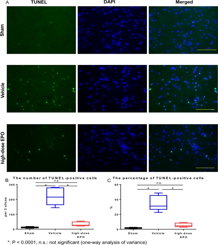
TUNEL-positive cells were significantly increased in the vehicle group compared with the other two groups (sham and high-dose EPO groups) (P < 0.0001: one-way ANOVA) (Fig 7A and 7B). We found no significant difference between the sham and high-dose EPO groups. The ratios of TUNEL-positive cells to DAPI-positive cells (%) were 1.72 ± 0.59% (sham group), 35.01 ± 9.17% (vehicle group), and 5.66 ± 2.27% (high-dose EPO group). The ratio in the vehicle group was significantly higher than that in the other two groups (P < 0.0001: one-way ANOVA), and we found no significant difference between the sham and high-dose EPO groups (Fig 7A and 7C).
Fig 7. Treatment experiment: TUNEL staining.
A: TUNEL staining was performed to detect apoptotic cells at 10 weeks after surgery. DAPI/TUNEL double staining is shown in each group (DAPI staining, TUNEL staining, DAPI/TUNEL staining, Bar = 100 μm). The vehicle group showed the highest number of TUNEL-positive cells. B: The number of TUNEL-positive cells was counted in each group. The number of TUNEL-positive cells in the vehicle group was significantly higher than in the other two groups (*P < 0.0001: one-way ANOVA), with no significant difference between the sham and high-dose EPO groups. C: The percentage of TUNEL-positive cells in each group. The percentage in the vehicle group was significantly higher than in the other two groups (*P < 0.0001: one-way ANOVA).
Caspase3 staining
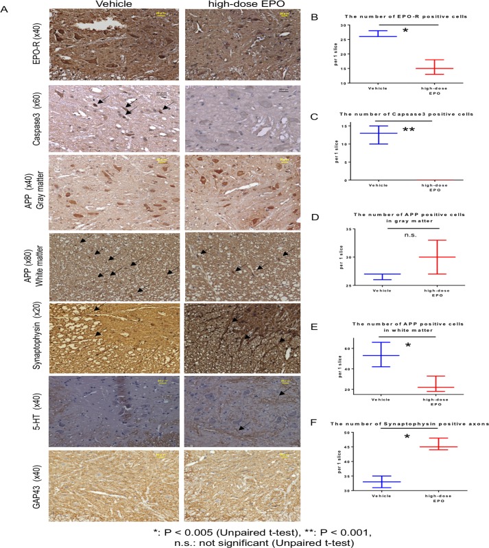
Caspase-3 staining was performed at 10 weeks after surgery. Although caspase-3-positive cells were detected in the vehicle group (12 ± 2.1 cells), we failed to observe any of these cells in the high-dose EPO group (P < 0.001, unpaired t-test) (Fig 8A and 8C).
Fig 8. Immunostaining at 10 weeks.
Top panels: The spinal cord at the C5 level was sliced into 5-μm-thick sections at 10 weeks after surgery and the gray matter was stained for EPO-R (original magnification ×40, scale bar = 20 μm). The expression of EPO-R in the anterior horn cells was clear in the vehicle group but weak in the EPO group. Second panels: Caspase-3 staining of spinal cord is shown. The arrows indicate caspase-3-positive nuclei, which were detected in the vehicle group but not in the EPO group. Third panels: APP staining of the gray matter of spinal cord is shown. Staining of anterior horn cells with APP was similar in both groups. Fourth panels: APP staining in the white matter of spinal cord is shown. Arrows show APP-positive cells. More APP-positive cells were observed in the vehicle group. Fifth panels: Synaptophysin staining of the white matter of spinal cord is shown. Arrows indicate synaptophysin-positive axons. In the EPO group, synaptophysin-positive axons extending from the gray matter to the white matter were clearly observed. Sixth panels: 5-HT staining of the gray matter of spinal cord is shown. Nerve fibers were clearly stained with 5-HT (indicated by arrows) in the gray matter from EPO group but not in that from vehicle group. Bottom panels: GAP-43 staining of gray matters of spinal cord is shown. The expression of positive cells was not clear in both the vehicle and EPO groups. B: The number of EPO-R-positive anterior horn cells was significantly higher in the vehicle group (P < 0.005, unpaired t-test). C: Caspase-3-positive cells were detected only in the vehicle group (P < 0.001, unpaired t-test). D: No significant difference was observed in the number of APP-positive cells in the gray matter between the two groups (P = 0.13, unpaired t-test). E: Significantly more APP-positive cells were detected in the white matter of the vehicle group (P < 0.05, unpaired t-test). F: The number of synaptophysin-positive axons was significantly higher in the high-dose EPO group (P < 0.005, unpaired t-test).
EPO-R staining
EPO-R staining was performed at 10 weeks after surgery. In all groups, EPO-R was mainly expressed in anterior horn cells. The number of EPO-R-positive anterior horn cells was significantly lower in the high-dose EPO group than in the vehicle group (vehicle: 26.6 ± 0.9 versus EPO: 15.3 ± 2.1 cells, P < 0.005, unpaired t-test) (Fig 8A and 8B).
APP staining
There was no significant difference in the number of APP positive cells in gray matter between the vehicle and the high dose EPO group at 10 weeks after surgery (26.7 ± 0.5 versus 30.0 ± 2.4) (Fig 8A and 8D). However, in white matter, the number of APP-positive cells was significantly higher in the vehicle group than in the high-dose EPO group (53.7 ± 9.8 versus 24.3 ± 6.3; P < 0.05, unpaired t-test) (Fig 8A and 8E).
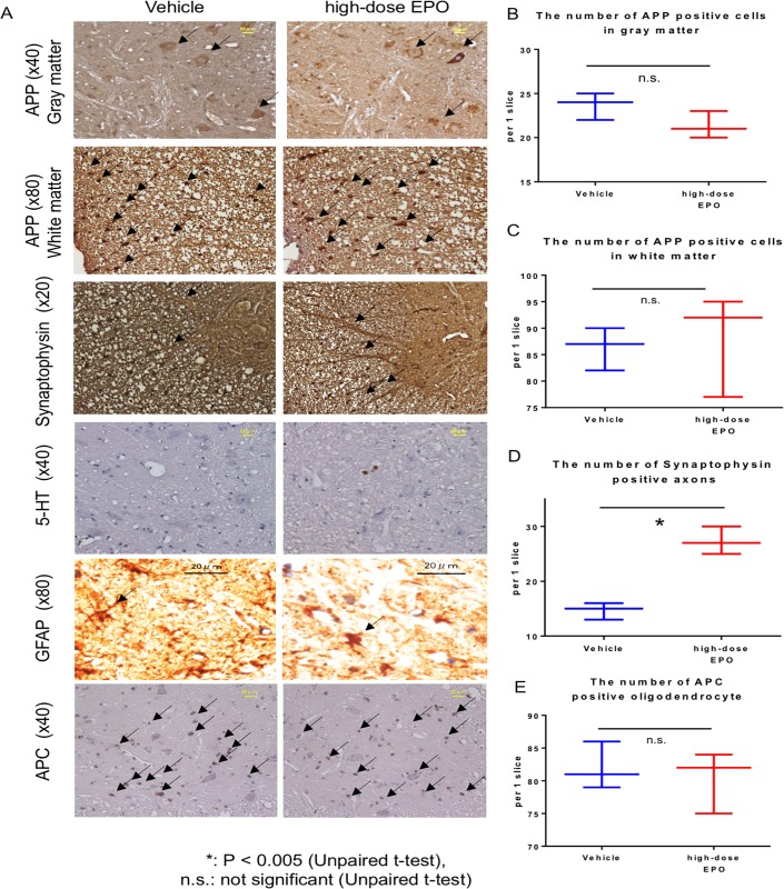
At 16 weeks after surgery, there was no significant difference in the number of APP-positive cells in the gray matter between the vehicle and high-dose EPO group (23.7 ± 1.3 versus 21.3 ± 1.3) (Fig 9A and 9B). However, APP staining intensity in anterior horn cells was attenuated in the EPO group (Fig 8A). In the white matter, there was no significant difference in the number of APP-positive cells between the vehicle and high-dose EPO group (86.3 ± 3.3 versus 88.0 ± 7.9) (Fig 9A and 9D).
Fig 9. Immunostaining at 16 weeks.
A Top panels: The spinal cord at the C5 levels was sliced into 5- μm thick sections at 16 weeks after surgery. APP staining of gray matter of spinal cord is shown (original magnification ×40, scale bar = 20 μm). Arrows show APP-positive cells. The staining intensity of APP in the anterior horn cells was lower in the vehicle group than that in the EPO group. Second panels: APP staining of the white matter of spinal cord is shown. Arrows show APP-positive cells. Third panels: Synaptophysin staining of the white matter of spinal cord is shown. The arrows indicate synaptophysin-positive axons. Synaptophysin-positive axons extending from the gray matter to the white matter were clearly observed in the EPO group. Fourth panels: 5-HT staining of gray matter of spinal cord is shown. The expression of 5-HT was not clear in both groups. Fifth panels: Astrocytes were occasionally observed in the gray matter of both groups. The arrows indicate astrocytes. Bottom panels: Oligodendrocytes were occasionally observed in the gray matter of both groups. The arrows indicate oligodendrocytes. B: There was no significant difference in the number of APP-positive cells in the gray matter between the two groups (P = 0.13, unpaired t-test). C: There was no significant difference in the number of APP-positive cells in the white matter between the two groups (P = 0.80, unpaired t-test). D: The number of synaptophysin-positive axons was significantly higher in the high-dose EPO group (P < 0.005, unpaired t-test). E: There was no significant difference in the number of APC-positive oligodendrocytes between the two groups (P = 0.74: unpaired t-test).
Synaptophysin staining
The number of synaptophysin-positive axons at 10 weeks after surgery was significantly higher in the high-dose EPO group than in the vehicle group (45.7 ± 1.7 versus 33.0 ± 1.6; P < 0.005, unpaired t-test) (Fig 8A and 8F).
The number of synaptophysin-positive axons at 16 weeks after surgery was also significantly preserved and found to be higher in the high-dose EPO group than in the vehicle group (27.3 ± 2.1 versus 14.7 ± 1.2; P < 0.005, unpaired t-test) (Fig 9A and 9D).
5-HT staining
Although the expression was unclear in both groups at 16 weeks after surgery (Fig 9A), clear expression was detected along the nerve fibers in the gray matter of the EPO group at 10 weeks after surgery (Fig 8A). The expression was weak in the vehicle group.
GAP-43 staining
Although GAP-43 staining was performed at 10 weeks after surgery, the expression in the anterior horn cells was not clear in both groups (Fig 8A).
GFAP staining
GFAP staining was performed 16 weeks after surgery. The results showed some astrocytes in the gray matter of both groups, but there was no significant difference between the two groups (Fig 9A).
APC staining
APC staining was performed 16 weeks after surgery. Oligodendrocytes in gray matter was 82.0 ± 2.9 cells in the vehicle group and 80.3 ± 3.9 cells in the high-dose EPO group, and there was no significant difference between the two groups (P = 0.74: unpaired t-test) (Fig 9A and 9E).
Hematological data
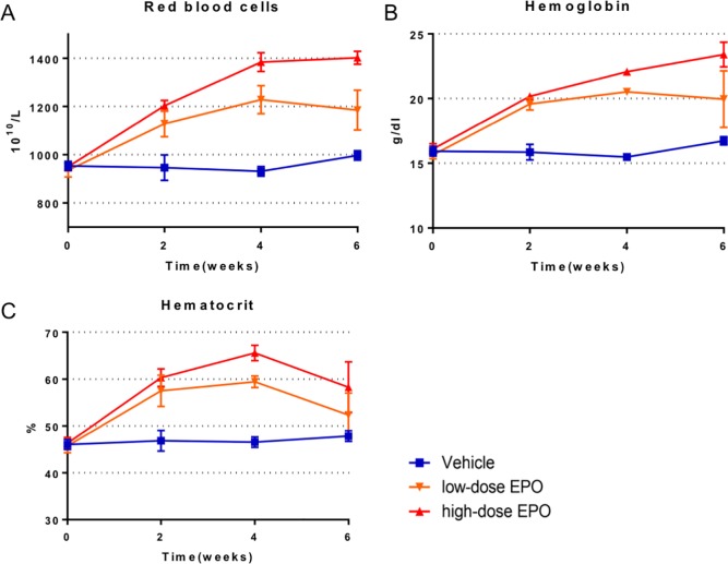
After administration of EPO, the RBC, Hb, and Ht values increased immediately in the EPO-administered groups (low-dose and high-dose EPO groups) (P < 0.0001: two-way ANOVA). The trend in RBC and Hb values showed a similar increasing tendency after EPO administration (Fig 10A and 10B). Eventually, the RBC and Hb values increased to approximately 1.2 and 1.4 times in the low-dose and high-dose EPO groups, respectively, compared to the baseline value (vehicle group). The values were significantly higher in both EPO-administered groups than the vehicle group until 6 weeks after administration (P < 0.0001: two-way ANOVA) (Fig 10A and 10B).
Fig 10. Treatment experiment: Hematological data.
A: Time course of red blood cells (RBCs). RBCs increased immediately in the low-dose and high-dose EPO groups (P < 0.0001: two-way ANOVA). From 4 weeks after administration, we found a significant difference between the low-dose and high-dose EPO groups (P < 0.0001: two-way ANOVA). Eventually, RBCs increased up to approximately 1.2 and 1.4 times in the low-dose and high-dose EPO groups, respectively, compared with the vehicle group. B: Time course of hemoglobin (Hb). The Hb value increased immediately in the low-dose and high-dose EPO groups (P < 0.0001: two-way ANOVA). The time course was similar to that of RBCs. Eventually, the Hb value increased up to approximately 1.2 and 1.4 times in the low-dose and high-dose EPO groups, respectively, compared with the vehicle group. C: Time course of hematocrit (Ht). The Ht value increased immediately in the low-dose and high-dose EPO groups (P < 0.0001: two-way ANOVA). The Ht value was the highest at 4 weeks, and then peaked. The maximum Ht value was approximately 1.3 and 1.4 times in the low-dose and high-dose EPO groups, respectively, compared with the vehicle group.
The Ht value in the EPO-administered groups was the highest at 4 weeks and increased to approximately 1.3 and 1.4 times in the low-dose and high-dose EPO groups, respectively, compared to the baseline value (P < 0.0001: two-way ANOVA). At 6 weeks after administration of EPO, the Ht value of the EPO-administered groups started to peak. The Ht value of the high-dose EPO group was significantly higher than that of the other two groups (P = 0.005: two-way ANOVA) at 6 weeks (Fig 10C).
rhEPO levels in spinal cord tissue
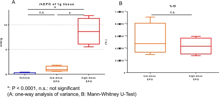
The rhEPO levels in the spinal cord 12 hours after subcutaneous injection of rhEPO was less than 0.10 mIU/g in the vehicle group, 1.07 ± 0.46 mIU/g in the low-dose EPO group, and 8.67 ± 2.33 mIU/g in the high-dose EPO group. The rhEPO levels was remarkably higher in the high-dose EPO group than in the other two groups (P < 0.0001: one-way ANOVA). In the low-dose EPO group, the rhEPO levels was slightly increased, but that of the low-dose EPO group did not show a significant difference compared with the vehicle group (Fig 11A).
Fig 11. Treatment experiment: ELISA of recombinant human EPO (rhEPO).
A: The amount of rhEPO per 1 g spinal cord tissue. The rhEPO levels was significantly higher in the high-dose EPO group compared to the other two groups (*P < 0.0001: one-way ANOVA). B: The tissue rhEPO levels for injected dose (%ID) in the low-dose and high-dose EPO groups. No significant difference was found in the %ID between the two groups.
The tissue % ID was 4.4 ± 1.2 (10−4%) in the high-dose EPO group and 5.4 ± 2.3 (10−4%) in the low-dose EPO group. We found no significant difference between the two groups (Fig 11B). This result shows that the rhEPO levels in the spinal cord was dose dependent.
Discussion
The present study demonstrated that EPO improved locomotor functions and preserved motor neurons and axons, even in developing myelopathy due to spinal cord compression. Furthermore, EPO was transferred into spinal cord tissue following subcutaneous EPO administration.
Some studies reported that EPO improves motor function in an experimental acute SCI model [21, 22, 33, 34]. EPO and the EPO receptor (EPO-R) are highly expressed in both the central and peripheral nervous systems [35]. The roles of EPO in these areas are in neuroprotection, angiogenesis, anti-apoptosis, and anti-inflammation [18, 24, 36]. Clinically, a preliminary randomized comparative trial was performed in patients with acute SCI. In this trial, the effect of EPO treatment was compared with high-dose methylprednisolone treatment. EPO had higher efficacy and fewer side effects than methylprednisolone, indicating a potential therapeutic effect for acute SCI patients [25].
On the other hand, there are few studies on neuroprotective agents for degenerative cervical myelopathy including spinal cord compression, compared to the studies in acute SCI. Among them, Riluzole, a sodium channel blocker, has shown efficacy in animal models of degenerative cervical myelopathy. Riluzole reduces glutamatergic excitotoxicity and improves neurological functional outcome [37, 38, 39].
The agent is currently approved by the US Food and Drug Administration for the treatment of amyotrophic lateral sclerosis. However, in the multicenter randomized control trial about the combined effect of riluzole in the surgery for degenerative cervical myelopathy, the additional effect has not been confirmed in neurological improvement [40].
Methylprednisolone has been also reported to be effective in decompression surgery for an animal model of degenerative cervical myelopathy. The addition of the agent to decompression surgery demonstrated a reduced inflammatory response, enhanced neuronal preservation and accelerated locomotor recovery without changes to the peripheral immune cell populations [41]. However, there are very few clinical studies on the role of corticosteroids in degenerative cervical myelopathy.
We have been investigating several neuroprotecitve agents for degenerative cervical myelopathy using a spinal cord compression model. In these studies, we have shown that Limaprost alfadex and Cilostazol have a preventive effect on compression-induced cervical myelopathy [8] [9]. Furthermore, we have confirmed that G-CSF not only has a preventive effect but also a therapeutic effect in the progressive phase of compression myelopathy [10]. However, in healthy people, G-CSF causes marked leukocytosis, which commonly results in fever, arthralgia, and rarely, thromboembolism and splenomegaly [11].
In contrast, EPO is used commonly and safely for renal anemia and preoperative autologous blood donation even in elderly patients or those with critical disease [13] [15].
On the other hand, the mechanism of neuroprotective effect of EPO for compression myelopathy remains unknown.
We previously demonstrated that blood flow in the compressed segment is markedly reduced, indicating the presence of local spinal cord ischemia in the chronic compression myelopathy model [35].
Consistent with our previous studies [9, 10], this study also demonstrated that chronic spinal cord compression induces apoptotic cell death. In hypoxic stress conditions, endogenous EPO is produced in response to low oxygen partial pressure and protects neurons [42]. Importantly, cell apoptosis induced by spinal cord compression is inhibited by high-dose EPO administration, indicating anti-apoptosis and anti-inflammatory effects of EPO [18, 24, 36]. Additionally, a rapid increase in RBC, Hb, and Ht values following EPO administration may improve the local oxygen supply and restore motor function (Fig 10). Liem et al. reported that blood transfusion for anemia improves cerebral oxygenation in newborn infants [43]. Although we could not directly evaluate local oxygen pressure, we speculate that improvement in cervical myelopathy is due to anti-apoptotic effects of EPO, preservation of motor neurons and axons, and improvement in local ischemia in the spinal cord with an increased oxygen supply.
Zhu et al. found that the expression of EPO-R increases following ischemia in the central nervous system, and that EPO treatment could reduce the expression of EPO-R by improving the ischemia [44]. In the present study, EPO-R expression in the anterior horn cells increased in the vehicle group but decreased in the EPO group. The results suggest that EPO improved local ischemia (Fig 9A and 9B).
In the current study, both high-dose and low-dose EPO increased hematopoietic values including RBC, Hb, and Ht. However, functional recovery was observed with high-dose EPO treatment in particular. High-dose EPO may have passed through the blood-spinal cord barrier. EPO is a high-molecular weight glycoprotein (30.4 kDa) [12]. In classic papers, the blood–brain barrier (BBB) was considered to be impermeable to large glycosylated molecules like EPO [45]. However, some recent studies have reported that EPO can pass through the BBB due to a high concentration and after BBB disruption such as that which follows brain and spinal cord contusion [46–48]. EPO can cross the BBB at 450 IU/kg or more in rats [46] and crosses the BBB in a dose-dependent manner in a rat brain contusion model [49]. In the current study, in fact, high-dose EPO was predominantly transferred into the spinal cord tissue 12 hours after EPO subcutaneous administration, probably resulting from passing through the blood-spinal cord barrier. Transfer of EPO into spinal cord tissue was dose dependent (Fig 11A), and the transfer activity was almost the same between the low-dose and high-dose EPO groups (Fig 11B). This finding demonstrates that the higher the dose of EPO that was administered, the more EPO can transfer into spinal cord tissue. This result indicates that EPO directly affected the spinal cord to provide neuronal protection and indirectly affected the cord by increasing RBC, Hb, and Ht values.
Dhillon et al. examined the changes in the expression of APP (marker of axonal injury), serotonin (axonal neurotransmitter), synaptophysin (synaptic markers), and GAP-43 (axonal growth marker) in a model of spinal cord compression [32]. They showed the increase in the expression of APP and the decrease in the expression of serotonergic neurons and synaptophysin upon spinal cord compression. On the other hand, surgical decompression of the spinal cord reduced the expression of APP, recovered serotonergic fibers and synaptophysin expression, and increased the expression of GAP-43[32]. In other words, surgical decompression led to the recovery and plasticity of axons. In the present study, we found that the expression of APP reduced in the white matter of the EPO group at 10 weeks after surgery (Fig 8A and 8E). The staining intensity of APP-positive cells decreased in the gray matter at 16 weeks after surgery (Fig 9A), suggestive of the partial inhibition of axonal injury. Although the expression of GAP-43 was not obvious (Fig 8A), 5-HT was abundantly expressed in the EPO group at 10 weeks after surgery (Fig 8A). Axons with synaptophysin expression were significantly preserved in the EPO group compared to the vehicle group at 10 and 16 weeks after surgery (Fig 8A, 8F and Fig 9A and 9D).
Considering the above changes in APP, 5-HT, and synaptophysin, EPO administration may improve motor function through the protection and plasticity of axons, including serotonergic axons. Zhao et al. [50] also reported the axonal protection effect of EPO, consistent with our findings.
Here, we also observed the changes of astrocytes and oligodendrocytes and found no significant difference between groups treated with or without EPO (Fig 9A and 9E).
To summarize the above findings, EPO administration for spinal cord compression can improve motor function through the inhibition of apoptosis of the anterior horn cells, preservation of motor neurons, and protection and plasticity of axons.
The dosage of EPO (500 IU/kg or 5000 IU/kg) in this study was decided based on previous reports in acute or subacute SCI with no side effects including hematological complications [21, 51–53].
EPO has been used in clinical practice for a long time, and knowledge of the hematopoietic effect, clinical safety, and side effects of EPO has accumulated. The possible side effects of EPO in humans include hypertension, coagulation disorders, and polycythemia [54]. However, no adverse effects occurred in brain injury patients treated with 10000 IU/kg for 7 consecutive days [55]. In a recent preliminary randomized comparative trial (EPO versus methylprednisolone) for human acute SCI, EPO (500 IU/kg) had a predominant effect and no adverse effects compared with high-dose methylprednisolone. Based on these data, EPO may be a clinically acceptable agent for progressive compressive myelopathy as well as a hematopoietic cytokine.
Polycythemia vera (erythemia) is defined as a Hb value more than 18.5 in males and 16.5 in females by WHO guidelines [56]. In practical clinical use, EPO should be used while monitoring of RBC, Hb, and Ht values, especially in hematologically healthy people. Administration of EPO is indicated for patients with anemia and those waiting for surgery and expecting preoperative hematopoietic effects.
The effect of EPO treatment gradually declined at 4 weeks after EPO administration in this rat model of cervical myelopathy, although the group given high-dose EPO was finally superior to the group given NS in terms of motor functions. Therefore, the best treatment period may be limited to several weeks after EPO administration, and surgical decompression may be considered during that period.
Certainly, continuous administration of EPO to patients with simple degenerative cervical myelopathy over a long period seems unrealistic considering the side effects and high costs. Practical clinical use of EPO may occur for a limited period, especially in patients with worsening symptoms of degenerative cervical myelopathy who have higher systemic risks such as severe anemia, older age, and diabetes mellitus, and those who live far from a hospital that performs spinal surgery. Furthermore, EPO may be effective against surgical complications such as compression myelopathy due to postoperative epidural hematoma and spinal alignment failure. EPO itself can be effective against degenerative cervical myelopathy, but may be more synergistic when combined with decompression surgery.
The detailed mechanisms of the neuroprotective effect of EPO for degenerative cervical myelopathy still remain to be elucidated. In addition, in this study, the changes in local blood flow and oxygen partial pressure in the spinal cord were not elucidated.
However, this study suggests that EPO may inhibit anterior horn cell apoptosis, preserve motor neurons, induce protection and plasticity of axons, and improve motor function. This study strongly suggests that EPO has potential for treating patients with degenerative cervical myelopathy, and may be worth reconsidering for clinical use to provide both neuroprotective and hematopoietic effects. Further investigations including larger randomized controlled trials with long-term follow-up surveys are required to establish the clinical efficacy of EPO treatment and elucidate therapy-related adverse events.
Conclusions
EPO improved motor function in rats with spinal cord compression-induced cervical myelopathy. EPO suppressed neuronal cell apoptosis, protected motor neurons, and induced axonal protection and plasticity. The neuroprotective effects were produced following transfer of EPO into the spinal cord tissue. These findings suggest that EPO has high potential as a treatment for degenerative cervical myelopathy.
Supporting information
(DOCX)
Acknowledgments
We thank Ms. Emi Hirata for technical assistance. Human Recombinant Erythropoietin was kindly provided by Chugai Pharmaceutical Co., Ltd., Osaka, Japan.
Data Availability
All relevant data are within the manuscript and its Supporting Information files.
Funding Statement
This work was supported in part by the General Insurance Association of Japan and a Grant-in-Aid for Scientific Research of Japan (No. 17K10903). The funders had no role in study design, data collection and analysis, decision to publish, or preparation of the manuscript.
References
- 1.Benoist M. Natural history of the aging spine. Eur Spine J. 2003;12 Suppl 2:S86–9. Epub 2003/09/10. 10.1007/s00586-003-0593-0 [DOI] [PMC free article] [PubMed] [Google Scholar]
- 2.Papadakis M, Sapkas G, Papadopoulos EC, Katonis P. Pathophysiology and biomechanics of the aging spine. The open orthopaedics journal. 2011;5:335–42. Epub 2011/10/04. 10.2174/1874325001105010335 [DOI] [PMC free article] [PubMed] [Google Scholar]
- 3.Kurokawa R, Murata H, Ogino M, Ueki K, Kim P. Altered blood flow distribution in the rat spinal cord under chronic compression. Spine. 2011;36(13):1006–9. Epub 2010/12/31. 10.1097/BRS.0b013e3181eaf33d . [DOI] [PubMed] [Google Scholar]
- 4.Holly LT, Matz PG, Anderson PA, Groff MW, Heary RF, Kaiser MG, et al. Clinical prognostic indicators of surgical outcome in cervical spondylotic myelopathy. J Neurosurg Spine. 2009;11(2):112–8. Epub 2009/09/23. 10.3171/2009.1.SPINE08718 . [DOI] [PubMed] [Google Scholar]
- 5.Fehlings MG, Wilson JR, Kopjar B, Yoon ST, Arnold PM, Massicotte EM, et al. Efficacy and safety of surgical decompression in patients with cervical spondylotic myelopathy: results of the AOSpine North America prospective multi-center study. The Journal of bone and joint surgery American volume. 2013;95(18):1651–8. Epub 2013/09/21. 10.2106/JBJS.L.00589 . [DOI] [PubMed] [Google Scholar]
- 6.Karadimas SK, Erwin WM, Ely CG, Dettori JR, Fehlings MG. Pathophysiology and natural history of cervical spondylotic myelopathy. Spine. 2013;38(22 Suppl 1):S21–36. Epub 2013/08/22. 10.1097/BRS.0b013e3182a7f2c3 . [DOI] [PubMed] [Google Scholar]
- 7.Kim P, Haisa T, Kawamoto T, Kirino T, Wakai S. Delayed myelopathy induced by chronic compression in the rat spinal cord. Ann Neurol. 2004;55(4):503–11. Epub 2004/03/30. 10.1002/ana.20018 . [DOI] [PubMed] [Google Scholar]
- 8.Kurokawa R, Nagayama E, Murata H, Kim P. Limaprost alfadex, a prostaglandin E1 derivative, prevents deterioration of forced exercise capability in rats with chronic compression of the spinal cord. Spine. 2011;36(11):865–9. Epub 2010/12/31. 10.1097/BRS.0b013e3181e878a1 . [DOI] [PubMed] [Google Scholar]
- 9.Yamamoto S, Kurokawa R, Kim P. Cilostazol, a selective Type III phosphodiesterase inhibitor: prevention of cervical myelopathy in a rat chronic compression model. J Neurosurg Spine. 2014;20(1):93–101. Epub 2013/11/12. 10.3171/2013.9.SPINE121136 . [DOI] [PubMed] [Google Scholar]
- 10.Yoshizumi T, Murata H, Yamamoto S, Kurokawa R, Kim P, Kawahara N. Granulocyte Colony-Stimulating Factor Improves Motor Function in Rats Developing Compression Myelopathy. Spine. 2016;41(23):E1380–e7. Epub 2016/04/28. 10.1097/BRS.0000000000001659 . [DOI] [PubMed] [Google Scholar]
- 11.Platzbecker U, Prange-Krex G, Bornhauser M, Koch R, Soucek S, Aikele P, et al. Spleen enlargement in healthy donors during G-CSF mobilization of PBPCs. Transfusion. 2001;41(2):184–9. Epub 2001/03/10. 10.1046/j.1537-2995.2001.41020184.x . [DOI] [PubMed] [Google Scholar]
- 12.Choi D, Kim M, Park J. Erythropoietin: physico- and biochemical analysis. Journal of chromatography B, Biomedical applications. 1996;687(1):189–99. Epub 1996/12/06. 10.1016/s0378-4347(96)00308-8 . [DOI] [PubMed] [Google Scholar]
- 13.Winearls CG, Oliver DO, Pippard MJ, Reid C, Downing MR, Cotes PM. Effect of human erythropoietin derived from recombinant DNA on the anaemia of patients maintained by chronic haemodialysis. Lancet (London, England). 1986;2(8517):1175–8. 10.1016/s0140-6736(86)92192-6 . [DOI] [PubMed] [Google Scholar]
- 14.Bokemeyer C, Honecker F, Wedding U, Spath-Schwalbe E, Lipp HP, Kolb G, et al. Use of hematopoietic growth factors in elderly patients receiving cytotoxic chemotherapy. Onkologie. 2002;25(1):32–9. 10.1159/000055200 . [DOI] [PubMed] [Google Scholar]
- 15.Qureshi R, Puvanesarajah V, Jain A, Hassanzadeh H. Perioperative Management of Blood Loss in Spine Surgery. Clinical spine surgery. 2017;30(9):383–8. Epub 2017/03/25. 10.1097/BSD.0000000000000532 . [DOI] [PubMed] [Google Scholar]
- 16.Cotena S, Piazza O, Tufano R. The use of erythtropoietin in cerebral diseases. Panminerva medica. 2008;50(2):185–92. Epub 2008/07/09. . [PubMed] [Google Scholar]
- 17.Velly L, Pellegrini L, Guillet B, Bruder N, Pisano P. Erythropoietin 2nd cerebral protection after acute injuries: a double-edged sword? Pharmacology & therapeutics. 2010;128(3):445–59. Epub 2010/08/25. 10.1016/j.pharmthera.2010.08.002 . [DOI] [PubMed] [Google Scholar]
- 18.Nekoui A, Blaise G. Erythropoietin and Nonhematopoietic Effects. The American journal of the medical sciences. 2017;353(1):76–81. Epub 2017/01/21. 10.1016/j.amjms.2016.10.009 . [DOI] [PubMed] [Google Scholar]
- 19.Hua W, Wu H, Zhou M, Liu W, Zhu J, Gu Y, et al. [Protective effects of recombinant human erythropoietin on oligodendrocyte after cerebral infarction]. Zhonghua bing li xue za zhi Chinese journal of pathology. 2015;44(5):323–8. Epub 2015/07/17. . [PubMed] [Google Scholar]
- 20.Peng W, Xing Z, Yang J, Wang Y, Wang W, Huang W. The efficacy of erythropoietin in treating experimental traumatic brain injury: a systematic review of controlled trials in animal models. Journal of neurosurgery. 2014;121(3):653–64. Epub 2014/07/19. 10.3171/2014.6.JNS132577 . [DOI] [PubMed] [Google Scholar]
- 21.Gorio A, Gokmen N, Erbayraktar S, Yilmaz O, Madaschi L, Cichetti C, et al. Recombinant human erythropoietin counteracts secondary injury and markedly enhances neurological recovery from experimental spinal cord trauma. Proceedings of the National Academy of Sciences of the United States of America. 2002;99(14):9450–5. Epub 2002/06/26. 10.1073/pnas.142287899 [DOI] [PMC free article] [PubMed] [Google Scholar]
- 22.Kaptanoglu E, Solaroglu I, Okutan O, Surucu HS, Akbiyik F, Beskonakli E. Erythropoietin exerts neuroprotection after acute spinal cord injury in rats: effect on lipid peroxidation and early ultrastructural findings. Neurosurgical review. 2004;27(2):113–20. Epub 2003/08/16. 10.1007/s10143-003-0300-y . [DOI] [PubMed] [Google Scholar]
- 23.Freitag MT, Marton G, Pajer K, Hartmann J, Walder N, Rossmann M, et al. Monitoring of Short-Term Erythropoietin Therapy in Rats with Acute Spinal Cord Injury Using Manganese-Enhanced Magnetic Resonance Imaging. J Neuroimaging. 2015;25(4):582–9. Epub 2014/12/17. 10.1111/jon.12202 . [DOI] [PubMed] [Google Scholar]
- 24.Marti HH, Bernaudin M, Petit E, Bauer C. Neuroprotection and Angiogenesis: Dual Role of Erythropoietin in Brain Ischemia. News in physiological sciences: an international journal of physiology produced jointly by the International Union of Physiological Sciences and the American Physiological Society. 2000;15:225–9. Epub 2001/06/08. 10.1152/physiologyonline.2000.15.5.225 . [DOI] [PubMed] [Google Scholar]
- 25.Costa DD, Beghi E, Carignano P, Pagliacci C, Faccioli F, Pupillo E, et al. Tolerability and efficacy of erythropoietin (EPO) treatment in traumatic spinal cord injury: a preliminary randomized comparative trial vs. methylprednisolone (MP). Neurol Sci. 2015;36:1567–74. Epub 2015/03/31. 10.1007/s10072-015-2182-5 . [DOI] [PubMed] [Google Scholar]
- 26.Meyer OA, Tilson HA, Byrd WC, Riley MT. A method for the routine assessment of fore- and hindlimb grip strength of rats and mice. Neurobehavioral toxicology. 1979;1(3):233–6. Epub 1979/01/01. . [PubMed] [Google Scholar]
- 27.Rexed B. SOME ASPECTS OF THE CYTOARCHITECTONICS AND SYNAPTOLOGY OF THE SPINAL CORD. Progress in brain research. 1964;11:58–92. Epub 1964/01/01. 10.1016/s0079-6123(08)64044-3 . [DOI] [PubMed] [Google Scholar]
- 28.Molander C, Xu Q, Rivero-Melian C, Grant G. Cytoarchitectonic organization of the spinal cord in the rat: II. The cervical and upper thoracic cord. The Journal of comparative neurology. 1989;289(3):375–85. Epub 1989/11/15. 10.1002/cne.902890303 . [DOI] [PubMed] [Google Scholar]
- 29.Oros J, Matsushita S, Rodriguez JL, Rodriguez F, Fernandez A. Demonstration of rat CAR bacillus using a labelled streptavidin biotin (LSAB) method. The Journal of veterinary medical science. 1996;58(12):1219–21. Epub 1996/12/01. 10.1292/jvms.58.12_1219 . [DOI] [PubMed] [Google Scholar]
- 30.Strittmatter SM, Vartanian T, Fishman MC. GAP-43 as a plasticity protein in neuronal form and repair. Journal of neurobiology. 1992;23(5):507–20. Epub 1992/07/01. 10.1002/neu.480230506 . [DOI] [PubMed] [Google Scholar]
- 31.Shigematsu K, McGeer PL. Accumulation of amyloid precursor protein in neurons after intraventricular injection of colchicine. The American journal of pathology. 1992;140(4):787–94. Epub 1992/04/01. [PMC free article] [PubMed] [Google Scholar]
- 32.Dhillon RS, Parker J, Syed YA, Edgley S, Young A, Fawcett JW, et al. Axonal plasticity underpins the functional recovery following surgical decompression in a rat model of cervical spondylotic myelopathy. Acta neuropathologica communications. 2016;4(1):89 Epub 2016/08/25. 10.1186/s40478-016-0359-7 [DOI] [PMC free article] [PubMed] [Google Scholar]
- 33.Okutan O, Solaroglu I, Beskonakli E, Taskin Y. Recombinant human erythropoietin decreases myeloperoxidase and caspase-3 activity and improves early functional results after spinal cord injury in rats. Journal of clinical neuroscience: official journal of the Neurosurgical Society of Australasia. 2007;14(4):364–8. Epub 2007/01/24. 10.1016/j.jocn.2006.01.022 . [DOI] [PubMed] [Google Scholar]
- 34.Zhang DX, Zhang LM, Zhao XC, Sun W. Neuroprotective effects of erythropoietin against sevoflurane-induced neuronal apoptosis in primary rat cortical neurons involving the EPOR-Erk1/2-Nrf2/Bach1 signal pathway. Biomedicine & pharmacotherapy = Biomedecine & pharmacotherapie. 2017;87:332–41. Epub 2017/01/09. 10.1016/j.biopha.2016.12.115 . [DOI] [PubMed] [Google Scholar]
- 35.Lykissas MG, Korompilias AV, Vekris MD, Mitsionis GI, Sakellariou E, Beris AE. The role of erythropoietin in central and peripheral nerve injury. Clin Neurol Neurosurg. 2007;109(8):639–44. 10.1016/j.clineuro.2007.05.013 . [DOI] [PubMed] [Google Scholar]
- 36.Jaquet K, Krause K, Tawakol-Khodai M, Geidel S, Kuck KH. Erythropoietin and VEGF exhibit equal angiogenic potential. Microvascular research. 2002;64(2):326–33. Epub 2002/09/03. 10.1006/mvre.2002.2426 . [DOI] [PubMed] [Google Scholar]
- 37.Fehlings MG, Wilson JR, Karadimas SK, et al. Clinical evaluation of a neuroprotective drug in patients with cervical spondylotic myelopathy undergoing surgical treatment: design and rationale for the CSM-Protect trial. Spine (Phila Pa 1976) 2013;38(22 Suppl 1):S68–75. [DOI] [PubMed] [Google Scholar]
- 38.Karadimas SK, Laliberte AM, Tetreault L, et al. Riluzole blocks perioperative ischemia-reperfusion injury and enhances postdecompression outcomes in cervical spondylotic myelopathy. Sci Transl Med. 2015;7:316ra194 10.1126/scitranslmed.aac6524 [DOI] [PubMed] [Google Scholar]
- 39.Satkunendrarajah K, Nassiri F, Karadimas SK, et al. Riluzole promotes motor and respiratory recovery associated with enhanced neuronal survival and function following high cervical spinal hemisection. Exp Neurol. 2016;276:59–71. 10.1016/j.expneurol.2015.09.011 [DOI] [PubMed] [Google Scholar]
- 40.Michael Fehlings BK, Badhiwala J, Ahn H, et al. The safety and efficacy of riluzole in enhancing clinical outcomes in patients undergoing surgery for cervical spondylotic myelopathy: results of the CSM-Protect double-blinded, multicentre randomized controlled trial in 300 patients. Can J Surg. 2019;62(4 Suppl 1):S46. [Google Scholar]
- 41.Vidal PM, Ulndreaj A, Badner A, et al. Methylprednisolone treatment enhances early recovery following surgical decompression for degenerative cervical myelopathy without compromise to the systemic immune system. J Neuroinflammation. 2018;15:222 10.1186/s12974-018-1257-7 [DOI] [PMC free article] [PubMed] [Google Scholar]
- 42.Masuda S, Okano M, Yamagishi K, Nagao M, Ueda M, Sasaki R. A novel site of erythropoietin production. Oxygen-dependent production in cultured rat astrocytes. The Journal of biological chemistry. 1994;269(30):19488–93. Epub 1994/07/29. . [PubMed] [Google Scholar]
- 43.Liem KD, Hopman JC, Oeseburg B, de Haan AF, Kollee LA. The effect of blood transfusion and haemodilution on cerebral oxygenation and haemodynamics in newborn infants investigated by near infrared spectrophotometry. European journal of pediatrics. 1997;156(4):305–10. Epub 1997/04/01. 10.1007/s004310050606 . [DOI] [PubMed] [Google Scholar]
- 44.Zhu L, Huang L, Wen Q, Wang T, Qiao L, Jiang L. Recombinant human erythropoietin offers neuroprotection through inducing endogenous erythropoietin receptor and neuroglobin in a neonatal rat model of periventricular white matter damage. Neuroscience letters. 2017;650:12–7. Epub 2017/04/01. 10.1016/j.neulet.2017.03.024 . [DOI] [PubMed] [Google Scholar]
- 45.Juul SE, Stallings SA, Christensen RD. Erythropoietin in the cerebrospinal fluid of neonates who sustained CNS injury. Pediatric research. 1999;46(5):543–7. Epub 1999/12/14. 10.1203/00006450-199911000-00009 . [DOI] [PubMed] [Google Scholar]
- 46.Brines ML, Ghezzi P, Keenan S, Agnello D, de Lanerolle NC, Cerami C, et al. Erythropoietin crosses the blood-brain barrier to protect against experimental brain injury. Proceedings of the National Academy of Sciences of the United States of America. 2000;97(19):10526–31. Epub 2000/09/14. 10.1073/pnas.97.19.10526 [DOI] [PMC free article] [PubMed] [Google Scholar]
- 47.Liu K, Sun T, Wang P, Liu YH, Zhang LW, Xue YX. Effects of erythropoietin on blood-brain barrier tight junctions in ischemia-reperfusion rats. Journal of molecular neuroscience: MN. 2013;49(2):369–79. Epub 2012/09/25. 10.1007/s12031-012-9883-5 . [DOI] [PubMed] [Google Scholar]
- 48.Wang R, Wu X, Liang J, Qi Z, Liu X, Min L, et al. Intra-artery infusion of recombinant human erythropoietin reduces blood-brain barrier disruption in rats following cerebral ischemia and reperfusion. The International journal of neuroscience. 2015;125(9):693–702. Epub 2014/09/17. 10.3109/00207454.2014.966354 . [DOI] [PubMed] [Google Scholar]
- 49.Statler PA, McPherson RJ, Bauer LA, Kellert BA, Juul SE. Pharmacokinetics of high-dose recombinant erythropoietin in plasma and brain of neonatal rats. Pediatric research. 2007;61(6):671–5. Epub 2007/04/12. 10.1203/pdr.0b013e31805341dc . [DOI] [PubMed] [Google Scholar]
- 50.Zhao Y, Zuo Y, Wang XL, Huo HJ, Jiang JM, Yan HB, et al. Effect of neural stem cell transplantation combined with erythropoietin injection on axon regeneration in adult rats with transected spinal cord injury. Genetics and molecular research: GMR. 2015;14(4):17799–808. Epub 2016/01/20. 10.4238/2015.December.22.4 . [DOI] [PubMed] [Google Scholar]
- 51.Ning B, Zhang A, Song H, Gong W, Ding Y, Guo S, et al. Recombinant human erythropoietin prevents motor neuron apoptosis in a rat model of cervical sub-acute spinal cord compression. Neuroscience letters. 2011;490(1):57–62. Epub 2010/12/21. 10.1016/j.neulet.2010.12.025 . [DOI] [PubMed] [Google Scholar]
- 52.Gorio A, Madaschi L, Di Stefano B, Carelli S, Di Giulio AM, De Biasi S, et al. Methylprednisolone neutralizes the beneficial effects of erythropoietin in experimental spinal cord injury. Proceedings of the National Academy of Sciences of the United States of America. 2005;102(45):16379–84. Epub 2005/11/02. 10.1073/pnas.0508479102 [DOI] [PMC free article] [PubMed] [Google Scholar]
- 53.Jin W, Ming X, Hou X, Zhu T, Yuan B, Wang J, et al. Protective effects of erythropoietin in traumatic spinal cord injury by inducing the Nrf2 signaling pathway activation. The journal of trauma and acute care surgery. 2014;76(5):1228–34. Epub 2014/04/22. 10.1097/TA.0000000000000211 . [DOI] [PubMed] [Google Scholar]
- 54.Lippi G, Franchini M, Favaloro EJ. Thrombotic complications of erythropoiesis-stimulating agents. Seminars in thrombosis and hemostasis. 2010;36(5):537–49. Epub 2010/07/16. 10.1055/s-0030-1255448 . [DOI] [PubMed] [Google Scholar]
- 55.Aloizos S, Evodia E, Gourgiotis S, Isaia EC, Seretis C, Baltopoulos GJ. Neuroprotective Effects of Erythropoietin in Patients with Severe Closed Brain Injury. Turk Neurosurg. 2015;25(4):552–8. Epub 2015/08/06. 10.5137/1019-5149.JTN.9685-14.4 . [DOI] [PubMed] [Google Scholar]
- 56.Barbui T, Thiele J, Gisslinger H, Finazzi G, Vannucchi AM, Tefferi A. The 2016 revision of WHO classification of myeloproliferative neoplasms: Clinical and molecular advances. Blood reviews. 2016;30(6):453–9. Epub 2016/06/28. 10.1016/j.blre.2016.06.001 . [DOI] [PubMed] [Google Scholar]
Associated Data
This section collects any data citations, data availability statements, or supplementary materials included in this article.
Supplementary Materials
(DOCX)
Data Availability Statement
All relevant data are within the manuscript and its Supporting Information files.


