Abstract
Anterior cruciate ligament (ACL) injury is one of the common musculoskeletal injuries. The most serious condition shall be managed by surgery, while the partial tear prefers conservative treatment, rehabilitation, exercise training, or platelet-rich plasma (PRP) injection. We describe the case of a 25-year-old female started to have right knee pain for a long time and the ACL partial tear was diagnosed through the magnetic resonance imaging (MRI). After three times of PRP injection through ultrasound guidance, the pain, instability, and enhancements of ACL tear in the postintervention MRI were decreased. This case confirms the effect of PRP combined with conservative treatment under the accuracy procedure and may provide another choice for the treatment of the ACL tear.
Keywords: Anterior cruciate ligament, platelet-rich plasma injection, ultrasound
INTRODUCTION
Anterior cruciate ligament (ACL) injury is one of the common musculoskeletal injuries. Partial tear prefers conservative treatment, rehabilitation, exercise training, or platelet-rich plasma (PRP) injection. We introduced a case of a 25-year-old female with ACL partial tear diagnosed by the magnetic resonance imaging (MRI). After three times of PRP injection through ultrasound-guidance, the clinical symptoms and post-intervention MRI both demonstrated improvement. This case confirms the effect of PRP combined with conservative treatment under the ultrasound-guidance, and may provide another choice for the treatment of the ACL tear.
CASE REPORT
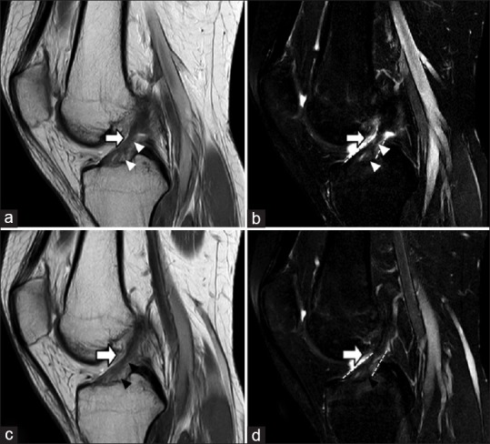
A 25-year-old female student started to have right knee pain while jumping and landing in flexed knee 3 months ago. Thermotherapy, electrotherapy, therapeutic ultrasound, therapeutic exercises, and medicine had been prescribed but not satisfactory. She still felt some pain, weakness, and give-away sensation while walking or climbing stairs at the time she came to our clinic. Physical examination demonstrated positive findings of Lachman test and anterior drawer test. Right knee magnetic resonance imaging (MRI) revealed anterior cruciate ligament (ACL) partial tear with poor collagen alignment but still consistent between femur and tibia insertion. Several small enhancements and irregular architecture over ACL tibial plateau insertion and its main trunk, which indicated partial ACL tear [Figure 1a and b].
Figure 1.

(a) T2 sagittal view: Anterior cruciate ligament showed poor collagen alignment, but still consistent between femur and tibia insertion. Several small enhancements and irregular architecture over anterior cruciate ligament tibial plateau insertion and its main trunk were observed. (b) T2 sagittal view with fat suppression view: Several small enhancements over anterior cruciate ligament tibial plateau insertion. The lesions were diagnosed as the anterior cruciate ligament tear were observed. (c) T2 sagittal view: Decreased enhancements of anterior cruciate ligament tear in tibial plateau area and its main trunk were observed. (d) T2 sagittal view with fat suppression view: Decreased enhancements of anterior cruciate ligament tear in tibial plateau area and its main trunk were observed. Less bony edema was noted. White arrow: Anterior cruciate ligament. White arrowhead: Anterior cruciate ligament tear. Black arrowhead: Healing site of anterior cruciate ligament tear. White small plots: Anterior cruciate ligament border
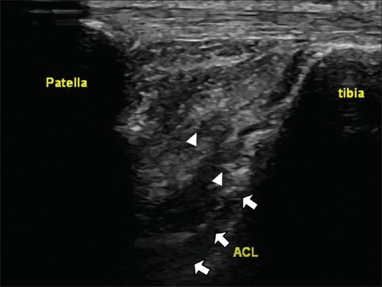
Four months after the insult, three times of Regenlab platelet-rich plasma (PRP) (Regen Lab SA, Switzerland) injection through ultrasound guidance were applied over ACL torn region, including ACL insertion site and its main trunk [Figure 2]. In each course, 2.5 cc PRP (3400 rpm centrifugation, 3–4 times concentrated platelets without the red blood cells) was prepared. The patient lied in the supine position with right knee flexion at 90°. The 10 MHz linear transducer was placed parallel to patellar tendon with the proximal end rotated counterclockwise in 30° to clarify the insertion of both anteromedial and posterolateral bundles over tibial plateau. The three times of injections were performed by the same physician through out-of-plane approach with frequency of once per 3 weeks [Video 1]. During intervention, the patient kept thermotherapy, electrotherapy, and exercise training. Six months after finishing PRP injection, postintervention MRI revealed decreased enhancements of ACL tear in the tibial plateau area and its main trunk [Figure 1c and d]. The patient felt less pain with visual analog scale of pain decreased from 10 to 2, and the stability was improved while walking.
Figure 2.

The transducer was located between the patella and tibia bone with proximal end of transducer pivoting to the lateral side of the knee joint. The needle tip reached anterior cruciate ligament tibial plateau insertion area under out-of-plane approach. White arrow: Anterior cruciate ligament; white arrowhead: needle tip
DISCUSSION
ACL is an intra-articular and extrasynovial structure. It has two separate bundles, anteromedial and posterolateral bundles, which maintain different tensions according to different degrees of knee flexion angle. People usually get ACL injury when weight-bearing and rotation with a slightly flexed knee. Owing to extrasynovial environment, impaired fibrin formation leads to poor healing process.[1] Most serious conditions such as complete tear shall be managed by surgery, while partial tear prefers conservative treatment, rehabilitation, and exercise training. Recently, PRP is used for augmentation of ACL partial tear healing, and the PRP can be precisely applied to the ACL torn region under ultrasound guidance.
PRP is known to contain platelets and several growth factors, involving platelet-derived growth factor (PDGF) and transforming growth factor (TGF)-beta.[2] Both PDGF and TGF-beta have been reported to be the most critical modulators in healing process by enhancing proliferation and collagen production. TGF-beta is a key regulator during embryologic tendon development and plays an important role in the early modulation of scar tissue during healing.[2] Seijas et al. reported a high return rate to sport of patients with ACL partial tear who treated with intraligamentous application of PRP.[3] In clinical practice, we inject PRP into torn region via needling technique. With the assistance of sonography, the architecture, quality, insertion, and origin of the soft tissue can be evaluated.[4,5,6] Moreover, we gain more confidence of identifying ACL torn region. In this patient, we combine PRP injection with thermotherapy, electrotherapy, and exercise training. The symptoms of pain and functional limitation improve compared to previous conservative treatment only. PRP injection under ultrasound guidance along with conservative rehabilitation program might be a treatment choice for ACL partial tear.
CONCLUSION
This manuscript highlights the effect of PRP combined with conservative treatment for ACL partial tear under accuracy and efficiency of ultrasound-guided injection with both improving of the symptoms and images.
Declaration of patient consent
The authors certify that they have obtained all appropriate patient consent forms. In the form the patient(s) has/have given his/her/their consent for his/her/their images and other clinical information to be reported in the journal. The patients understand that their names and initials will not be published and due efforts will be made to conceal their identity, but anonymity cannot be guaranteed.
Financial support and sponsorship
Nil.
Conflicts of interest
There are no conflicts of interest.
Video available on: www.jmuonline.org
REFERENCES
- 1.Rość D, Powierza W, Zastawna E, Drewniak W, Michalski A, Kotschy M, et al. Post-traumatic plasminogenesis in intraarticular exudate in the knee joint. Med Sci Monit. 2002;8:CR371–8. [PubMed] [Google Scholar]
- 2.Meaney Murray M, Rice K, Wright RJ, Spector M. The effect of selected growth factors on human anterior cruciate ligament cell interactions with a three-dimensional collagen-GAG scaffold. J Orthop Res. 2003;21:238–44. doi: 10.1016/S0736-0266(02)00142-0. [DOI] [PubMed] [Google Scholar]
- 3.Seijas R, Ares O, Cuscó X, Alvarez P, Steinbacher G, Cugat R, et al. Partial anterior cruciate ligament tears treated with intraligamentary plasma rich in growth factors. World J Orthop. 2014;5:373–8. doi: 10.5312/wjo.v5.i3.373. [DOI] [PMC free article] [PubMed] [Google Scholar]
- 4.Wu WT, Chang KV, Mezian K, Naňka O, Lin CP, Özçakar L, et al. Basis of shoulder nerve entrapment syndrome: An ultrasonographic study exploring factors influencing cross-sectional area of the suprascapular nerve. Front Neurol. 2018;9:902. doi: 10.3389/fneur.2018.00902. [DOI] [PMC free article] [PubMed] [Google Scholar]
- 5.Chang KV, Wu WT, Huang KC, Jan WH, Han DS. Limb muscle quality and quantity in elderly adults with dynapenia but not sarcopenia: An ultrasound imaging study. Exp Gerontol. 2018;108:54–61. doi: 10.1016/j.exger.2018.03.019. [DOI] [PubMed] [Google Scholar]
- 6.Chang KV, Wu WT, Han DS, Özçakar L. Static and dynamic shoulder imaging to predict initial effectiveness and recurrence after ultrasound-guided subacromial corticosteroid injections. Arch Phys Med Rehabil. 2017;98:1984–94. doi: 10.1016/j.apmr.2017.01.022. [DOI] [PubMed] [Google Scholar]
Associated Data
This section collects any data citations, data availability statements, or supplementary materials included in this article.


