Abstract
Mutations in Fragile X mental retardation gene (Fmr1) cause the most common inherited human autism spectrum disorder. FMR1 influences translation, but identifying functional targets has been difficult. We analyzed quiescent Drosophila oocytes, which like neural synapses depend heavily on translating stored mRNA. Ribosome profiling revealed that FMR1 enhances rather than represses the translation of mRNAs that overlap previously identified FMR1 targets, and acts preferentially on large proteins. Human homologs of at least twenty targets are associated with dominant intellectual disability, and thirty others with recessive neurodevelopmental dysfunction. Stored oocytes lacking FMR1 usually generate embryos with severe neural defects, unlike stored wild type oocytes, suggesting that translation of multiple large proteins using stored mRNAs is defective in FMR1 and possibly other autism spectrum disorders.
Keywords: FMR1, mental retardation, autism, oocyte, translational regulation
FMR1 is a polysome-associated RNA-binding protein required for the nervous system and ovary to develop and function normally in humans, mice and Drosophila (1, 2). Both tissues translationally control stored mRNAs associated with FMR1-containing ribonucleoprotein particles (RNPs) (3–6), suggesting that FMR1 specifically functions in utilizing stored mRNAs. However, the challenge of obtaining highly enriched FMR1-containing RNPs from neural tissue may have contributed to difficulties in defining FMR1 target genes (7–9). We reasoned that FMR1 function could be studied in a physiologically-relevant context using mature Drosophila oocytes, which lack transcription and depend entirely on ongoing translation.
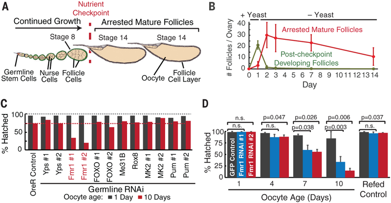
Drosophila oogenesis is highly amenable for such studies, since each female can hold up to thirty mature oocytes per ovary for several weeks (Fig. 1A). In the ovary, each oocyte is surrounded by 800 somatic cells constituting a follicle. We found a reliable method (See Supplement) to maintain completed follicles in the ovary for a known period of time in the absence of new follicle maturation (Fig. 1B). Ovulation and fertilization could then be induced by adding males to test the stored oocyte’s ability to support embryogenesis.
Figure 1. Fmr1 is specifically required during the storage of mature, quiescent stage 14 oocytes in the ovary.
(A) Schematic of a Drosophila ovariole with immature pre-checkpoint follicles and two stored mature stage 14 follicles. (B) Plot shows arrested mature follicle stability (red) following feeding protocol as described (Supplemental). (C) Fmr1 knockdown (line #1 and line #2), but not knockdowns of controls or other indicated genes, specifically reduces 10-day stored (red) but not 1-day stored (black) oocytes from developing into hatching larvae. (D) Fmr1 germline RNAi during storage progressively reduces the fraction of mature oocytes competent after 1, 4, 7 or 10 days of storage to support development. Re-feeding females to promote maturation of fresh stage 8 follicles restores full developmental potential.
We tested the function of specific genes during oocyte storage by depleting their transcripts using germline-specific GAL4-driven RNAi, which is produced throughout oogenesis starting in the germline stem cell. Disrupting a gene required during oocyte storage would cause oocytes to at first develop normally, but to lose developmental capacity more rapidly than wild type during further storage. To screen for such genes, we depleted Fmr1 mRNA or several other candidate gene transcripts and analyzed oocytes after a storage period of one day or ten days (Fig. 1C). In most gene knockdown lines and in wild type controls the hatch rate was nearly 100% after one day of storage, and after 10 days only dropped to 80%. In contrast, Fmr1-depleted oocytes hatched normally at first but developed only 20–30% of the time following ten days of storage (Fig. 1C). Germline Fmr1 RNAi drastically reduced Fmr1 mRNA levels, and antibody staining confirmed that FMR1 protein was effectively depleted throughout oogenesis specifically in germ cells but not in somatic cells (Fig. S1).
We validated the Fmr1 requirement in stored oocytes by showing that oocyte viability drops continuously over time without Fmr1 (Fig. 1D). Importantly, the defects were specific to the old oocytes, themselves. Re-feeding the mother caused the remaining stored oocytes to be laid, and new oocytes to mature which were fully functional (Fig. 1D). Thus, germline Fmr1 RNAi does not significantly impair stem cells, follicle development, or any non-autonomous aspects of female germline function. This differs from Fmr1 mutants which lose stem cells and produce fewer follicles because mutants affect niche cells and other somatic cells in addition to germ cells (10).
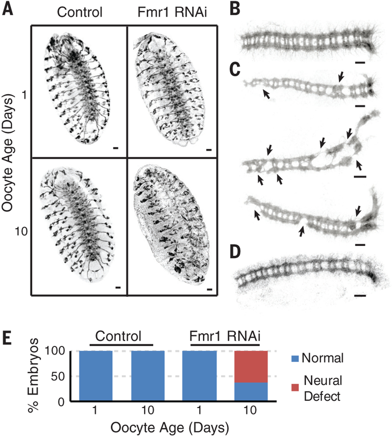
We analyzed embryos derived from control and FMR1-deficient oocytes to investigate what processes are impacted by FMR1 depletion. Embryos from control oocytes developed a normal nervous system regardless of prior storage, as shown by staining using broadly expressed neural markers (Fig. 2A,B). The same was true of embryos derived from Fmr1 RNAi oocytes after one day of storage (Fig. 2A). However, in embryos derived from follicles lacking FMR1 during ten days of storage ventral nerve cord-specific labeling showed missing commissures and breaks in the longitudinal connectives (Fig. 2C), in contrast to wild type. These neuronal defects were not due to a generalized deterioration of the FMR1-depleted oocyte’s ability to support embryonic development (Fig. S2). We never observed comparable neural defects in embryos derived from wild type oocytes even when they were stored for fourteen days when less than 20% of their embryos developed to hatching (Fig. 2D). In contrast, more than 50% of embryos derived from FMR1-deficient oocytes stored in the ovary for ten days developed a severely abnormal nervous system (Fig. 2E). Thus, disrupting FMR1 function while oocytes are fully dependent on translational regulation specifically compromised their ability to support neural development compared to controls.
Figure 2. Stored FMR1-depleted oocytes frequently generate embryos with neural defects.
(A) Control oocytes stored in vivo for 1 day or 10 days support normal embryonic nervous system development (22C10 antibody). Fmr1 RNAi oocytes stored in vivo for 1 day support normal development but after storage for 10 days produce highly abnormal nervous system development. (B) Normal ventral nerve cord (BP102 antibody) from embryo developed from 10 day control oocyte (C), abnormal ventral nerve cords including broken or fused connectives (arrows) from three embryos developed from 10 day Fmr1 RNAi oocytes (D) Normal nerve cord from a control oocyte stored 14 days. (E) Summary of nervous system development in embryos from Control (GFP) and Fmr1 RNAi oocytes. Bar = 20μm.
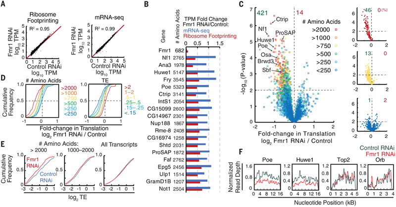
To determine how FMR1 disruption affects oocyte translation and to identify Fmr1 translational target genes that may be important for sustaining nervous system development, we developed a ribosome profiling protocol based on (11) (Supplemental Methods) to quantify oocyte translation in an unbiased, genome-wide manner. Flies induced for oocyte storage were analyzed after only 1–2 days to identify initial translational changes in healthy Fmr1 knockdown oocytes prior to viability reduction. Although whole ovaries were needed to get enough material for profiling, most of the analyzed ribosomes should still derive from stored stage 14 follicles, because they are larger than the total size of earlier follicles. Interestingly, the ribosome footprints and transcript levels of most mRNAs were unaffected by germline-Fmr1 knockdown (Fig. 3A), arguing that FMR1 does not generally control translation or mRNA stability.
Figure 3. FMR1 stimulates the translation during storage of transcripts from multiple intellectual disability and autism genes.
(A) Translational profile (log10TPM; transcripts per million) and mRNA abundance profile (mRNA-seq, log10TPM) are highly similar between control vs. Fmr1-RNAi oocytes (stored 1–2d). (B) Top genes translationally reduced by Fmr1 RNAi from 11 ribosome footprinting experiments do not show significant changes in mRNA levels. (C) Significance vs. fold change plot reveals 421 candidate targets translationally stimulated by FMR1 (p<0.01; t-test). Protein size class indicated by color. (D) Cumulative plot of translation (Fmr1 RNAi/control) as a function of protein size (left), or translational efficiency (TE, right) defined as ribosome footprinting TPM(Fmr1 RNAi)/mRNA-seq TPM. (E) Translation of large mRNAs in Fmr1 RNAi vs. controls is reduced independent of TE. (F) Normalized read depth is plotted for two FMR1 targets (Poe and Huwe1) and two non-targets (Orb and Top2). In Fmr1 RNAi oocytes, target gene footprints are reduced at all positions along the mRNA.
From eleven independent, highly reproducible (Fig. S3), ribosome profiling experiments we identified 421 genes whose germline expression significantly declined and 14 genes that significantly increased expression in Fmr1 RNAi (Fig. 3B,C and Table S1, S2). Except for Fmr1 itself, translation of the significantly altered targets generally decreased 1.3–2.5-fold, which we verified by Western blotting (Fig. S4), while their RNA levels were unchanged (Fig. 3B). Many downregulated genes, at least 56/421, are orthologs of human genes that have been implicated in human neuro-developmental syndromes (Fig. 3C, Table S3) a fraction greater than expected by chance (p=1.1E-9, Fig. S5A). For example, the neurofibromatosis gene, Nf1, which is associated with cognitive and behavioral disorders and neural tumors (12), was reduced 2.5-fold. Several E3 ubiquitin ligases, including Ctrip, Poe, and Huwe1, whose human homologs are associated with intellectual disability, autism, early onset dementia, and schizophrenia (13–15), were reduced about 2.0-fold. In total, homologs of at least 20 dominant autism/intellectual disability genes were significantly reduced (Table S3). Since mutations in these genes are dominant (16), a two-fold reduction in expression has potential consequences, even for a single target.
In order to determine whether FMR1 acted on target transcripts through direct binding, we compared our candidate FMR1 targets with previous reports, which used proximity-based strategies to cross-link mRNAs in brain tissue before immunoprecipitation of FMR1 (7). We found significant overlap between both datasets (p=1.2E-25, Fig. S5B), suggesting that FMR1 directly binds many affected transcripts. However, our results differed from prior studies in two important ways. First, the great majority of targets decreased in expression, indicating that FMR1 usually enhances rather than represses translation, in contrast to most (17, 18) but not all (9, 19) previous reports. This difference might arise because multiple Fmr1 targets act negatively on protein stability, translation, or cell growth, including nine ubiquitin ligases, Nf1, and Not1 (Table S1). Downregulation of these negative regulators following Fmr1 loss might substantially increase protein levels, simulating the direct effects of a translational repressor.
Second, almost all of the affected proteins are much larger in size than the average Drosophila protein. Dividing mRNAs undergoing translation into size classes showed that FMR1 strikingly affects translation in proportion to protein size (Fig. 3C,D) and to some extent UTR length (Fig. S6A), but not by transcript level (Fig. S6D). The translation of nearly half (46%) of expressed proteins >2,000 amino acids, 13% of proteins 750–1000 amino acids, but only 1% of proteins <250 amino acids was significantly reduced with Fmr1 knockdown (Fig. 3C). Fmr1 RNAi did not always impair the translation of large proteins, as there was a broad response to Fmr1 knockdown (Fig. 3C), and the translation of many large proteins was only minimally affected. Fmr1 targets had low translation efficiencies (TEs) in oocytes (Fig. 3D), similar to long transcripts generally (Fig. S6B). Fmr1, however, boosted the translation of affected long transcripts regardless of TE (Fig. 3E), indicating size and not low TE as the predominant factor. The size effect was not due to reduced processivity, since we observed a uniform reduction in footprints across the entire coding sequence of target mRNAs (Fig. 3F). The preferential effect on large mRNAs is likely mediated by direct FMR1 binding, since the average size of target proteins common to both this study and in (7) was 1,841 amino acids. Furthermore, genes linked to autism as a group are significantly longer than average (16, 20).
The Poe/Ubr4 gene, encoding one of the longest Drosophila proteins (5,322 amino acids), was investigated as an FMR1 target with potentially large effects, something not previously identified. Both Poe and Fmr1 mutant Drosophila are male sterile, cannot fly, and show increased neuromuscular junction synaptic excitability (21–23). Stored Poe mutant oocytes lost developmental competence at the same rate as Fmr1 germline RNAi oocytes (Fig. 4A) and these embryos also developed a high frequency of neural defects (Fig. 4B; Fig. S7A). Beginning in nearly mature follicles, POE protein formed 0.5–2 micron spherical particles in germ cells (Fig. 4C), that are distinct from RNP granules (Fig. S8). Poe antibody staining was lost in Poe germline RNAi and Poe mutants (Fig. 4D). As predicted, POE protein levels were reduced and particles were reduced or eliminated in Fmr1-germline RNAi and in Fmr1-null egg chambers (Fig. 4D). These observations demonstrate that Poe is a major functional target of Fmr1, and that Poe is itself essential to maintain the oocytes ability to support neural development. However, over-expressing POE using a duplication in an Fmr1 RNAi background restored POE granule expression (Fig. 4D), but did not rescue Fmr1 RNAi lethality upon storage (Fig. S7F).
Figure 4. Poe is required for oocyte storage and neural development.
(A) Poe mutation accelerates oocyte decline during storage. (B) Poe mutant oocytes frequently fail to support normal neural development after prolonged storage. (C) POE antibody staining (see Methods) during follicle development, showing germline granules that arise in maturing follicles. Bar = 3μm. (D) Many POE granules are seen in wild type stage 10 follicles, but not in Poe RNAi, Poe01659, Fmr1 RNAi, or null Fmr13/Δ50 follicles. Fmr1 RNAi combined with POE overexpression recovers POE granules. Bar = 20μm.
FMR1 function in maintaining an oocyte’s ability to support neural development may only be revealed during oocytes storage because some FMR1 targets act catalytically. For example, E3 ligases whose levels are reduced in FMR1 deficient oocytes might no longer be able to prevent the over accumulation of some of their target proteins which might eventually reach levels in the stored oocytes that interfere with neural development.
Fmr1 regulates translation in both the ovary and during neural development as part of RNP granules that may either stimulate or repress translation. Studies of these granules suggest a potential explanation for size effect we observed on target proteins. Mammalian and yeast mRNAs that are large and/or inefficiently translated preferentially associate with stress granules where their translation is repressed (24). FMR1 may function to counteract the inherent tendency of specific large mRNAs to be segregated into inactive RNP particles. Alternatively, Fmr1 might directly or indirectly promote translation initiation in association with RNPs, or it might affect mRNA transport along microtubules to sites of active initiation.
In conclusion, we propose that maintaining the translation of large mRNAs in the translational environments experienced far from the nucleus at synapses in many neurons represents a general challenge that underlies Fragile X syndrome, and may be relevant to other neurodevelopmental conditions. The same challenges likely exist in oocytes, spermatocytes and in non-neural somatic cells that require regulated translation from stored mRNAs. Since the challenges of translating large proteins are only likely to increase in adult neurons under the influence of aging, the pathways and targets assayed here may contribute to adult-onset neural impairments such as schizophrenia and dementia. Improved knowledge of how FMR1 preserves target translation, and the identities of major target genes such as Poe/Ubr4 open new opportunities to monitor susceptible cells, and to intervene to mitigate declining levels of the most critical targets. Small molecule agents that counteract the tendency of large mRNAs to be segregated into inactive granules represent potentially valuable therapeutics. Continued study of these highly conserved pathways in Drosophila represents one powerful and efficient means to further address both the fundamental and applied implications of these findings.
Supplementary Material
Acknowledgements:
We thank Allison Pinder for help generating RNA-seq libraries. We are grateful to Joshua Dunn (UCSF) for technical advice for our ribosome footprinting experiments. We thank Eric Wagner (UTMB) and Thomas Jongens (UPenn) for generously providing the IntS1 antibody and the Fmr13 mutant fly strain respectively.
Funding: This work was supported by funding through the Jane Coffin Childs Memorial Fund (E.G.) and the Howard Hughes Medical Institute (A.C.S.).
Footnotes
Data Availability: Ribosome profiling and mRNA-seq data reported here are available from the NIH BioProject Website under accession PRJNA466150.
REFERENCES
- 1.Hagerman RJ et al. , Nat Rev Dis Primers. 3, 17065 (2017). [DOI] [PubMed] [Google Scholar]
- 2.Drozd M, Bardoni B, Capovilla M, Front Mol Neurosci 11, 124 (2018). [DOI] [PMC free article] [PubMed] [Google Scholar]
- 3.Barbee SA et al. , Neuron 52, 997–1009 (2006). [DOI] [PMC free article] [PubMed] [Google Scholar]
- 4.Rosario R et al. , PLoS ONE. 11, e0163987 (2016). [DOI] [PMC free article] [PubMed] [Google Scholar]
- 5.Christie SB, Akins MR, Schwob JE, Fallon JR, J. Neurosci 29, 1514–1524 (2009). [DOI] [PMC free article] [PubMed] [Google Scholar]
- 6.Costa A et al. , Developmental Cell. 8, 331–342 (2005). [DOI] [PubMed] [Google Scholar]
- 7.Darnell JC et al. , Cell. 146, 247–261 (2011). [DOI] [PMC free article] [PubMed] [Google Scholar]
- 8.Ascano M et al. , Nature. 492, 382–386 (2012). [DOI] [PMC free article] [PubMed] [Google Scholar]
- 9.Tabet R et al. , Proc. Natl. Acad. Sci. U.S.A 113, E3619–28 (2016). [DOI] [PMC free article] [PubMed] [Google Scholar]
- 10.Yang L et al. , Hum. Mol. Genet 16, 1814–1820 (2007). [DOI] [PubMed] [Google Scholar]
- 11.Dunn JG, Foo CK, Belletier NG, Gavis ER, Weissman JS, Elife. 2, e01179 (2013). [DOI] [PMC free article] [PubMed] [Google Scholar]
- 12.Morris SM et al. , JAMA Psychiatry. 73, 1276–1284 (2016). [DOI] [PMC free article] [PubMed] [Google Scholar]
- 13.Zhang J et al. , Hum. Genet 136, 377–386 (2017). [DOI] [PMC free article] [PubMed] [Google Scholar]
- 14.Monies D et al. , Hum. Genet 136, 921–939 (2017). [DOI] [PMC free article] [PubMed] [Google Scholar]
- 15.Bosshard M et al. , Sci Rep 7, 15050 (2017). [DOI] [PMC free article] [PubMed] [Google Scholar]
- 16.Pereanu W et al. , Nucleic Acids Res 46, D1049–D1054 (2017). [DOI] [PMC free article] [PubMed] [Google Scholar]
- 17.Zalfa F et al. , Cell. 112, 317–327 (2003). [DOI] [PubMed] [Google Scholar]
- 18.Laggerbauer B, Ostareck D, Keidel EM, Ostareck-Lederer A, Fischer U, Hum. Mol. Genet 10, 329–338 (2001). [DOI] [PubMed] [Google Scholar]
- 19.Bechara EG et al. , PLoS Biol 7, e16 (2009). [DOI] [PMC free article] [PubMed] [Google Scholar]
- 20.King IF et al. , Nature. 501, 58–62 (2013). [DOI] [PMC free article] [PubMed] [Google Scholar]
- 21.Richards S, Hillman T, Stern M, Genetics. 142, 1215–1223 (1996). [DOI] [PMC free article] [PubMed] [Google Scholar]
- 22.Fabrizio JJ, Hime G, Lemmon SK, Bazinet C, Development. 125, 1833–1843 (1998). [DOI] [PubMed] [Google Scholar]
- 23.Zhang YQ et al. , Cell. 107, 591–603 (2001). [DOI] [PubMed] [Google Scholar]
- 24.Khong A et al. , Mol. Cell 68, 808–820.e5 (2017). [DOI] [PMC free article] [PubMed] [Google Scholar]
- 25.Ingolia NT, Brar GA, Rouskin S, McGeachy AM, Weissman JS, Nat Protoc 7, 1534–1550 (2012). [DOI] [PMC free article] [PubMed] [Google Scholar]
- 26.Dunn JG, Weissman JS, BMC Genomics. 17, 958 (2016). [DOI] [PMC free article] [PubMed] [Google Scholar]
- 27.Trapnell C et al. , Nature Biotechnology. 28, 511–515 (2010). [DOI] [PMC free article] [PubMed] [Google Scholar]
Associated Data
This section collects any data citations, data availability statements, or supplementary materials included in this article.


