

Abstract
Context:
Both gingivitis and periodontitis are due to the detrimental effects of the microbe-laden biofilm. The mainstay of periodontal treatment is, therefore, the disruption of this biofilm by scaling and root planing (SRP). Other treatment protocols such as systemic antimicrobials have been administered as adjuvants after scaling and root planning. However, due to antimicrobial resistance, as well as a shift of the flora from a symbiotic to a dysbiotic one, this mode of treatment has its shortcomings. Thus, local drug delivery has gained prominence as a therapeutic tool.
Aims:
The aim of this study is to compare the efficacy of subgingivally delivered probiotics as a monotherapy, in combination with tetracycline fibers, and tetracycline fibers alone after SRP.
Settings and Design:
This study was a parallel arm, randomized clinical and microbiological study. Thirty patients with chronic periodontitis aged between 20 and 50 years were selected from the outpatient ward of a tertiary referral care hospital in Hyderabad and equally divided into three groups.
Materials and Methods:
This study was conducted from January 2017 to February 2017 and ethical clearance was obtained from the institutional ethical committee.
Statistical Analysis Used:
Mean values and standard deviations were calculated for Plaque Index, Sulcular Bleeding Index (SBI), probing depth (PD), and microbial colony-forming units, for all the three groups at different time intervals. Paired “t-test” was used for intragroup comparison and Student's “t-test” for intergroup comparison. Results were regarded as statistically significant when P < 0.05.
Results:
Intragroup comparison yielded significant improvement in all the variables (P < 0.0001). However, intergroup comparison showed statistically significant differences pertaining to the PD (P < 0.001) and SBI only (P < 0.001), between Group A and Group B and Group B and Group C respectively.
Conclusions:
Group A and Group C showed better results than Group B.
Keywords: Clinical parameters, colony-forming units, periodontitis, probiotics, tetracycline fibers
INTRODUCTION
Periodontitis is a polymicrobial disease which is initiated by dental plaque. Plaque is a complex biofilm, which becomes more toxic to the host as it matures. This is because the initial colonizers, Gram-positive cocci, and rods are replaced by the secondary colonizers which are Gram-negative and more putative in nature. As the disease progresses, it causes more attachment and bone loss, finally culminating in tooth loss if left untreated.[1]
Scaling and root planing (SRP) remains the cornerstone of periodontal therapy;[2] however, some microorganisms such as Aggregatibacter actinomycetemcomitans and Porphyromonas gingivalis (Pg) can invade the host tissues with ease due to the virulence factors they possess. Thus, to prevent these organisms from gaining entry, systemic antimicrobials are often prescribed after SRP. Although this mode of treating periodontitis is beneficial, it has its limitations, such as development of drug resistance, due to the high doses administered to maintain minimum inhibitory concentrations in the tissues. Furthermore, there is a shift in the microflora from a symbiotic to a dysbiotic state, and other side effects such as gastric disturbances, nausea, and vomiting on prolonged use of antibiotics. To overcome these shortcomings, local drug delivery (LDD) into the periodontal pockets has been advocated.[3] These LDD's have been administered in the form of gels, fibers, chips, etc.
Background
Probiotics are being used to help in the recolonization of beneficial bacteria as well as to modulate the immune response. These agents have been administered in the form of tablets, gums, lozenges, and mouthrinses to name a few. However, they have not been used as local delivery systems due to their inability to be retained for long at the site administered. Thus, this study was done to assess the retentive capacity of a probiotic when mixed with tragacanth powder (gum) and placed in periodontal pockets.
MATERIALS AND METHODS
Thirty patients with chronic periodontitis were divided equally into:
Group A – Included 10 patients with 10 sites, treated by SRP, and subgingivally delivered probiotics (Inlife Pharma, Pre and Probiotic capsules comprising of Lactobacillus acidophilus and Lactobacillus rhamnosus 0.60 billion colony-forming units (CFU) each, Bifidobacterium bifidus and Bifidobacterium longum 0.60 billion CFU) mixed with tragacanth gum (TG) powder (0.04 mg of probiotic powder which was mixed with 0.02 mg of TG powder plus 0.2 ml of saline) [Figure 1]
Group B – Included 10 patients with 10 sites, treated by SRP and subgingivally delivered tetracycline fibers (PerioColTM-TC, Eucare Pharma) (10 mg of tetracycline fibers only mixed with 0.2 ml of saline)
Group C – Included 10 patients with 10 sites, treated with SRP along with subgingivally delivered probiotics (Inlife Pharma) mixed with TG powder and tetracycline fibers (PerioColTM-TC, Eucare Pharma) (10 mg of tetracycline fibers coated with 0.04 mg of probiotic and 0.02 mg of TG powder mixed with 0.2 ml of saline) [Figure 2].
Figure 1.
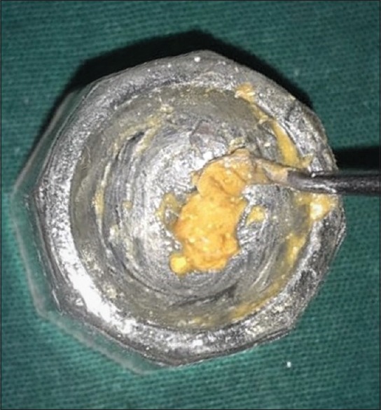
Probiotic powder, mixed with tragacanth gum and saline
Figure 2.
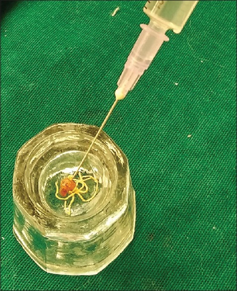
Tetracycline fibers mixed with probiotic powder, tragacanth gum, and saline
Inclusion criteria
Patients having at least 20 teeth, with periodontal pockets which bled on probing with probing depths (PDs) ≥5 mm, were included in the study.
Exclusion criteria
Smokers, systemically compromised individuals, patients taking medications, such as corticosteroids or calcium channel blockers, which are known to interfere with periodontal wound healing, patients allergic to medications, pregnant or lactating women, and patients who underwent periodontal therapy in the past 6 months were excluded from the study.
Clinical parameters
The Plaque Index (PI), Sulcular Bleeding Index (SBI), and PDs were assessed at baseline and after 45 days, using a Williams probe.
Microbiological analysis
A sterile Gracey curette, was used to obtain the plaque which was taken from pockets with depths ≥5 mm. Plaque samples were taken preoperatively and 45 days after SRP and LDD administration. The plaque samples were transferred into test tubes containing transport medium (Thioglycolate broth) and microbiological analysis after culture (Blood agar medium) for CFU was done both at baseline and 45 days [Figure 3].
Figure 3.
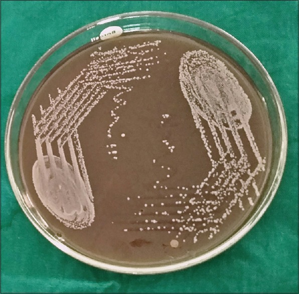
Blood agar culture medium with bacterial growth
Method of local drug delivery administration
Each patient was given careful instructions on proper oral hygiene measures. A full-mouth supragingival and subgingival SRP procedure were performed under local anesthesia following which the LDD was introduced subgingivally into the pockets with the help of ball end of a community periodontal index of treatment needs (CPITN) probe [Figures 4 and 5]. For Group A and Group C patients, the probiotic powder within the capsule was made use of for the study. By applying firm pressure with fingers at the entrance of the pocket, the material was stabilized within the pocket, after which a periodontal dressing was given for a week [Figures 6 and 7]. Patients were instructed not to disturb the area with tongue, finger or toothpick, not to chew any hard, or sticky food for at least 1 week, and postpone brushing and flossing on the treated site for 1 week. At the first postoperative visit, patients were examined for any adverse signs or symptoms related to the LDD administration. All the patients were recalled after a period of 45 days.
Figure 4.
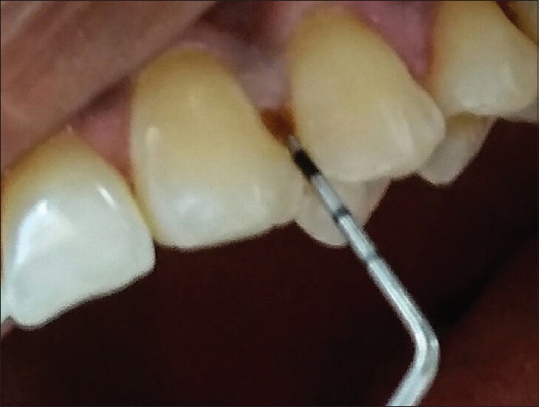
Probiotic being introduced with ball end of CPITN probe
Figure 5.
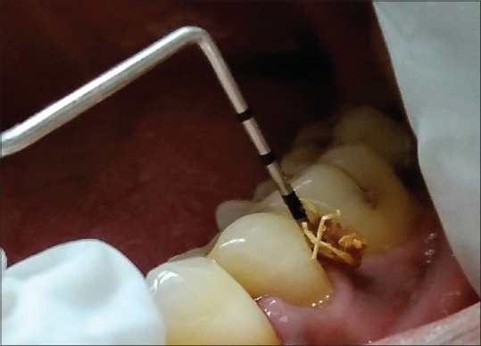
Tetracycline fibers with probiotic being placed with ball end of CPITN probe
Figure 6.
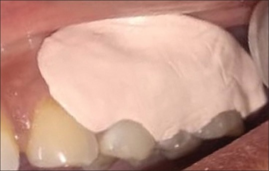
Periodontal dressing after probiotic placement
Figure 7.
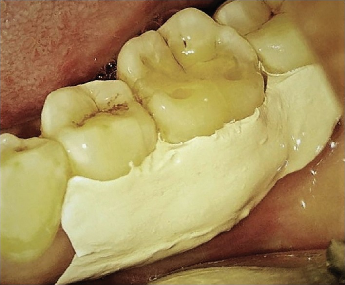
Periodontal dressing after tetracycline with probiotic placement
Outcome measures
The primary outcome measures assessed were the clinical parameters, whereas the CFU/ml was the secondary outcome measure evaluated.
Statistical analysis
Substituting the values for primary outcome measure with power at 95% and level of significance 5%, ten patients per group were analyzed to be sufficient. Mean values and standard deviations were calculated for PI, SBI, PD, and CFU for all the three groups at different time intervals. Paired “t-test” was used for comparison of values within the group and Student “t-test” was used for intergroup comparison. Results were regarded as statistically significant when P < 0.05.
RESULTS
Intragroup comparison
There was a significant improvement in both the clinical parameters and CFU from baseline to 45 days in all the three groups (P < 0.001) [Tables 1–3].
Table 1.
Intragroup comparison in Group A (scaling and root planing and subgingivally delivered probiotics mixed with tragacanth gum powder)
| Parameters | Group A | n | Mean±SD | P |
|---|---|---|---|---|
| PI | Baseline | 10 | 2.44±0.437 | <0.0001** |
| After 45 days | 10 | 0.77±0.279 | ||
| SBI | Baseline | 10 | 3.7±0.483 | <0.0001** |
| After 45 days | 10 | 1.4±0.516 | ||
| PD | Baseline | 10 | 6.7±0.483 | <0.0001** |
| After 45 days | 10 | 3.7±0.823 | ||
| CFU | Baseline | 10 | 4.6±0.516 | <0.0001** |
| After 45 days | 10 | 2.3±0.483 |
**Statistically significant, P < 0.05 statistically significant. n – Sample size; SD – Standard deviation; PI – Plaque Index; SBI – Sulcular Bleeding Index; PD – Probing depth; CFU – Colony-forming units; P – Probability Value
Table 3.
Intragroup comparison in Group C (scaling and root planing along with subgingivally delivered probiotics mixed with tragacanth gum powder and tetracycline fibers [PerioColTM - TC, Eucare Pharma])
| Parameters | Group C | n | Mean±SD | P |
|---|---|---|---|---|
| PI | Baseline | 10 | 2.45±0.368 | <0.0001** |
| After 45 days | 10 | 0.79±0.255 | ||
| SBI | Baseline | 10 | 3.5±0.527 | <0.0001** |
| After 45 days | 10 | 1.0±0.001 | ||
| PD | Baseline | 10 | 7.0±0.942 | <0.0001** |
| After 45 days | 10 | 3.3±0.483 | ||
| CFU | Baseline | 10 | 4.7±0.483 | <0.0001** |
| After 45 days | 10 | 2.1±0.316 |
** Statistically significant, P<0.05 statistically significant, n – Sample size; SD – Standard deviation; PI – Plaque Index; SBI – Sulcular Bleeding Index; PD – Probing depth; CFU – Colony-forming units; P – Probability Value
Table 2.
Intragroup comparison in Group B (scaling and root planing and subgingivally delivered tetracycline fibers [PerioColTM - TC, Eucare Pharma])
| Parameters | Group B | n | Mean±SD | P |
|---|---|---|---|---|
| PI | Baseline | 10 | 2.35±0.34 | <0.0001** |
| After 45 days | 10 | 0.73±0.29 | ||
| SBI | Baseline | 10 | 3.1±0.567 | 0.0038* |
| After 45 days | 10 | 1.9±0.994 | ||
| PD | Baseline | 10 | 7.2±0.918 | 0.0007* |
| After 45 days | 10 | 5.6±0.843 | ||
| CFU | Baselines | 10 | 4.7±0.483 | <0.0001** |
| After 45 days | 10 | 2.3±0.483 |
**Statistically significant, P < 0.05 statistically significant. n – Sample size; SD – Standard deviation; PI – Plaque Index; SBI – Sulcular Bleeding Index; PD – Probing depth; CFU – Colony-forming units; P – Probability Value
Intergroup comparison
When a comparison was made between Group A and Group B, it was observed that there were statistically significant changes pertaining to the PDs and SBI, from baseline to 45 days in Group A (P < 0.001) [Table 4].
Table 4.
Intergroup comparison between Group A and Group B
| Parameters after 45 days | Groups | n | Mean±SD | P |
|---|---|---|---|---|
| PI | Group A | 10 | 0.77±0.279 | 0.352 (NS) |
| Group B | 10 | 0.73±0.29 | ||
| SBI | Group A | 10 | 1.4±0.516 | <0.0001** |
| Group B | 10 | 1.9±0.994 | ||
| PD | Group A | 10 | 3.7±0.823 | <0.0001** |
| Group B | 10 | 5.6±0.843 | ||
| CFU | Group A | 10 | 2.3±0.483 | 0.931 (NS) |
| Group B | 10 | 2.3±0.483 |
**Statistically significant, P < 0.05 statistically significant. n – Sample size; SD – Standard deviation; PI – Plaque Index; SBI – Sulcular Bleeding Index; PD – Probing depth; CFU – Colony-forming units; NS – Not significant; P – Probability value
On comparing Group A and Group C, it was found that there was a significant difference in relation to PD only, after 45 days in Group C (P < 0.001) [Table 5].
Table 5.
Intergroup comparison between Group A and Group C
| Parameters after 45 days | Groups | n | Mean±SD | P |
|---|---|---|---|---|
| PI | Group A | 10 | 0.77±0.279 | 0.872 (NS) |
| Group C | 10 | 0.79±0.255 | ||
| SBI | Group A | 10 | 1.4±0.516 | 0.246 (NS) |
| Group C | 10 | 1.0±0.001 | ||
| PD | Group A | 10 | 3.7±0.823 | 0.0011** |
| Group C | 10 | 3.3±0.483 | ||
| CFU | Group A | 10 | 2.3±0.483 | 0.198 (NS) |
| Group C | 10 | 2.1±0.316 |
** Statistically significant, P < 0.05 statistically significant, n – Sample size; SD – Standard deviation; PI – Plaque Index; SBI – Sulcular Bleeding Index; PD – Probing depth; CFU – Colony-forming units; NS – Not significant; P – Probability value
Similarly, when a comparison was made between Group B and Group C, there was a significant improvement in the SBI as well as the PDs from baseline to 45 days in Group C (P < 0.001) [Table 6].
Table 6.
Intergroup comparison between Group B and Group C
| Parameters after 45 days | Groups | n | Mean±SD | P |
|---|---|---|---|---|
| PI | Group B | 10 | 0.73±0.29 | 0.319 (NS) |
| Group C | 10 | 0.79±0.255 | ||
| SBI | Group B | 10 | 1.9±0.994 | <0.0001** |
| Group C | 10 | 1.0±0.001 | ||
| PD | Group B | 10 | 5.6±0.843 | <0.0001** |
| Group C | 10 | 3.3±0.483 | ||
| CFU | Group B | 10 | 2.3±0.483 | 0.308 (NS) |
| Group C | 10 | 2.1±0.316 |
**Statistically significant, P < 0.05 statistically significant. n – Sample size; SD – Standard deviation; PI – Plaque Index; SBI – Sulcular Bleeding Index; PD – Probing depth; CFU – Colony-forming units; NS – Not significant; P – Probability value
DISCUSSION
The severity of periodontitis is measured based on the pocket PDs, the attachment loss, and bone loss. Higher the number of sites with increased pocket PDs, greater is the extent of the disease. Therefore, measures to reduce the PDs and clinical attachment loss form the mainstay of periodontal treatment. SRP is the primary step in the treatment protocol. However, if there is no reduction in the PDs after repeated scaling and root planning, then other interventions such as LDD or surgery is planned. LDD helps in reducing the microbial load of the pockets, thus helping in the repair of the periodontal tissues affected. Thus, this study was done to compare the beneficial effects of probiotics[4] tetracycline fibers and a combination of probiotics and tetracycline fibers in patients with periodontitis.
Probiotics work well as host-modulating agents, as they lower the salivary PH, thus preventing the formation of dental plaque as well as calculus. They also produce antioxidants which neutralize the free electrons which play a pivotal role in plaque formation as well as stain buildup.[5,6]
The ability of the probiotic microorganisms to thrive and colonize in the oral cavity is still questioned. Studies employing the traditional culture methods have not been able to show the adherence of probiotic strains in the plaque or saliva of patients who have consumed the probiotics for a long period of time.[7,8,9]
A study was conducted to assess if three probiotic bacteria present in the milk product Cultura dofilus naturell could be seen in saliva and on oral mucosal surfaces and if they colonized dental surfaces in situ of 8 caries inactive individuals after 8 daily exposures to the milk product for up to 3 days. Bacteria were identified by fluorescence in situ hybridization and confocal laser scanning microscopy. While probiotic bacteria were detected sporadically in the oral cavity, on mucosal surfaces and in the saliva after 3 days of use of the probiotic milk, they were not detected on dental surfaces.[10]
In another study, 59 patients with gingivitis were divided into: 20 patients receiving Lactobacillus reuteri 1 formulation (LR1) at a dose of 2 × 10(8) CFU/day, 21 patients receiving LR2 formulation at a dose of 2 × 10(8) CFU/day, and 18 patients receiving a placebo for a 2 week period, to assess the efficacy of the probiotic strain on gingival and plaque indices which were measured at baseline and after 2 weeks. Both the test and placebo were incorporated in chewing gums. At baseline, the clinician cleaned all the surfaces both in the test and control group, after which the patients were instructed about oral hygiene maintenance and were asked to use the chewing gum thrice daily after brushing. After a 2-week period on evaluation, the gingival index significantly improved in all the three groups (P < 0.0001), however LR-1, but not LR-2 improved more than placebo (P < 0.0001). The PI also fell significantly in LR-1 (P < 0.05), and in LR-2 (P < 0.01) between day 0 and day 14, but there was no significant change in the placebo group. Thus, it was concluded that LR was efficacious in reducing both gingivitis and plaque in patients with moderate-to-severe gingivitis.[11]
Lactobacillus acidophilus strains have been observed to inhibit the growth of a wide array of bacterial species due to the production of bacteriocins and are thus the most widely used probiotic therapeutic agents.[12]
In another study done to assess the effects of L. reuteri (Prodentis) in combination with SRP in patients with periodontitis, 30 patients were enrolled in a split mouth study design, wherein all the patients received SRP in two quadrants only, either right or left. 15 patients received Prodentis lozenges (1 × 10⁸ CFU DSM17938 + 1 × 10⁸ CFU ATCC PTA 5289) twice daily from day 21 to 42 and 15 patients received placebo lozenges twice daily for the same time period. The indices assessed were the PI, Gingival Index, Gingival Bleeding Index, probing pocket depth, clinical attachment level (CAL), and microbiological levels of the pathogens A. actinomycetemcomitans, Pg, and Prevotella intermedia. These parameters were assessed at baseline, day 21, and day 42, respectively. It was observed that all the clinical and microbiological parameters significantly improved in the patients administered with Prodentis after SRP.[13]
To improve the retentive capacity of the probiotic TG was used as a carrier in both Group A (SRP + Probiotic) and Group C (SRP + Tetracycline fibers + Probiotic). Tragacanth gum (TG) is a natural product obtained from Astragalus gummifer Labillardiere and other species of Astragalus. The gum comprised of a mixture of water-insoluble and water-soluble polysaccharides. Bassorin, which constitutes 60%–70% of the gum, is the main water-insoluble portion, while the remainder of the gum consists of the water-soluble material, tragacanth. TG is often used in the food industry as an additive with E number as “E 413.” In addition, TG has a very wide area of applications, it can be utilized as a thickening agent as well as a binder and even stabilizer and in industries such as paper making, cosmetics, and textile industry. It was affirmed as GRAS (generally recognized as safe) when used as a food additive by the Food and Drug Administration.[14,15,16]
In recent years, considerable research has focused on the development of safe and efficient drug delivery systems with the use of polymers as vehicles for the controlled release of drugs from various types of formulated products such as tablets, hydrogel membranes, microspheres, and adhesive strips. TG is a natural polymer and the release of drugs, absorbed or encapsulated by polymer, involves their slow and controlled diffusion from or through polymeric material. Therefore, TG, when used as a vehicle, follows the zero order of release which guarantees a constant rate of drug release over time.[17,18]
This study was done to assess the effect of probiotic on clinical parameters when mixed with TG. However, the pharmacologic release of the probiotic from TG was not assessed.
In this study, it was observed that Group A (SRP + Probiotic) and Group C (SRP + Tetracycline fibers + Probiotic) showed significant improvement in the SBI and pocket PDs when compared to Group B (SRP + Tetracycline fibers).
The tetracyclines are a group of broad-spectrum antimicrobial agents that are primarily bacteriostatic and are effective against many Gram-negative species such as A. actinomycetemcomitans. They are of importance in treating periodontitis due to their anti-inflammatory and anticollagenase properties. Moreover, they inhibit bone resorption and promote the attachment of fibroblasts to the root surface.
Some researchers, observed that tetracycline filled hollow fibers placed in the gingival sulcus had a dramatic effect both on the periodontal flora and clinical manifestation of disease. A single placement of tetracycline fibers virtually eradicated spirochetes from the pockets and prevented their recolonization.[19]
Tetracycline (Periocol–TC) was used in this study as an LDD. Periocol-TC vial contains Type I, fibrillar collagen of fish origin of approximately 25 mg, impregnated with approximately 2.0 mg of tetracycline hydrochloride IP, sterilized by gamma radiation with shelf life of 2 years. It releases tetracycline and it gets dissolved in the period of 8–12 days (Eucare Pharmaceuticals, Chennai.).
Other researchers conducted a study on 30 patients with periodontitis who were equally divided into Group A and Group B. Patients in Group A received SRP alone, whereas those, in Group B received SRP with tetracycline fibers as an LDD. It was observed that the number of sites with bleeding on probing (BOP) was more in Group A and the PDs also remarkably decreased in Group B when compared to Group A, 30- and 90-day postintervention, thus highlighting the beneficial role of tetracycline fibers in periodontitis.[20]
In another study, a total of 113 patients receiving regular supportive periodontal therapy were treated with whole mouth SRP. Two nonadjacent sites were selected in each patient for monitoring based on criteria that the pockets were 5–8 mm deep and had BOP. The chosen sites were randomly assigned to one of the two treatment groups scaling and root planning only or SRP with adjunctive use of tetracycline fibers. PD, BOP, and CAL were measured at baseline and 1, 3, and 6 months, respectively. At 1, 3, and 6 months, adjunctive fiber therapy was significantly better in reducing PD (P < 0.05) and reducing BOP (P < 0.05) than SRP alone. At 6 months, also, fiber therapy was significantly better in promoting clinical attachment gain (P < 0.05) than scaling and root planning alone.[21]
This study was in accordance with the previous studies in that, patients on tetracycline fibers after scaling and root planning (Group B) showed a significant improvement in all the clinical parameters. However, when an intergroup comparison was made, it was observed that patients in Group A (SRP + Probiotic) and Group C (SRP + Tetracycline fibers + Probiotic) showed better results than Group B.
Limitations of the study
This study should have been conducted for a longer period of time. The release kinetics of probiotic from TG polymer was not assessed.
CONCLUSION
This study has reiterated the importance of probiotics in periodontal therapy. The probing pocket depths have significantly reduced in Group C when compared to Group A and Group B, showing the benefits of administering both tetracycline fibers and probiotic synergistically to treat periodontitis. In the future, Nano-emulsion systems,[22] which have better thermodynamic stability than the emulsions, may make the delivery and sustenance of the LDD agents more effective.
Financial support and sponsorship
Nil.
Conflicts of interest
There are no conflicts of interest.
REFERENCES
- 1.Page RC, Offenbacher S, Schroeder HE, Seymour GJ, Kornman KS. Advances in the pathogenesis of periodontitis: Summary of developments, clinical implications and future directions. Periodontol 2000. 1997;14:216–48. doi: 10.1111/j.1600-0757.1997.tb00199.x. [DOI] [PubMed] [Google Scholar]
- 2.Apatzidou DA, Kinane DF. Nonsurgical mechanical treatment strategies for periodontal disease. Dent Clin North Am. 2010;54:1–2. doi: 10.1016/j.cden.2009.08.006. [DOI] [PubMed] [Google Scholar]
- 3.Siddharth T, Nikhil S, Rahul C. Local drug delivery: A current concept in periodontology. Mod Appl Bioequiv Availab. 2017;1:555552. [Google Scholar]
- 4.Teughels W, Newman MG, Coucke W, Haffajee AD, Van Der Mei HC, Haake SK, et al. Guiding periodontal pocket recolonization: A proof of concept. J Dent Res. 2007;86:1078–82. doi: 10.1177/154405910708601111. [DOI] [PubMed] [Google Scholar]
- 5.Dhawan R, Dhawan S. Role of probiotics on oral health: A randomized, double-blind, placebo-controlled study. J Interdiscip Dent. 2013;3:71–8. [Google Scholar]
- 6.Ishihara K, Miyakawa H, Hasegawa A, Takazoe I, Kawai Y. Growth inhibition of Streptococcus mutans by cellular extracts of human intestinal lactic acid bacteria. Infect Immun. 1985;49:692–4. doi: 10.1128/iai.49.3.692-694.1985. [DOI] [PMC free article] [PubMed] [Google Scholar]
- 7.Bunting RW, Nickerson G, Hard DG, Crowley M. Further studies of the relation of Bacillus acidophilus to dental caries. J D Cosmos. 1928;70:1–8. [Google Scholar]
- 8.Busscher HJ, Mulder AF, van der Mei HC. In vitro adhesion to enamel and in vivo colonization of tooth surfaces by lactobacilli from a bio-yoghurt. Caries Res. 1999;33:403–4. doi: 10.1159/000016541. [DOI] [PubMed] [Google Scholar]
- 9.Caglar E, Topcuoglu N, Cildir SK, Sandalli N, Kulekci G. Oral colonization by Lactobacillus reuteri ATCC 55730 after exposure to probiotics. Int J Paediatr Dent. 2009;19:377–81. doi: 10.1111/j.1365-263X.2009.00989.x. [DOI] [PubMed] [Google Scholar]
- 10.Ravn I, Dige I, Meyer RL, Nyvad B. Colonization of the oral cavity by probiotic bacteria. Caries Res. 2012;46:107–12. doi: 10.1159/000336960. [DOI] [PubMed] [Google Scholar]
- 11.Krasse P, Carlsson B, Dahl C, Paulsson A, Nilsson A, Sinkiewicz G. Decreased gum bleeding and reduced gingivitis by the probiotic Lactobacillus reuteri. Swed Dent J. 2006;30:55–60. [PubMed] [Google Scholar]
- 12.Silva M, Jacobus NV, Deneke C, Gorbach SL. Antimicrobial substance from a human Lactobacillus strain. Antimicrob Agents Chemother. 1987;31:1231–3. doi: 10.1128/aac.31.8.1231. [DOI] [PMC free article] [PubMed] [Google Scholar]
- 13.Vivekananda MR, Vandana KL, Bhat KG. Effect of the probiotic Lactobacilli reuteri (Prodentis) in the management of periodontal disease: A preliminary randomized clinical trial. J Oral Microbiol. 2010;2:5344. doi: 10.3402/jom.v2i0.5344. [DOI] [PMC free article] [PubMed] [Google Scholar]
- 14.Balaghi S, Mohammadifar MA, Zargaraan A, Gavlighi HA, Mohammadi M. Compositional analysis and rheological characterization of gum tragacanth exudates from six species of Iranian Astragalus. Food Hydrocoll. 2011;25:1775–84. [Google Scholar]
- 15.Balaghi S, Mohammadifar MA, Zargaraan A. Physicochemical and rheological characterization of gum tragacanth exudates from six species of Iranian Astragalus. Food Biophys. 2010;5:59–71. [Google Scholar]
- 16.Eastwood MA, Brydon WG, Anderson DM. The effects of dietary gum tragacanth in man. Toxicol Lett. 1984;21:73–81. doi: 10.1016/0378-4274(84)90226-1. [DOI] [PubMed] [Google Scholar]
- 17.Andreopoulos AG, Tarantili PA. Xanthan gum as a carrier for controlled release of drugs. J Biomater Appl. 2001;16:34–46. doi: 10.1106/XBFG-FYFX-9TW9-M83U. [DOI] [PubMed] [Google Scholar]
- 18.Nagarjuna G, Kumara Babu P, Maruthi Y, Parandhama A, Madhavi C, Subha MC, et al. Sodium alginate/tragacanth gum blend hydrogel membranes for controlled release of verapamil hydrochloric acid. Indian J Adv Chem Sci. 2016;4:469–77. [Google Scholar]
- 19.Goodson JM, Haffajee A, Socransky SS. Periodontal therapy by local delivery of tetracycline. J Clin Periodontol. 1979;6:83–92. doi: 10.1111/j.1600-051x.1979.tb02186.x. [DOI] [PubMed] [Google Scholar]
- 20.Panwar M, Gupta SH. Local drug delivery with tetracycline fiber: An alternative to surgical periodontal therapy. Med J Armed Forces India. 2009;65:244–6. doi: 10.1016/S0377-1237(09)80014-2. [DOI] [PMC free article] [PubMed] [Google Scholar]
- 21.Newman MG, Kornman KS, Doherty FM. A 6-month multi-center evaluation of adjunctive tetracycline fiber therapy used in conjunction with scaling and root planing in maintenance patients: Clinical results. J Periodontol. 1994;65:685–91. doi: 10.1902/jop.1994.65.7.685. [DOI] [PubMed] [Google Scholar]
- 22.Halnor VV, Pande VV, Borawake DD, Nagare HS. Nanoemulsion: A novel platform for drug delivery system. J Mat Sci Nanotechnol. 2018;6:104. [Google Scholar]