Abstract
Plant-parasitic, root-knot nematodes (Meloidogyne spp.) are a serious problem in agri- and horticultural crops worldwide. Understanding their complex host recognition process is essential for devising efficient and environmental-friendly management tactics. In this study, the authors report a new, simple, inexpensive, efficient, and quantitative method to analyze the chemotaxis of M. incognita second-stage juveniles (J2s) using a combination of pluronic gel and agar in a petri dish. The authors quantitatively defined the concentration gradient formation of acid fuchsin on the assay plate. Using this novel assay method, the authors have accurately measured the nematode response (attraction or repulsion) to various volatile (isoamyl alcohol, 1-butanol, benzaldehyde, 2-butanone, and 1-octanol) and non-volatile (root exudates of tomato, tobacco, and marigold) compounds. Isoamyl alcohol, 1-butanol, and 2-butanone were attractive to J2s through a broad range of concentrations. On the contrary, J2s were repelled when exposed to various concentrations of 1-octanol. Despite being attractive at lower concentrations, undiluted benzaldehyde was repulsive to J2s. Tomato and tobacco root exudates were attractive to J2s while marigold root exudates repelled J2s. The present quantitative assay method could be used as a reference to screen and identify new candidate molecules that attract or repel nematodes.
Keywords: Agarose, Attraction, Diluents, Odorants, Pluronic gel, Repulsion, Root exudates
Plant-parasitic nematodes (PPNs) especially root-knot (Meloidogyne spp.) and cysts cause substantial economic damage to vascular plants worldwide (Palomares-Rius et al., 2017). During coevolution with the host plant, these phytoparasites have developed the capacity to recognize and orientate toward chemical stimuli of host origin (Curtis, 2008). Immediately after hatching, the infective second-stage juveniles (J2s) of Meloidogyne spp. locate and invade host roots, migrate intercellularly toward the vascular tissue and establish an intimate nutritional relationship with their host via the development of multinucleate feeding site, i.e., giant cell (Jones et al., 2013). J2s recognize an array of chemical cues emanating from the host roots via their sensory organs and modulate their complex behavioral patterns accordingly, in order to reach the preferred site in host tissue starting from their migration in soil (Perry, 2005; Reynolds et al., 2011). Understanding the complex network of plant-nematode interactions during the host finding process is necessary in order to identify the vulnerable points in nematodes which could be targeted to disrupt PPN host recognition.
Since data of laboratory bioassays can be extrapolated to understand the actual nematode behavior in natural soil and around the host root, researchers have frequently used agar and sand as assay media to test different hypotheses (Robinson, 2000; Spence et al., 2008; Farnier et al., 2012). Plenty of modifications were adopted for agar-and sand-based assays because both of them allow good nematode dispersion and stimulus diffusion (Rasmann et al., 2005; Ali et al., 2010). However, in the rigid agar medium nematodes move on their sides in two-dimensions which is quite unlike of their movement in natural soil environment. On agar surfaces nematodes can be trapped in water films. In addition, the opaque nature of sand renders the observation of nematode movement toward the concentration gradient of a test compound in sand column difficult (Spence et al., 2008). Of late, due to its non-rigid texture and high transparency, pluronic gel (PF-127) has emerged as an ideal medium to investigate the short-distance attraction of PPNs to host roots and their accumulation around certain sites (Wang et al., 2009a, 2009b, 2010; Dutta et al., 2011; Reynolds et al., 2011; Kumari et al., 2016; Dash et al., 2017). Nematodes move in three-dimensions in PF-127 medium which allows more realistic evaluations of nematode-host interactions.
Other three-dimensional behavioral assays include the use of micro-molded substrates (to study the effect of pore structure on nematode migration; Eo et al., 2007, 2008) and gel-filled micro channel array (M. incognita chemotaxed to KNO3 in a complicated micro maze; Hida et al., 2015). Although these assays may quantitatively analyze the chemotactic responses of PPNs along a concentration gradient, they are technically challenging to execute which is in sharp contrast with the demand for simple assays to measure accurate behavioral responses of PPNs to various chemicals.
The extremely simple in vitro assays such as PF-127 assays provide sufficient traction to allow PPN movement toward a chemical gradient (Wang et al., 2009b, 2010). However, high spatial resolution (due to reduced convection) property of PF-127 gel sometimes results in tight clump formation by PPNs (Wang et al., 2009a, 2010) and numerous sinusoidal tracks of PPN movement on the gel surface (Fig. 1), which inhibits the quantitative analyses of PPN chemotaxis toward the concentration gradient of a test chemical. In order to overcome this problem, in the present study, agar was combined with PF-127 gel (poured in separate areas) in a petri plate. Test compounds were applied in the wells created in agar, adjacent to the interface with PF-127 gel. J2s of M. incognita were inoculated at the center of the petri plate in PF-127 gel (Fig. 2). A chemical gradient from the point source was established along the PF-127 medium and J2s responded by accumulating toward the point source and ultimately being trapped at the interface of PF-127 gel and agar. At the end of the assay, all the J2s could be easily collected from the interface and counted under the microscope. The live J2s recovered in our assay facilitates further downstream experiments involving those J2s in contrast to previous assays where PPNs were immobilized on assay plate using various anesthetizing agents such as sodium azide and potassium cyanide (Wuyts et al., 2006; Wang et al., 2010).
Figure 1.
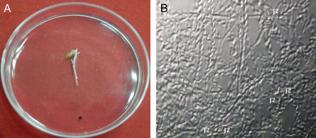
Attraction of M. incognita J2s toward tomato root tip in PF-127 medium in a petri dish. (A) 50 J2s were inoculated at 1.5 cm distance (marked by black dot) from the tomato root tip. (B) Numerous sinusoidal tracks inscribed on PF-127 medium due to J2 locomotion towards tomato root were documented.
Figure 2.
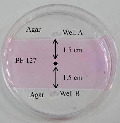
Actual photograph of an in vitro chemotaxis assay plate used in the present study. Due to the premixing of pink colored acid fuchsin stain with PF-127 gel, areas containing agar and PF-127 can be easily demarcated. Nematode inoculation point (*) is 1.5 cm equidistant from agar-PF-127 junction. Agar-PF-127 junction is 1 cm distant from the nearest edge of the petri plate. In well A, different test compounds were applied to observe either nematode attraction or repulsion. In well B, diluents (in which test compounds were dissolved) were applied as control.
Materials and methods
Nematode cultures
A pure culture of M. incognita race 1 was multiplied on eggplant (cv. Pusa Purple Long) in a greenhouse. Egg masses were collected from eight-week-old infected roots using sterilized forceps and kept for hatching in a modified Baermann assembly (Whitehead and Hemming, 1965). Freshly hatched J2s were used for all the experiments.
Collection of root exudates
Seeds of tomato (cv. Pusa Ruby), tobacco (cv. Petite Hawana), and marigold (cv. Arpit) were surface sterilized and germinated via standard methods (Papolu et al., 2013; Dutta et al., 2015). Each of the five- to six-day-old seedlings was hydroponically grown in half strength Hoagland solution (Hoagland and Arnon, 1950) in 15 ml Falcon conical centrifuge tubes (Sigma-Aldrich) for 15 d followed by removal and insertion of the roots of the plantlets (post washing) in sterile distilled water in a fresh Falcon tube for another 5 d. The whole assembly (covered in aluminium foil) was maintained in a growth chamber at 28°C, 70% RH, and 14 hr: 10 hr light: dark photoperiod. Sterile water containing the root exudates were pooled from 50 plants for each of the hosts, filtered through Whatman No. 1 filter paper and concentrated to 5 ml suspension (from initial volume of 50 ml) for each of the hosts in Eppendorf tubes via vacuum evaporation in a Speed Vac Concentrator (Labconco) using a vacuum of 100 to 500 mTorr.
Preparation of agar-pluronic gel assay plate
Pluronic F-127 (PF-127) (Sigma-Aldrich) gel was prepared as described previously (Wang et al., 2009a). To prepare 0.8% agarose gel, 0.8 g of agarose powder (Sigma-Aldrich) was dissolved in 100 ml of sterile distilled water in a microwave oven. Six ml of agarose gel was poured onto a 50×10 mm petri plate (Tarsons, part number - 461010) and allowed to solidify. Two parallel imaginary lines, each equidistant of 1.5 cm from the center of the petri plate, were drawn (Fig. 2). Agar was scooped out from the area between these two lines using sterilized forceps and was filled with 3 ml of 23% PF-127 gel and allowed to solidify at room temperature (i.e. >15°C). Small wells of 1.5 mm diameter were created (by scooping out the agar halfway through the plate with rear end of 1 ml pipette tip) on the agar surface adjacent to agar-PF-127 junction (almost 1 cm distant from the nearest edge of the petri plate) for application of test compounds (Fig. 2).
Chemical gradient assay
To quantitatively define the concentration gradient of a test compound in the assay plate, a colorimetric assay using acid fuchsin was carried out. PF-127 hardly adsorbs acid fuchsin which aids in rapid analysis of their concentration distribution in PF-127 gel by measuring the color intensity. Also, 20 µl of acid fuchsin (stock solution: 3.5 g acid fuchsin, 250 ml acetic acid, 750 ml distilled water) was pipetted into a well in assay plate and incubated at room temperature. After 40 min, 50 µl each of the test samples was collected from PF-127 gel at 2, 5, 10, and 15 mm distance from the application point of acid fuchsin and liquefied on ice. To each sample, distilled water was added to make up the volume to 1 ml for measurement of their absorbance at 550 nm in a standard spectrophotometer (Eppendorf Biophotometer Plus). Experimental samples were measured against the average of three controls containing PF-127 gel and distilled water. A standard curve was generated by plotting the various concentrations of acid fuchsin in PF-127 gel vs the absorbance measurements of different concentrations of acid fuchsin that were fit to a linear correlation model (R2 = 0.98; Fig. 3).
Figure 3.
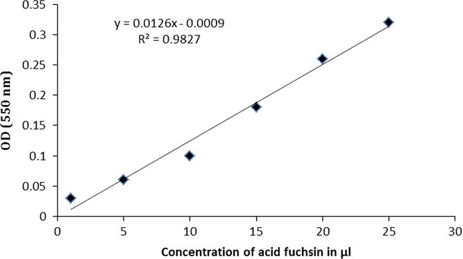
A standard curve displays absorbance (550 nm) calibration values plotted against different concentrations of acid fuchsin (1, 5, 10, 15, 20, and 25 µl) in PF-127 gel which were fit to a linear correlation model (R 2 = 0.98).
Chemotaxis assay
To measure the attraction or repulsion of J2 in response to test chemicals, various volatile compounds such as isoamyl alcohol, 1-butanol, benzaldehyde, 2-butanone, and 1-octanol (previously used in Caenorhabditis elegans chemotaxis experiment on agar plate; Bargmann et al., 1993) were screened at six different concentrations (100, 10−1, 10−2, 10−3, 10−4, and 10−5). First, each of the compounds (obtained from Sigma-Aldrich) was dissolved in sterile ethanol (0.05% v/v) which is designated as neat concentration (100). Subsequently, neat concentration of the compound was serially diluted (10 times each in sterile distilled water) until 10−5 concentration was achieved. Also, 5 µl aliquots of each of the odorants was pipetted in one well of the assay plate and 5 µl of ethanol (diluent) was placed at the opposite well of the same plate. The set up (petri plate with closed lid) was kept undisturbed for 40 min at room temperature to allow the test chemicals to form a concentration gradient. After that, approximately 100 J2s were added (by pipetting the concentrated 5 µl suspension containing J2s) to the center of the plate and incubated for 20 min at room temperature. J2s accumulating near the agar-PF-127 junction at either odorant or diluent side were easily pipetted out by liquefying the PF-127 gel (careful transfer of the assay plate to ice block led to the liquefaction of PF-127 gel since the gel has inherent property of becoming liquid at <15°C temperature) and counted under the microscope. The chemotaxis index (CI) was calculated as [(Number of J2s at odorant side−Number of J2s at diluent side)/Total number of J2s in assay] in which the index varies from 1.0 (perfect attraction) to −1.0 (perfect repulsion) (Bargmann et al., 1993). The number of nematodes that preferably did not move toward odorant/diluent side (from the application point) was not included in the index (although their number was counted to tally with total number applied). The negative control constituted the deionized water.
In order to measure the attraction or repulsion of J2 in response to host root exudates, 10 µl of neat exudates of tomato, tobacco, and marigold were separately screened in the assay plate as described above. The diluent was water in this case.
All the experiments were carried out with at least three technical replicates and were repeated at least thrice. Data were initially checked for normality and compared using one-way analysis of variance with Tukey’s HSD tests in SAS statistical package. All individual treatments were statistically compared to negative controls, as stated in the figure legends.
Results
Distribution of test chemical in the assay plate
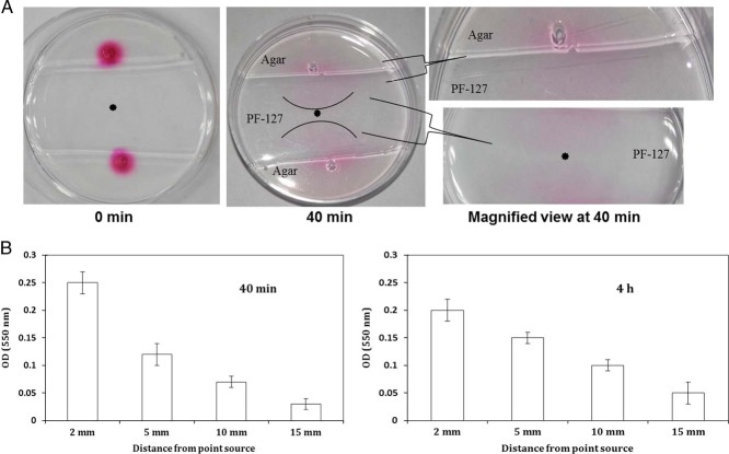
We measured the change in color intensity of acid fuchsin stain in assay plate. Qualitatively, it was ascertained that acid fuchsin can diffuse through the PF-127 medium from higher concentration to lower concentration within 40 min to establish equilibrium (Fig. 4A). We estimated that the concentration gradient from the point source to nematode inoculation point remained in a steady state up to 4 hr. Acid fuchsin concentration measured (in terms of absorbance at 550 nm) at indicated distances from the point source suggested the gradual decrease of test compound concentration from the point source (Fig. 4B). This suggests that similar to acid fuchsin our assay plate could also establish the concentration gradient of different volatile and non-volatile test compounds. However, we assume that all of the volatile compounds may not behave the same since the chemical structure and molecular weight of volatiles differ considerably than that of acid fuchsin.
Figure 4.
Qualitative (A) and quantitative (B) analyses of establishment of concentration gradient of a test chemical (acid fuchsin) in chemotaxis assay plate. Intensity of color suggests that acid fuchsin had diffused from its point source to nematode inoculation point by 40 min. Concentration of acid fuchsin (absorbance at 550 nm) attained an equilibrium at 40 min and remained in steady-state up to 4 hr at different indicated distances from the point source. Error bars indicate the standard error between three biological and technical replicates.
Chemotaxis response of M. incognita J2 to volatile odorants
M. incognita J2 exposed to a selection of volatile odorants (at increasing dilutions) was gauged by either an attraction or repellent phenotype relative to negative control (deionized water). The diluent (ethanol) itself did not have any behavioral effect on J2s. Notably, isoamyl alcohol, 1-butanol, and 2-butanone were attractive through a broad range of concentrations. J2s showed significant attraction to all the concentrations of isoamyl alcohol compared to negative control. The CI was recorded as 0.9 ± 0.1 (p < 0.001, on an average 95 J2s were counted at odorant side and 5 at diluent side), 0.8 ± 0.2 (p < 0.001, 85 at odorant and 5 at diluent), 0.6 ± 0.1 (p < 0.01, 75 at odorant and 15 at diluent), 0.5 ± 0.08 (p < 0.01, 70 at odorant and 20 at diluent), 0.5 ± 0.06 (p < 0.01, 72 at odorant and 22 at diluent), and 0.4 ± 0.1 (p < 0.05, 64 at odorant and 24 at diluent) at 100, 10−1, 10−2, 10−3, 10−4, and 10−5 concentrations of isoamyl alcohol, respectively (Fig. 5A). Similarly, J2s exposed to increasing dilutions of 2-butanone exhibited gradual decrease in attraction. The CI was recorded as 0.8 ± 0.07 (p < 0.001, 89 J2s at odorant and 9 at diluent), 0.7 ± 0.09 (p < 0.01, 78 at odorant and 8 at diluent), 0.6 ± 0.1 (p < 0.01, 76 at odorant and 16 at diluent), 0.5 ± 0.15 (p < 0.01, 68 at odorant and 18 at diluent), 0.4 ± 0.2 (p < 0.05, 66 at odorant and 26 at diluent), and 0.3 ± 0.12 (p > 0.05, 65 at odorant and 35 at diluent) at 100, 10−1, 10−2, 10−3, 10−4, and 10−5 concentrations of 2-butanone, respectively (Fig. 5A).
Figure 5.
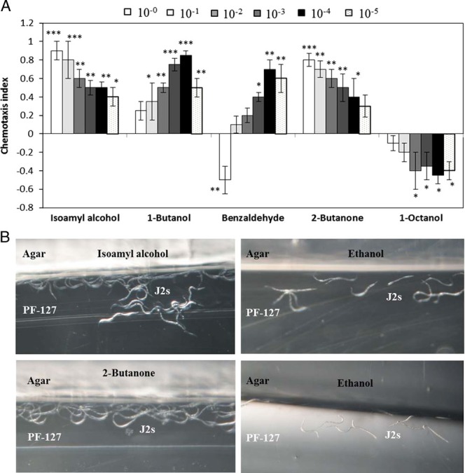
The chemotactic response of M. incognita J2 to volatile test compounds. (A) Chemotaxis bioassays show attraction (positive index) or repulsion (negative index) values. Statistical analysis performed using one-way ANOVA with Tukey’s HSD (p values: *p < 0.05; **p < 0.01; ***p < 0.001) tests comparing with water as negative control. Five µl of test compounds were screened at six different dilutions (100, 10−1, 10−2, 10−3, 10−4, and 10−5) against approximately 100 J2s. Error bars represent standard error of three biological and three technical replicates. (B) A close-up view of the assay plate shows that greater number of J2s was accumulated at agar-PF-127 junction in the odorant side compared to lesser number in the diluent side while exposed to isoamyl alcohol and 2-butanone.
By contrast, upon 1-butanol exposure, attraction increased gradually from neat to 10−4 dilution, suggesting 1-butanol was attractive at lower concentrations. The CI was documented as 0.25 ± 0.1 (p > 0.05, 51 J2s at odorant and 26 at diluent), 0.35 ± 0.2 (p < 0.05, 54 at odorant and 19 at diluent), 0.5 ± 0.05 (p < 0.01, 65 at odorant and 15 at diluent), 0.75 ± 0.07 (p < 0.001, 80 at odorant and 5 at diluent), 0.85 ± 0.05 (p < 0.001, 90 at odorant and 5 at diluent), and 0.5 ± 0.1 (p < 0.01, 72 at odorant and 22 at diluent) at 100, 10−1, 10−2, 10−3, 10−4, and 10−5 concentrations of 1-butanol, respectively (Fig. 5A). Somewhat similar trend was observed for benzaldehyde to which J2s were attractive at low concentrations but repulsive while undiluted. The CI was documented as −0.5 ± 0.15 (p < 0.01, 23 at odorant and 73 at diluent), 0.1 ± 0.09 (p > 0.05, 50 at odorant and 40 at diluent), 0.2 ± 0.08 (p > 0.05, 55 at odorant and 35 at diluent), 0.4 ± 0.05 (p < 0.05, 65 at odorant and 25 at diluent), 0.7 ± 0.1 (p < 0.01, 80 at odorant and 10 at diluent), and 0.6 ± 0.15 (p < 0.01, 72 at odorant and 12 at diluent) at 100, 10−1, 10−2, 10−3, 10−4, and 10−5 concentrations of 1-butanol, respectively (Fig. 5A).
Surprisingly, 1-octanol was not attractive at any of the concentrations and was repulsive at high concentrations. CI was observed as −0.1 ± 0.08 (p > 0.05, 40 at odorant and 50 at diluent), −0.2 ± 0.1 (p > 0.05, 35 at odorant and 55 at diluent), −0.4 ± 0.2 (p < 0.05, 23 at odorant and 63 at diluent), −0.35 ± 0.15 (p < 0.05, 20 at odorant and 55 at diluent), −0.45 ± 0.09 (p < 0.05, 20 at odorant and 65 at diluent), and −0.4 ± 0.1 (p < 0.05, 18 at odorant and 58 at diluent) at 100, 10−1, 10−2, 10−3, 10−4, and 10−5 concentrations of 1-octanol, respectively (Fig. 5A).
Photographs showed that in response to neat concentrations of isoamyl alcohol and 2-butanone exposure, greater number of J2s was accumulated at the agar-PF-127 junction at the odorant side and lesser number of J2s was accumulated at the agar-PF-127 junction at the diluent side (Fig. 5B).
Chemotaxis response of M. incognita J2 to host root exudates
The diluted host root exudates did not elicit any significant chemotactic response of M. incognita J2s with respect to negative control (deionized water) in our assay plate. Therefore, only neat concentrations of root exudates were tested in this assay. Upon exposure to 10 µl neat exudates of tomato and tobacco, J2s exhibited significant attraction response; whereas, exudates of marigold elicited strong repulsion response in J2s. CI was calculated as 0.75 ± 0.2 (p < 0.001, 82 at exudate and 7 at control side), 0.65 ± 0.15 (p < 0.01, 77 at exudate and 12 at control), and −0.55 ± 0.2 (p < 0.01, 20 at exudate and 75 at control) toward the exudates of tomato, tobacco, and marigold, respectively (Fig. 6).
Figure 6.
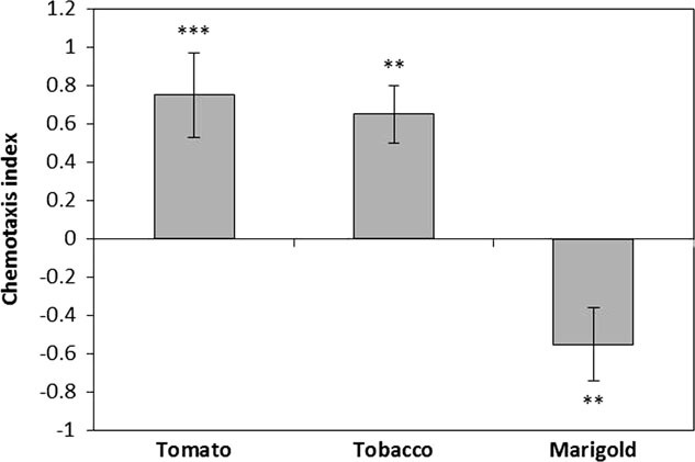
The chemotactic response of M. incognita J2 to host root exudates. Chemotaxis bioassays show attraction (positive index) or repulsion (negative index) values. Statistical analysis performed using one-way ANOVA with Tukey’s HSD (p values: *p < 0.05; **p < 0.01; ***p < 0.001) tests comparing with water as negative control. Ten microliters of neat exudates was tested against approximately 100 J2s. Error bars represent standard error of three biological and three technical replicates.
Discussion
Compared to the free-living worm, C. elegans, little is known about the olfactory behavior of PPNs (Rengarajan and Hallem, 2016) likely because of their obligate parasitic nature and lack of simple in vitro bioassays for quantitative analyses of PPN chemotaxis. Herein, we propose a simple, efficient, and reproducible new assay method to quantitatively analyze the chemotactic behavior of a PPN, i.e., M. incognita to a range of test compounds on combined agar and PF-127 medium in a petri dish.
One of the major technical issues with agar plate assays is that nematodes venture around both on the surface of the gel and within the gel matrix. This makes it difficult to track and quantify the nematode behavior accurately. Although PF-127 gel allows nematode movement in three-dimensional plane, same is true for PF-127-based assays as well (Hida et al., 2015). In line with the C. elegans in vitro assays, Wuyts et al. (2006) assessed the chemotactic behavior of M. incognita, Radopholus similis, and Pratylenchus penetrans toward different phytochemicals based on the tracks developed due to nematode migration (instead of nematode themselves) on the agar plate. The inference drawn based on only nematode tracks may be indicative of their chemotactic response, but not conclusive. On the contrary, our assay demonstrates the accurate quantitation of attraction/repulsion of M. incognita J2s to various test compounds due to the preferential accumulation of J2s at agar and PF-127 interface (agarose and PF-127 gel has differential texture). Unlike other in vitro assays, our assay does not require killing/anaesthetizing the nematodes using chemical reagents at the end of the assay for better assessment of the chemotactic behavior. All the live J2s recovered in our assay can be used for further downstream applications such as expression analysis of nematode chemosensory genes in response to various environmental cues via quantitative real time PCR (qRT-PCR) or RNAseq/transcriptomics-based investigation of nematode olfactory genes upon exposure to various chemicals.
We confirmed experimentally the concentration gradient formation of a chemical compound in our assay plate. This was achieved by monitoring and quantifying the changes in the color intensity of acid fuchsin over time from the point source in PF-127 medium. We assume that different volatile and non-volatile test compounds may establish concentration gradient in our assay plate. However, this may not hold true for all the volatile compounds because chemical structure and molecular weight of volatiles differ considerably than that of acid fuchsin. It has been suggested that nematodes can perceive chemical gradients while migrating in PF-127 gel matrix which mimics the three-dimensional perception and migration that occurs in natural soil (Wang et al., 2009a). Using our assay plate we demonstrate that M. incognita responds to a selective number of volatile odorants (in different concentrations) in quantitative and reproducible manner besides responding to the host root exudates. In accordance with the findings for C. elegans (Bargmann et al., 1993), in our assay, J2s of M. incognita positively chemotaxed to isoamyl alcohol, 2-butanone (preferably at higher concentrations), 1-butanol, and benzaldehyde (preferably at lower concentrations); whereas, J2s negatively chemotaxed to all the tested concentrations of 1-octanol and undiluted concentration of benzaldehyde. As expected, undiluted tomato and tobacco root exudates elicited attraction response and marigold exudates repelled M. incognita J2s in our assay plate. Considering that PF-127 exerts minimal toxicity to test organisms (Gardener and Jones, 1984; Ko and Van Gundy, 1988) it can be speculated that the inherent properties of PF-127 gel do not contaminate our test results. The results obtained in our in vitro assay can be extrapolated to field conditions for better understanding of nematode chemotaxis.
In conclusion, the new in vitro chemotaxis assay designed in the present study has allowed the easy and time-effective screening of various test compounds (both volatile and non-volatile), in a way that yields accurate representations of nematode responses to test compounds in a very short time (40 min for setting up the chemical gradient and 20 min for nematode behavioral response). Our assay could be very useful for screening a number of bioactive compounds for nematode behavior analysis on a large-scale. The results obtained may become useful for predicting the long-term fate of negative or positive interactions of nematodes with various test chemicals in field conditions. The application of this in vitro assay can also be extended to study the response of PPNs to various nematicides or drugs. Overall, our study provides a promising new tool for future investigation on PPN chemotaxis.
Acknowledgments
Tagginahalli N. Shivakumara, PhD Student, acknowledges his co-guide Dr Visakha Raina, School of Biotechnology, KIIT, Bhubaneswar, India.
References
- Ali J., Alborn H., and Stelinski L.. 2010. Subterranean herbivore induced volatiles released by citrus roots upon feeding by Diaprepes abbreviatus recruit entomopathogenic nematodes. Journal of Chemical Ecology 36: 361-368. [DOI] [PubMed] [Google Scholar]
- Bargmann C. I., Hartwieg E., and Horvitz H. R.. 1993. Odorant-selective genes and neurons mediate olfaction in C. elegans. Cell 74: 515-527. [DOI] [PubMed] [Google Scholar]
- Curtis R. H. C. 2008. Plant-nematode interactions: Environmental signals detected by the nematode’s chemosensory organs control changes in the surface cuticle and behaviour. Parasite 15: 310-316. [DOI] [PubMed] [Google Scholar]
- Dash M., Dutta T. K., Phani V., Papolu P. K., Shivakumara T. N., and Rao U.. 2017. RNAi-mediated disruption of neuropeptide genes, nlp-3 and nlp-12, cause multiple behavioral defects in Meloidogyne incognita. Biochemical and Biophysical Research Communications 490: 933-940. [DOI] [PubMed] [Google Scholar]
- Dutta T. K., Powers S. J., Kerry B. R., Gaur H. S., and Curtis R. H. C.. 2011. Comparison of host recognition, invasion, development and reproduction of Meloidogyne graminicola and M. incognita on rice and tomato. Nematology 13: 509-520. [Google Scholar]
- Dutta T. K., Papolu P. K., Banakar P., Choudhary D., Sirohi A., and Rao U. 2015. Tomato transgenic plants expressing hairpin construct of a nematode protease gene conferred enhanced resistance to root-knot nematodes. Frontiers in Microbiology 6: 260. [DOI] [PMC free article] [PubMed] [Google Scholar]
- Eo J., Nakamoto T. N., Otobe K., and Mizukubo T. M.. 2007. The role of pore size on the migration of Meloidogyne incognita juveniles under different tillage systems. Nematology 9: 751-758. [Google Scholar]
- Eo J., Otobe K., and Mizukubo T.. 2008. Absence of geotaxis in soil-dwelling nematodes. Nematology 10: 147-149. [Google Scholar]
- Farnier K., Bengtsson M., Becher P. G., Witzell J., Witzgall P., and Manduríc S.. 2012. Novel bioassay demonstrates attraction of the white potato cyst nematode Globodera pallida (Stone) to non-volatile and volatile host plant cues. Journal of Chemical Ecology 38: 795-801. [DOI] [PubMed] [Google Scholar]
- Gardener S., and Jones J. G.. 1984. A new solidifying agent for culture media which liquefies on cooling. Journal of General Microbiology 130: 731-733. [Google Scholar]
- Hida H., Nishiyama H., Sawa S., Higashiyama T., and Arata H.. 2015. Chemotaxis assay of plant-parasitic nematodes on a gel-filled microchannel device. Sensors and Actuators B: Chemical 221: 1483-1491. [Google Scholar]
- Hoagland D. R., and Arnon D. I.. 1950. The water-culture method for growing plants without soil, University of California, College of Agriculture, Agricultural Experiment Station, Berkeley, CA. [Google Scholar]
- Jones J. T., Haegeman A., Danchin E. G. J., Gaur H. S., Helder J., Jones M. G. K., Kikuchi T., Manzanilla-Lopez R., Palomares-Rius J. E., Wesemael W. M. L., and Perry R. N.. 2013. Top 10 plant-parasitic nematodes in molecular plant pathology. Molecular Plant Pathology 14: 946-961. [DOI] [PMC free article] [PubMed] [Google Scholar]
- Ko M. P., and Van Gundy S. D.. 1988. An alternative gelling agent for culture and studies of nematodes, bacteria, fungi, and plant tissues. Journal of Nematology 20: 478-485. [PMC free article] [PubMed] [Google Scholar]
- Kumari C., Dutta T. K., Banakar P., and Rao U.. 2016. Comparing the defence related gene expression changes upon root-knot nematode attack in susceptible versus resistant cultivars of rice. Scientific Reports 6: 22846. [DOI] [PMC free article] [PubMed] [Google Scholar]
- Palomares-Rius J. E., Escobar C., Cabrera J., Vovlas A., and Castillo P.. 2017. Anatomical alterations in plant tissues induced by plant-parasitic nematodes. Frontiers in Plant Science 8: 1987. [DOI] [PMC free article] [PubMed] [Google Scholar]
- Papolu P. K., Gantasala N. P., Kamaraju D., Banakar P., Sreevathsa R., and Rao U.. 2013. Utility of host delivered RNAi of two FMRFamide like peptides, flp-14 and flp-18, for the management of root-knot nematode, Meloidogyne incognita. PLoS One 8: e80603. [DOI] [PMC free article] [PubMed] [Google Scholar]
- Perry R. N. 2005. An evaluation of types of attractants enabling plant-parasitic nematodes to locate plant roots. Russian Journal of Nematology 13: 83-88. [Google Scholar]
- Rasmann S., Kollner T. G., Degenhardt J., Hiltpold I., Toepfer S., Kuhlmann U., Gershenzon J., and Turlings T. C. J.. 2005. Recruitment of entomopathogenic nematodes by insect-damaged maize roots. Nature 434: 732-737. [DOI] [PubMed] [Google Scholar]
- Rengarajan S., and Hallem E. A.. 2016. Olfactory circuits and behaviors of nematodes. Current Opinion in Neurobiology 41: 136-148. [DOI] [PMC free article] [PubMed] [Google Scholar]
- Reynolds A. M., Dutta T. K., Curtis R. H. C., Powers S. J., Gaur H. S., and Kerry B. R.. 2011. Chemotaxis can take plant-parasitic nematodes to the source of a chemoattractant via the shortest possible routes. Journal of the Royal Society Interface 8: 568-577. [DOI] [PMC free article] [PubMed] [Google Scholar]
- Robinson A. F. 2000. Techniques for studying nematode movement and behavior on physical and chemical gradients. Society of Nematologists Ecology Committee.
- Spence K. O., Lewis E. E., and Perry R. N.. 2008. Host finding and invasion by entomopathogenic and plant-parasitic nematodes: evaluating the ability of laboratory bioassays to predict field results. Journal of Nematology 40: 93-98. [PMC free article] [PubMed] [Google Scholar]
- Wang C. L., Lower S., and Williamson V. M.. 2009a. Application of pluronic gel to the study of root-knot nematode behaviour. Nematology 11: 453-464. [Google Scholar]
- Wang C., Bruening G., and Williamson V. M.. 2009b. Determination of preferred pH for root-knot nematode aggregation using pluronic F-127 gel. Journal of Chemical Ecology 35: 1242-1251. [DOI] [PMC free article] [PubMed] [Google Scholar]
- Wang C., Lower S., Thomas V. P., and Williamson V. M.. 2010. Root-knot nematodes exhibit strain-specific clumping behavior that is inherited as a simple genetic trait. PLoS One 5: e15148. [DOI] [PMC free article] [PubMed] [Google Scholar]
- Whitehead A. G., and Hemming J. R.. 1965. A comparison of some quantitative methods of extracting small vermiform nematodes from soil. Annals of Applied Biology 55: 25-38. [Google Scholar]
- Wuyts N., Swennen R., and De Waele D.. 2006. Effects of plant phenylpropanoid pathway products and selected terpenoids and alkaloids on the behavior of the plant-parasitic nematodes Radopholus similis, Pratylenchus penetrans and Meloidogyne incognita. Nematology 8: 89-101. [Google Scholar]


