Abstract
Here, we describe an in vitro epithelial wound-healing assay using Electric Cell-Substrate Impedance Sensing (ECIS) technology. The ECIS technology is a real time cell growth assay based on a small (250 μm diameter) active gold electrode which resistance is measured continuously. When intestinal epithelial cells reach confluency on the gold electrode, resistances reach a plateau. For the wound-healing assays, confluent intestinal epithelial monolayers are subjected to a current of 40 kHz frequency, 1,400 μA amplitude, and 30-second duration. This kills the cells around the small active gold electrode, causing detachment and generating a wound that is healed by surrounding cells that have not been submitted to the current pulse. Wound healing is then assessed by continuous resistance measurements for approximately 30 h after wound. Both cell wounding and measurements of the subsequent healing process are carried out under computer control that takes online measurements each 30 s and stores the data. ECIS technology can be used to study the underlying causes for impaired mucosal healing and to test the efficacy of drugs in mucosal healing.
Keywords: Electric cell-substrate Impedance sensing, Wound-healing, Intestinal epithelial cell Monolayers, Ulcerative colitis (UC), Caco2-BBE cells
Background
Damage and impairment of the intestinal surface barrier are observed in the course of various diseases including Ulcerative Colitis (UC). Such characteristics may result in an increased penetration and absorption of toxic and immunogenic factors into the body leading to inflammation, uncontrolled immune response, and disequilibrium of the homeostasis of the host. Wound-healing assays in tissue culture have been used for many years to estimate the migration and proliferation rates of different cells under various culture conditions. These assays involve using instruments such as razor blades to “wound” a confluent monolayer of cells. The main disadvantages of these assays include a lack of reproducibility and an inability to precisely quantify wound healing. Here, we describe the ECIS technology, a non-invasive technique which monitors live cells in situ and in real time, which is an ideal cell-based system for the screening of potential drugs capable of achieving higher mucosal healing rates that provide significant clinical benefit for Ulcerative Colitis patients.
Materials and Reagents
75 cm2 Tissue culture flasks (Greiner Bio-One, catalog number: 658170)
2- and 10-ml plastic pipettes (Sigma-Aldrich, catalog number: CLS4101)
Pipet Tip, Pipetman, PK1000 (Grainger, catalog number: 52jy20)
InvitrogenTM CountessTM Cell Counting Chamber Slides (Thermo Fisher Scientific, Invitrogen, catalog number: C10312)
Epithelial cell line Caco2-BBE or C2BBe1 (ATCC, catalog number: ATCC® HTB-37TM)
DMEM (1:1) 1x medium (Thermo Fisher Scientific, Gibco, catalog number: 12634010)
Fetal Bovine Serum (FBS) Premium Heat Inactivated at 56 °C (Atlanta Biologicals, catalog number: S11050H)
Penicillin-Streptomycin (Pen/Strep) (Thermo Fisher Scientific, Gibco, catalog number: 15140122)
Phosphate Buffered Saline (PBS 1x) (Thermo Fisher Scientific, Gibco, catalog number: 10010049)
0.25% Trypsin-EDTA (1x) (Thermo Fisher Scientific, Gibco, catalog number: 25200056)
InvitrogenTM Trypan Blue Stain (0.4%) (Thermo Fisher Scientific, Invitrogen, catalog number: T10282)
Recombinant Human IL-22 Protein (R&D Systems, catalog number: 782-IL10)
Cell culture medium (see Recipes)
IL22 stock (see Recipes)
Equipment
PIPETMAN Classic P1000 (Gilson, catalog number: FA10006M)
Drummond Scientific Pipet-Aid® Portable XP w/110 V recharger (Drummond Scientific, catalog number: 4-000-100)
Laminar Flow Hood (LabGard Class II type A2 Biological Safety Cabinet, Nuaire, model: NU-540)
Thermo Scientific 3110 CO2 Water Jacketed Incubator (Thermo Fisher Scientific, model: 4120)
Digital Water Bath (VWR, catalog number: 89501-460)
Nikon Eclipse TS100 inverted microscope (Nikon, model: Eclipse TS100)
CountessTM automated cell counter (Thermo Fisher Scientific, Invitrogen, catalog number: AMQAF1000)
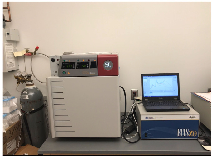
ECIS Zθ(Theta and 16 W array station (Applied Bio Physics, https://www.biophysics.com/products.php) (Figure 1)
CO2 Compact Tissue Culture Incubator (Shel Lab, Model SCO5A) (Figure 1)
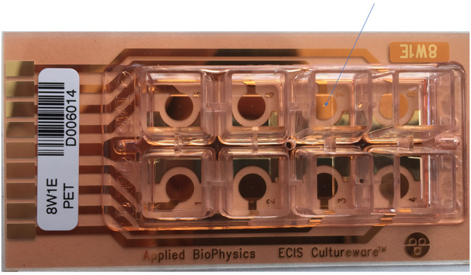
8W10E+ Cultureware (Ibidi, catalog number: 72001) (Figure 2)
OptiPlex 7760 All-in-One (6 Cores/9MB/6T/up to 4.1GHz/65W) (Dell)
Figure 1. ECIA Zq (Theta) and 16 W array station (inside the CO2 Compact tissue culture incubator) .
Figure 2. 8 Well arrays “8W1E”.
Each of the 8 wells contains a single circular 250 μm diameter active electrode (dot at the end of the blue arrow). Each well has a substrate area of 0.8 cm2 and a maximum volume of 500 μl.
Software
ECIS Z software (https://www.biophysics.com/products-ecisz0.php)
Microsoft Office Excel (Microsoft)
Procedure
Caco-2-BBE cells are routinely maintained in tissue culture flasks in Dulbecco’s Modified Eagle Medium (DMEM) medium containing 10% Inactivated Fetal Bovine Serum (FBS) and 1% penicillin-streptomycin and are put into a cell incubator set at 37 °C, in 5% CO2, and 90% humidity.
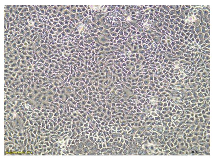
Observe confluency of Caco2-BBE cells in a 75 cm2 flask under the inverted microscope (Nikon Eclipse TS100) (Figure 3).
Under a laminar flow hood, rinse the 75 cm2 flask containing Caco2-BBE cells with PBS 1x (3 times each time with 5 ml PBS).
Add 2 ml of 0.25% Trypsin-EDTA.
Put the 75 cm2 flask containing Caco2-BBE cells with 2 ml of 0.25% Trypsin-EDTA into the cell incubator for 5 min.
Observe cell dissociation under the inverted microscope (Nikon Eclipse TS100).
If well dissociated, add 10 ml of DMEM supplemented with 10% FBS and 1% P/S media into the flask to inactivate trypsin and thoroughly pipette up and down (using 2 ml plastic pipettes) to break the cell clumps.
Place the 75 cm2 flask containing Caco2-BBE cells under the inverted microscope (Nikon Eclipse TS100) and observe that Caco2-BBE cells are a single cell suspension (no cell clumps). If not go back to Step 7.
Count the cells using Invitrogen CountessTM automated counter. The CountessTM automated cell counter is a benchtop counter designed to measure cell count and viability (live, dead, and total cells) accurately and precisely, using the standard trypan blue technique.
Plate Caco2-BBE cells at 0.3 x 10⁶ cells per well into two 8W10E+ Cultureware arrays with culture medium volume of 500 μl/well (one array contains 8 wells).
The 2 Culturewares (16 wells) in the ECIS array station allow the observer to design experiments with duplicates/triplicates with the appropriate controls.
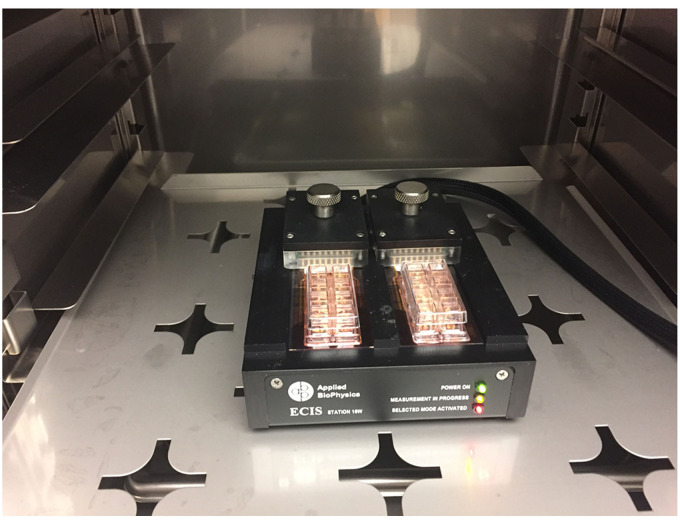
Put the 2 Culturewares in the ECIS array containing Caco2-BBE cells within the incubator station (see Figure 4) and close the incubator door. Incubator is set at 37 °C, in 5% CO2 and 90% humidity.
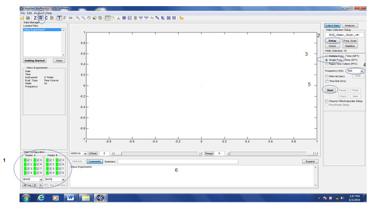
Chose “8W10E” as well configuration.
Click the “Set up” button to verify that arrays are properly connected to the system (Figure 5).
Wells that are connected to the system show a green color (Figure 5). Wells not connected to the system show a red color. If the array diagram shows red, adjust the position of the Cultureware slightly and click “Check” to make sure that each well of the Cultureware is connected properly to the system.
Select “The well to measure”.
Select “Single frequency” (For Caco2-BBE, the optimal single frequency is 500 Hz) ( Charrier et al., 2005 ) (Figure 5).
Interval (sec): uncheck. (maximum data acquisition) (Figure 5).
Click “Start” and specify where the data are stored (Figure 5).
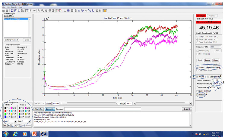
When Caco2-BBE reach confluency after 20-30 h growth (Resistance plateau) check “Wound/electroporation Setup” (Figure 6).
Select the “Wells to wound” in the “Well Configuration” (Figure 6).
Select “Wound time” (30 s [Figure 6], optimal for Caco2-BBE) ( Charrier et al., 2005 ; Zhang et al., 2016 ; Xiao et al., 2018 ).
Select “Wound Current” (1,400 μA [Figure 6], optimal for Caco2-BBE) ( Charrier et al., 2005 ; Zhang et al., 2016 ; Xiao et al., 2018 ).
Select “Frequency” (40k Hz [Figure 6], optimal for Caco2-BBE) ( Charrier et al., 2005 ; Zhang et al., 2016 ; Xiao et al., 2018 ).
Click “Activate” (Figure 6).
After wound infliction, verify that the R value drop to a value close to an electrode without cells. If that is not the case, repeat the wounding.
After the application of wound to the epithelium, treatments (drug-addition) may be applied to designated wells in order to assess the pro-healing or the anti-healing of the added drug.
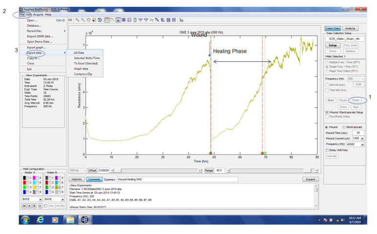
When the healing is done, R return to the resistance value before the wound, stop the experiment by click “Finish” (Figure 7).
Go to file and select “Export data” (Figure 7).
As an example, we have demonstrated (Figure 7 in Xiao et al., 2018 ) that wounded epithelial layers treated with IL-22 (0, 50 and 100 ng/ml as final concentration) showed significant and dose-dependent increase in recovery, in comparison with untreated wounded epithelial layers. These results clearly demonstrate that IL-22 can enhance the healing of a wounded colonic epithelial layer. In another study (Figure 6 in Charrier et al., 2005 ), we have demonstrated that after the wound, the resistance of Caco2-BBE/ADAM-15 cells (Caco2-BBE over-expressing ADAM-15) did not increase to reach the resistance values of the non-wounded cells (Figure 6A in Charrier et al., 2005 ). By contrast, the resistance of Caco2-BBE/Vect cells (endogenous ADAM-15 expression in Caco2-BBE) increased over time and reached the resistance values of the non-wounded cells (Figure 6A in Charrier et al., 2005 ). These results are in correlation with the observations made at the end of the experiments where the wounds were completely healed in Caco2-BBE/Vect cells but were unable to heal in Caco2-BBE/ADAM-15 cells (Figure 6B in Charrier et al., 2005 ).
Figure 3. Micrograpic image of confluent Caco2-BBE monolayers in 75 cm2 flasks.
Magnification: 10x.
Figure 4. Two Culturewares in the ECIS array station within the incubator.
Figure 5. ECIS software and set-up procedure to measure epithelial electrical resistance (R).
Select “8W1E” as well arrays (1), select all wells in “Holder A and Holder B” (1), click the “Setup” button (2) to check if arrays are properly connected to the system. Wells that are connected to the system show a green color (1). Select “Single Freq.” (3) as 500 Hz (4) and click “Start” (5) and specify where the data are stored. Experiment commments may be documented (6).
Figure 6. Wound to Caco-2-BBE monolayers.
Select the “wells to wound” in the “Well Configuration” (1), select “Wound/Electroporate Setup” (2), select “Wound” (3), select “30 sec” as “Wound Time” (4), select “1400 μA” as “Wound Current” (5), select “40k Hz” as “Frequency” (6) and click “Activate” (7).
Figure 7. Wound Healing of Caco-2-BBE monolayers.
When healing is done, click “Finish” (1), go the “File” (2), select “Export data” (3).
Data analysis
Recipes
-
Cell culture medium
Dulbecco’s Modified Eagle Medium (DMEM) medium containing 10% Inactivated Fetal Bovine Serum (FBS) and 1% penicillin-streptomycin
-
IL22 stock solution
10 μg/ml in cell culture medium
Acknowledgments
EV is a recipient of the Career Development Award from the Crohn’s and Colitis Foundation. This protocol was adapted from published work by Charrier et al. (2005), Zhang et al. (2016) and Xiao et al. (2018).
Competing interests
The authors declare no conflicts of interest to this work.
Citation
Readers should cite both the Bio-protocol article and the original research article where this protocol was used.
References
- 1. Charrier L., Yan Y., Driss A., Laboisse C. L., Sitaraman S. V. and Merlin D.(2005). ADAM-15 inhibits wound healing in human intestinal epithelial cell monolayers. Am J Physiol Gastrointest Liver Physiol 288(2): G346-353. [DOI] [PubMed] [Google Scholar]
- 2. Xiao B., Chen Q., Zhang Z., Wang L., Kang Y., Denning T. and Merlin D.(2018). TNFα gene silencing mediated by orally targeted nanoparticles combined with interleukin-22 for synergistic combination therapy of ulcerative colitis. J Control Release 287:235-246. [DOI] [PMC free article] [PubMed] [Google Scholar]
- 3. Zhang M., Viennois E., Prasad M., Zhang Y., Wang L., Zhang Z., Han M. K., Xiao B., Xu C., Srinivasan S. and Merlin D.(2016). Edible ginger-derived nanoparticles: A novel therapeutic approach for the prevention and treatment of inflammatory bowel disease and colitis-associated cancer. Biomaterials 101: 321-340. [DOI] [PMC free article] [PubMed] [Google Scholar]


