Abstract
Background
Sepsis combined with myocardial injury is an important cause of septic shock and multiple organ failure. However, the molecular mechanism of sepsis-induced myocardial dysfunction has not yet been thoroughly studied. Resveratrol has been an important research topic due its organ-protection function, but the specific mechanism is unclear. The purpose of this study was to explore the mechanism of organ injury in sepsis and to investigate the molecular mechanism of resveratrol in myocardial protection in sepsis.
Material/Methods
A classical Sprague-Dawley rat model of sepsis peritonitis was constructed for further experiments. The PI3K inhibitor LY294002 and resveratrol were used to intervene in a rat model of cardiomyopathy. HE staining was used to observe pathological changes. Cardiomyocyte apoptosis was detected by TUNEL assay. Western blot analysis was used to detect the level of maker proteins.
Results
The PI3K inhibitors could promote cardiac abnormalities and apoptosis, but resveratrol showed the opposite effect. The upregulation function of the PI3K inhibitor on the expression of NF-κB, IL-6, IL-1β, and TLR4 in LPS rats was not obvious, but the expression of TNF-α in LPS+LY294002 rats was increased by 22.85% compared with that in LPS rats (P<0.05). Compared with the LPS group, the expression of NF-κB, TNF-α, IL-6, IL-1β, and TLR4 in the LPS+resveratrol group was decreased. The expression of p-PI3K, p-AKT, and p-mTOR in LPS+LY294002 was reduced. The expression p-PI3K, p-AKT, and p-mTOR in the myocardium of the LPS+resveratrol group was increased.
Conclusions
Resveratrol can protect the myocardium in sepsis by activating the PI3K/AKT/mTOR signaling pathway and inhibiting the NF-κB signaling pathway and related inflammatory factors.
MeSH Keywords: Class III Phosphatidylinositol 3-Kinases, Myocardial Injury, Receptor Activator of Nuclear Factor-kappa B, Sepsis
Background
Sepsis is a common complication of severe burns, shock, and infection, as well as in surgical patients. It is a systemic inflammatory response syndrome (SIRS) caused by an imbalance of the host’s response to infection, and SIRS can develop into multiple organ failure [1]. It a common cause of death of critically ill patients without heart disease [2]. Sepsis has a high incidence rate, complicated condition, rapid progression, and poor prognosis, and its mortality rate is 30~70%, seriously affecting the life quality and safety of patients [3]. Over 18 million cases of severe sepsis are diagnosed annually worldwide, and more than 70% of sepsis-related deaths were attributed to organ failure [1]. Numerous studies have shown that inflammation of the vessels and endocardium and the persistent activation of inflammatory signals are the starting links of cardiac dysfunction in sepsis. Therefore, the study of the role and molecular mechanism of inflammatory signal transduction in myocardial injury in sepsis is of great value to elucidate the molecular mechanism underlying this disease.
Nuclear factor-κB (NF-κB), which belongs to the Rel family protein, is an important nuclear transcription factor. It plays a key role in the gene expression of various sepsis immunoregulatory factors, including cytokines, chemokines, adhesion molecules, apoptosis factors, and acute-phase protein. Moreover, it participates in the biological processes at the early stage of the immune response and various stages of inflammation, mediating the pathological process of various inflammation-related diseases [4–8]. Because NF-κB is a central regulator of the inflammatory response and has a close relationship with myocardial injury in sepsis, NF-κB may be a new potential target for sepsis therapy.
Recent studies have shown that altered levels of activated caspases are found in septic cardiomyocytes [9], and some studies have shown that cardiac myocyte apoptosis in septic rats increases and the level of the apoptotic effector Caspase-3 increases, revealing that apoptosis also plays an important role in myocardial injury in sepsis [10]. The PI3K/AKT/mTOR signaling pathway is a classical cell signaling pathway related to apoptosis. Recently, many studies have confirmed that this signaling pathway not only inhibits apoptosis but also plays an active role in inhibiting the inflammatory response and reducing mortality due to sepsis [11–13]. Therefore, the possible cross-regulation between the PI3K/AKT/mTOR signaling pathway and NF-κB signaling pathway provides a new direction to study the pathogenesis of sepsis.
Resveratrol (Res) is a nonflavonoid polyphenol compound that has demonstrated anti-inflammatory [14], antiviral [15], bacterial and fungi inhibition [16], and cardiovascular and antitumor protection effects. In recent years, its organ-protection effect in sepsis has also attracted much attention, but the specific mechanisms and targets are not yet clear.
Therefore, this study explored the function of and relationship among resveratrol, PI3K/AKT/mTOR, NF-κB, and inflammation using an LPS rat model treated with resveratrol and a PI3K inhibitor. We used 2 approaches – molecular biology and pathophysiology – to further explore the pathogenesis of septic myocardial inhibition and the organ-protection mechanism of resveratrol in sepsis, as well as to provide effective therapeutic targets and new research directions for myocardial injury in sepsis.
Material and Methods
Establishment and grouping of animal models
A classical LPS intraperitoneal injection method was used to construct a Sprague-Dawley rat model of sepsis peritonitis. Forty SD rats were randomly divided into 4 groups with 10 rats in each group: a control group, an LPS model group, an LPS+Resveratrol group, and an LPS+LY294002 group. First, rats in the LPS group, LPS+Resveratrol group, and LPS+LY294002 group were administered LPS (20 mg/kg body weight; dissolved in sterile saline) by intraperitoneal injection, while rats in the control group were given saline via intraperitoneal injection. Then, 1 h after LPS administration, the LPS+Resveratrol group and LPS+LY294002 group rats were given intraperitoneal injection of resveratrol (60 mg/kg body weight; dissolved in dimethyl sulfoxide (DMSO) and then in saline containing 0.01% DMSO) and an intraperitoneal injection of LY294002 (5 mg/kg body weight; dissolved in DMSO, and then in saline containing 0.01% DMSO), respectively. At the end of the experiment, the rats were sacrificed, and their blood and hearts were harvested for subsequent analysis. The protocol of the present study was approved by the Animal Care and Use Committee of Fujian Provincial Hospital. All the methods were performed in accordance with the United States Public Health Services Guide for the Care and Use of Laboratory Animals, and all efforts were made to minimize the suffering and number of animals used in this study.
Histopathological observation of damaged myocardium tissue
The hearts of each rat were taken immediately after blood collection and then were placed into 10% formaldehyde solution. The tissue was dehydrated and paraffin-embedded, and pathological sections were made. HE staining was used to observe pathological changes in the myocardium under a light microscope.
Serum creatine kinase-MB (CK-MB), cardiac troponin I (cTnI), and lactate dehydrogenase (LDH) measurements
Standard techniques were used to determine the serum levels of cK-MB, cTnI, and LDH by using commercial kits, and each procedure was conducted according to the manufacturer’s protocol.
Detection of cardiomyocyte apoptosis by the TUNEL assay
The myocardial tissue specimens fixed with formaldehyde solution were removed. After dewaxing and dehydration, microwave antigen repair was carried out, the sliced routines were treated with freshly prepared 3% H2O2 in room temperature for 10 min, followed by rinsing in PBS for 3 min×2 times, digestion by 20 μg/mL of proteinase K, and rinsing by PBS for 3 min×2 times. After adding the TUNEL mixture, the samples were placed in the incubating box at 37°C for 60 min, followed by rinsing with PBS for 3 min×3 times. After adding 50 μL of alkaline phosphatase antibody per slice, the samples were place in an incubation box at 37°C for 30 min, followed by rinsing with PBS for 3 min×3 times. Two drops of BCIP/NBT was added per piece, followed by incubation for 30 min at room temperature, rinsing with PBS for 3 min×3 times, mounting with water-based sealant, and then drying at 60°C. Under a normal light microscope, normal cells were not labeled, and the nuclei showed blue-black as positive cells. The average number of positive apoptotic cells in each high-magnification visual field was counted randomly in each slice, and the percentage of the number of positive cells and total cells was the apoptotic index, reflecting the apoptosis rate of each group.
Detection of the expression of related proteins by Western blotting
The protein extract RIPA was used to extract protein from cardiac tissue, and BCA was used to quantify the protein. Next, they were separated through 10% SDS-polyacrylamide gel electrophoresis and polyvinylidene fluoride (PVDF) membranes. Then, the membranes were incubated with 5% skim milk for 3 h and incubated with the primary antibodies (1: 1000 dilutions with PBST) at 4°C overnight. After 2 washes with 1X PBS, the membranes were incubated with a horseradish peroxidase-conjugated secondary antibody (1: 1000 dilutions with PBST) for 1 h at room temperature. Blots were developed using the ECL kit.
Statistical analysis
Data are presented as mean±SD. SPSS 17.0 statistical software was used for statistical analysis. One-way analysis of variance was followed by Tukey’s post hoc test for multiple group comparisons. The results were considered significant at P<0.05.
Results
Sepsis causes changes in systemic reactions
The rat models of sepsis were constructed with specific treatment groups. The mental status, body temperature, breathing, blood pressure, and white blood cell count were recorded and evaluated in all experiment groups. The results showed that the rats in the normal control group were generally in good condition, with free movement, stable breathing, no abnormal diet and drinking water, and normal response to external stimuli. However, after LPS modeling, the respiratory rate of rats was significantly accelerated, and there were symptoms of apathy, piloerection, lethargy, oozing blood of the eyes and nose, and loose stools, as well as weakened response to external stimuli. Body temperature, MAP, and WBC were significantly decreased in the LPS and LPS+LY294002 groups compared with those in the control group, whereas the LPS+resveratrol group showed a significant increase compared with that in the LPS group (Table 1).
Table 1.
Temperature, MAP and WBC in each group.
| Temperature (°C) | MAP (mmHg) | WBC (×109/L) | |
|---|---|---|---|
| Control | 38.2±0.1 | 103±8 | 4.5±0.5 |
| LPS | 37.3±0.2* | 75±4* | 2.1±0.4* |
| LPS+LY294002 | 37.0±0.3* | 77±6* | 2.0±0.3* |
| LPS+Resveratrol | 37.9±0.2# | 98±7# | 3.8±0.4# |
P<0.05 vs. Control group;
P<0.05 vs. LPS group.
Sepsis causes changes in cardiac function
The rat models of sepsis were constructed with different treatment groups. Cardiac functions were evaluated by ultrasonic cardiogram in all experimental groups. The results showed that heart rate (HR), left ventricular internal diameter at end-diastole (LVIDd), left ventricular internal diameter at end-systole (LVIDs), and cardiac output (CO) were significantly increased in the LPS and LPS+LY294002 groups, whereas these effects were reversed in the LPS+resveratrol group (Table 2). Additionally, the left ventricular ejection fraction (LVEF) was greatly decreased in the LPS and LPS+LY294002 groups, whereas it was partially recovered in the LPS+resveratrol group (Table 2), suggesting that LPS and LPS+LY294002 rats had decreased cardiac function, and resveratrol could attenuate this effect in LPS rat hearts.
Table 2.
Values of the hemodynamic parameters in each group.
| Parameters | Control | LPS | LPS+LY294002 | LPS+Resveratrol |
|---|---|---|---|---|
| LVIDd (mm) | 4.85±0.23 | 5.93±0.14* | 6.05±0.12* | 5.64±0.15 |
| LVIDs (mm) | 2.55±0.21 | 3.41±0.27* | 3.69±0.28* | 2.83±0.17 |
| IVSd (mm) | 1.54±0.07 | 1.61±0.05 | 1.6±0.04 | 1.59±0.04 |
| IVSs (mm) | 2.63±0.16 | 2.71±0.13 | 2.73±0.16 | 2.69±0.17 |
| LVPWd (mm) | 1.65±0.05 | 1.75±0.06 | 1.74±0.04 | 1.70±0.06 |
| LVPWs (mm) | 2.52±0.26 | 2.53±0.21 | 2.54±0.19 | 2.53±0.21 |
| HR (beat/min) | 352±27 | 453±21* | 469±13* | 447±17 |
| FS (%) | 48.56±2.14 | 40.15±2.26 | 41.37±2.78 | 42.18±4.29 |
| CO (L/min) | 0.11±0.04 | 0.20±0.04* | 0.21±0.03* | 0.18±0.07 |
| LVEF (%) | 84.27±2.13 | 46.35±3.21* | 40.27±4.17* | 67.17±2.52 |
P<0.05 vs. Control group.
Sepsis causes damage to the heart muscle
The levels of troponin cTnT, creatine kinase CK-MB, and lactate dehydrogenase LDH in the plasma of rats each experimental group were examined, and the results are shown in Figure 1. The results showed that cTnT and LDH were significantly increased in the LPS and LPS+LY294002 groups compared with those in the normal control group, whereas these effects were reversed in the LPS+resveratrol group (Figure 1) and were significantly different from the LPS group. However, there was no significant change in CK-MB in each group.
Figure 1.
cTnT, CK-MB, and LDH levels in serum samples. (A) cTnT levels. (B) CK-MB levels. (C) LDH levels. * P<0.05 versus control group; # P<0.05 versus LPS-treated group
Morphological results of effects of the PI3K inhibitor and resveratrol on myocardial injury
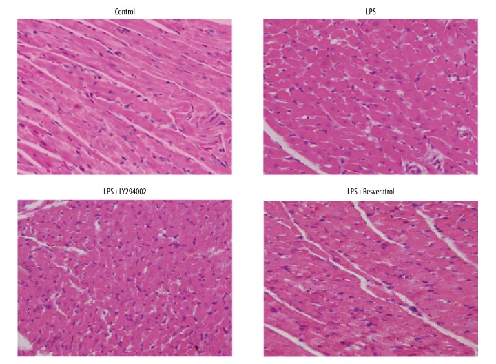
As we mainly focused on pathological changes in the heart, HE staining was judges to be sufficient to draw a conclusion that resveratrol decreases myocardial necrosis and results in well-arrange myocardial fibers. HE staining was performed to observe morphological results of effects of the PI3K inhibitor and resveratrol on myocardial injury. The HE staining results of pathological sections of the 4 tested groups of rats are shown in Figure 2. The results showed that, compared with the control group, in the LPS group, the cardiac muscle cell boundaries were not homogeneous, myocardial cells were disorderly and scattered in myofibrosis, and there were myocardial abnormalities and inflammatory cell infiltration. The PI3K inhibitor LY294002-treated group LPS+LY294002 showed more abnormalities than the LPS group. In the LPS+resveratrol group, the rat cardiac myocyte space was uniform, the boundary was clear, the myocardial fibers were clear and arranged, and no obvious inflammatory cells or fibrosis were found. Nuclear staining in the model rats was blue, indicating that the PI3K inhibitor can promote myocardial injury in septic rats, and resveratrol reduces myocardial injury in septic rats.
Figure 2.
Histopathology analyzed following resveratrol and PI3K inhibitor treatment in LPS-treated myocardial sepsis injury as detected by HE staining. Scale bar, 200 μM.
TUNEL detection results of the effect of the PI3K inhibitor and resveratrol on cardiomyocyte apoptosis
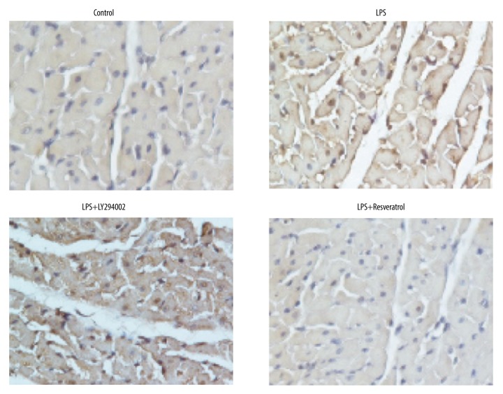
The apoptosis of cardiac myocytes was detected by TUNEL assay in the 4 tested groups of rats. The results (Figure 3) showed that, compared with the control group, the number of apoptotic cells with a positive color in the LPS group was obviously increased, and the number of apoptotic cells in LPS rats treated with the PI3K inhibitor LY294002 was further increased and was significantly higher than that in the LPS group. In the resveratrol-treated LPS rats (LPS+resveratrol), there were significantly fewer apoptotic cells with a positive color than in the LPS group, and no significant difference was found in the number of apoptotic cells compared with the normal control group. Thus, the PI3K inhibitor LY294002 can promote apoptosis of cardiac myocytes in sepsis, while resveratrol can inhibit the apoptosis of cardiac myocytes in sepsis.
Figure 3.
Cardiomyocyte apoptosis assessed following resveratrol and PI3K inhibitor treatment in LPS-treated myocardial sepsis injury by TUNEL assay. Scale bar, 200 μM.
Effects of the PI3K inhibitor and resveratrol on apoptosis pathway-related proteins
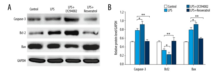
The expression of apoptosis pathway-related proteins in the myocardium were detected by Western blotting in the 4 groups of rats. The results (Figure 4) showed there was upregulated expression of capase-3 and Bax in the myocardium of LPS rats treated with the PI3K inhibitor. After resveratrol treatment, the expression levels of capase-3 and Bax in the myocardium of the LPS+resveratrol group rats were decreased significantly (by 32.51% and 25.34%, respectively) compared with those of the LPS group. However, the expression of Bcl2 in the myocardium of LPS rats treated with the PI3K inhibitor was downregulated. After resveratrol treatment, the expression of Bcl2 in the myocardium of the LPS+resveratrol group rats was significantly increased (by 39.20%) compared with that in the LPS group.
Figure 4.
Effect of resveratrol and the PI3K inhibitor on the expression of apoptosis pathway-related proteins in LPS-treated myocardial sepsis injury. (A) The expression levels of capase-3, Bax, and Bcl2 were analyzed by Western blotting. (B) Statistical analysis. The data are represented as means±SD of measurements. * P<0.05; ** P<0.01.
Effects of the PI3K inhibitor and resveratrol on NF-κB signaling pathway-related proteins
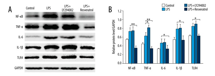
The effects of the PI3K inhibitor and resveratrol on the expression of NF-κB protein and inflammatory factors TNF-α, IL-6, IL-1β, and TLR4 were detected by Western blotting. The results are shown in Figure 5. The expression levels of NF-κB and inflammatory cytokines TNF-α, IL-6, IL-1β, and TLR4 in the myocardium of septic rats were significantly higher than those in the normal control group. The effect of the PI3K inhibitor on the expression levels of NF-κB, IL-6, IL-1β, and TLR4 in the myocardium of LPS rats was not obvious, but the expression of the inflammatory factor TNF-α was significantly increased, and the expression of TNF-α in LPS+LY294002 rats increased by 22.85% compared with that in the LPS group. Resveratrol significantly reduced the expression levels of NF-κB, TNF-α, IL-6, IL-1β, and TLR4 in the myocardium of LPS rats. Compared with the LPS group, the expression of NF-κB in the LPS+resveratrol group rats was decreased by 52.27%, the expression of TNF-α was decreased by 46.50%, the expression of IL-6 was decreased by 29.43%, the expression of IL-1β was decreased by 32.09%, and the expression of TLR4 was decreased by 26.09%. Moreover, after treatment with resveratrol, the expression of NF-κB and TNF-α in the myocardium of sepsis rats was lower than that of the normal control group, revealing the strong anti-inflammatory and myocardial-protective effects of resveratrol.
Figure 5.
Effect of resveratrol and the PI3K inhibitor on the expression of NF-κB signaling pathway-related proteins in LPS-treated myocardial sepsis injury. (A) The expression levels of NF-κB, IL-6, TNF-α, IL-1β, TLR4 were analyzed by Western blotting. (B) Statistical analysis. The data are represented as means±SD of measurements. * P<0.05; ** P<0.01.
Effects of the PI3K inhibitor and resveratrol on PI3K/Akt/mTOR signaling pathway-related proteins
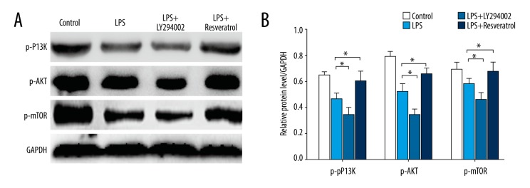
Western blotting was used to detect the expression of PI3K/AKT/mTOR signaling pathway-related proteins in the myocardium of the 4 groups of rats. The results are shown in Figure 6. The expression levels of PI3K, AKT, and mTOR in the myocardium of LPS rats treated with PI3K inhibitors was upregulated, but the difference was not obvious. After resveratrol treatment, the expression levels of PI3K, AKT, and mTOR in the myocardium of the LPS+resveratrol group was increased compared with that of the LPS group, but the difference was not significant. However, the expression levels of p-PI3K, p-AKT, and p-mTOR in the myocardium of LPS rats treated with the PI3K inhibitor were downregulated. After resveratrol treatment, the expression levels of p-PI3K, p-AKT, and p-mTOR in the myocardium of the LPS+resveratrol group rats were significantly increased (by 23.24%, 21.71%, and 14.43%, respectively) compared with those of the LPS group.
Figure 6.
Effect of resveratrol and the PI3K inhibitor on the expression of PI3K/Akt/mTOR signaling pathway-related proteins in LPS-treated myocardial sepsis injury. (A) The expression levels of PI3K, AKT, mTOR, p-PI3K, p-AKT, and p-mTOR were analyzed by Western blotting. (B) Statistical analysis. The data are represented as means±SD of measurements. * P<0.05.
Discussion
Sepsis is a life-threatening organ dysfunction caused by a host’s imbalance in response to infection, and the heart is one of the main target organs. Early clinical studies have confirmed that early sepsis is associated with a high incidence of organic damage to the myocardium, and these patients often have hypotension, heart failure, and arrhythmia [17]. At the same time, myocardial inhibition is one of the main indicators of a poor prognosis in patients with sepsis. Epidemiological research has shown that 40% of patients with sepsis are complicated with myocardial injury, and approximately 60% of patients with septic shock in the ICU have clinical manifestations of myocardial injury, and the mortality rate of sepsis patients can reach 70–90% [18]. The clinical treatment of SIMD is difficult and the prognosis is poor, but there is no special treatment for SIMD, perhaps because the molecular mechanism underlying the myocardial inhibition in sepsis has not yet been thoroughly studied. Resveratrol has been an important research topic in recent years because of its organ-protection function. However, its specific mechanism is unclear, limiting its clinical application.
Many studies have shown that inflammatory cytokines play an important role in the process of myocardial injury, and TNF-α and IL-6 have been proven to play a core role in the development of sepsis. Moreover, recent studies have revealed that SIMD is also caused by myocardial cell apoptosis [8,10]. However, several signaling pathways are involved in inflammation and cell apoptosis. One of these signaling pathways is the NF-κB pathway. Normally, NF-κB is inactive combined with its inhibitory factor. In sepsis, due to the stimulation of LPS, the signal is transduced into the cell under the synergy of CD14 and MD2, and NF-κB is rapidly activated and then quickly moved to the nucleus, activating various inflammatory factors such as TNF-α, IL-6, and IL-1β and resulting in cell apoptosis and damage to tissues and organs [19]. Additionally, NF-κB not only stimulates the expression of cytokines but is also activated by the stimulation of these cytokines, resulting in a sustained or amplified inflammatory reaction [5]. The PI3K/AKT/mTOR signaling pathway is another classical pathway related to myocardial protection. Some studies have shown that activation of the pathway can play a role in myocardial protection by activating endothelial nitric oxide synthase, mediating an anti-apoptosis effect, and regulating the opening of mitochondrial permeability transition pores [20–22]. Recent studies have confirmed that the PI3K/AKT/mTOR signaling pathway plays a positive inhibitory role in the pathogenesis of sepsis [11–13]; however, there has been little research on whether there is crosstalk between these 2 signaling pathways in the process of myocardial injury in sepsis.
In this study, the PI3K inhibitor LY294002 and resveratrol were used to intervene in the rat model of cardiomyopathy. The relationship between the PI3K/AKT/mTOR signaling pathway and NF-κB signaling pathway in the pathogenesis of myocardial injury in sepsis was investigated. This study also assessed whether the protective mechanism of resveratrol on sepsis-related myocardial inhibition is related to promotion of the PI3K/AKT/mTOR signal transduction pathway and inhibition of the NF-κB signal transduction pathway; identical results were obtained from Western blotting.
Our results showed that, when the LPS model rats were treated with resveratrol, the myocardial tissue injury was obviously inhibited, and the number of apoptotic cells was decreased significantly, even returning to the normal control level. At the same time, resveratrol significantly promoted the expression of p-PI3K, p-AKT, and p-mTOR protein in the myocardium of LPS rats and significantly reduced the expression of NF-κB, TNF-α, IL-6, IL-1β, and TLR4. Thus, resveratrol plays a protective role in sepsis by activating the PI3K/Akt/mTOR signaling pathway and inhibiting the NF-κB signaling pathway and related inflammatory factors, but the specific cascade reaction and target need to be further studied. When the level of PI3K was suppressed, the degree of damage to myocardial tissue in LPS rats significantly increased and the number of apoptotic cardiomyocytes increased significantly. Meanwhile, the expression of p-PI3K, p-AKT, and p-mTOR protein in the PI3K/AKT/mTOR signaling pathway was significantly lower than that in the LPS group without LY294002 treatment. In the NF-κB signaling pathway, the PI3K inhibitor had little effect on the expression levels of N-κB, IL-6, IL-1β, and TLR4, but significantly increased the expression level of TNF-α. Thus, the PI3K/AKT/mTOR signaling pathway may be activated in the process of myocardial injury in sepsis.
This study preliminarily explored the relationship between the PI3K/AKT/mTOR signaling pathway and NF-κB signaling pathway during myocardial injury in sepsis and revealed the mechanism of the inhibition of myocardial injury by resveratrol, providing a theoretical basis for the study of the pathogenesis of sepsis organ damage and a new direction for adjuvant therapy of resveratrol in sepsis-induced organ injury.
Conclusions
The PI3K/AKT/mTOR signaling pathway may be activated in the process of myocardial injury in sepsis. Resveratrol can protect the myocardium in sepsis by activating the PI3K/AKT/mTOR signaling pathway and inhibiting the NF-κB signaling pathway, as well as by related inflammatory factors.
Footnotes
Source of support: Natural Science Foundation of Fujian [Grant number (2015) 269] and Fujian Province Intensive Care Medical Center Construction Project grants from Fujian Provincial Hospital [Grant number (2017)510#]
Conflict of interest
None.
References
- 1.Potz BA, Sellke FW, Abid MR. Endothelial ROS and impaired myocardial oxygen consumption in sepsis-induced cardiac dysfunction. J Intensive Crit Care. 2016;2 doi: 10.21767/2471-8505.100020. pii: 20. [DOI] [PMC free article] [PubMed] [Google Scholar]
- 2.Balik M, Matousek V, Maly M, Brozek T. Management of arrhythmia in sepsis and septic shock. Anaesthesiol Intensive Ther. 2017;49:419–29. doi: 10.5603/AIT.a2017.0061. [DOI] [PubMed] [Google Scholar]
- 3.Rudiger A, Singer M. Mechanisms of sepsis-induced cardiac dysfunction. Crit Care Med. 2007;35:1599–608. doi: 10.1097/01.CCM.0000266683.64081.02. [DOI] [PubMed] [Google Scholar]
- 4.Li T, Zhang J, Feng J, et al. Resveratrol reduces acute lung injury in a LPS-induced sepsis mouse model via activation of Sirt1. Mol Med Rep. 2013;7:1889–95. doi: 10.3892/mmr.2013.1444. [DOI] [PubMed] [Google Scholar]
- 5.D’Ignazio L, Bandarra D, Rocha S. NF-κB and HIF crosstalk in immune responses. FEBS J. 2016;283:413–24. doi: 10.1111/febs.13578. [DOI] [PMC free article] [PubMed] [Google Scholar]
- 6.Larrosa M, Azorín-Ortuño M, Yañez-Gascón MJ, et al. Lack of effect of oral administration of resveratrol in LPS-induced systemic inflammation. Eur J Nutr. 2011;50:673–80. doi: 10.1007/s00394-011-0178-3. [DOI] [PubMed] [Google Scholar]
- 7.Gacar N, Gocmez S, Utkan T, et al. Effects of resveratrol on ileal smooth muscle reactivity in polymicrobial sepsis model. J Surg Res. 2012;174:339–43. doi: 10.1016/j.jss.2010.12.015. [DOI] [PubMed] [Google Scholar]
- 8.Ye W, Tang X, Yang Z, et al. Plasma-derived exosomes contribute to inflammation via the TLR9-NF-κB pathway in chronic heart failure patients. Mol Immunol. 2017;87:114–21. doi: 10.1016/j.molimm.2017.03.011. [DOI] [PubMed] [Google Scholar]
- 9.Buerke U, Carter JM, Schlitt A, et al. Apoptosis contributes to septic cardiomyopathy and is improved by simvastatin therapy. Shock. 2008;29:497–503. doi: 10.1097/shk.0b013e318142c434. [DOI] [PubMed] [Google Scholar]
- 10.Lancel S, Joulin O, Favory R, et al. Ventricular myocyte caspases are directly responsible for endotoxin-induced cardiac dysfunction. Circulation. 2005;111:2596–604. doi: 10.1161/CIRCULATIONAHA.104.490979. [DOI] [PubMed] [Google Scholar]
- 11.Jiang S, Zhu W, Li C, et al. α-Lipoic acid attenuates LPS-induced cardiac dysfunction through a PI3K/Akt-dependent mechanism. Int Immunopharmacol. 2013;16:100–7. doi: 10.1016/j.intimp.2013.03.024. [DOI] [PubMed] [Google Scholar]
- 12.Zhang WJ, Wei H, Hagen T, Frei B. Alpha-lipoic acid attenuates LPS-induced inflammatory responses by activating the phosphoinositide 3-kinase/Akt signaling pathway. Proc Natl Acad Sci USA. 2007;104:4077–82. doi: 10.1073/pnas.0700305104. [DOI] [PMC free article] [PubMed] [Google Scholar]
- 13.Liu XY, Zhou XY, Hou JC, et al. Ginsenoside Rd promotes neurogenesis in rat brain after transient focal cerebral ischemia via activation of PI3K/Akt pathway. Acta Pharmacol Sin. 2015;36:421–28. doi: 10.1038/aps.2014.156. [DOI] [PMC free article] [PubMed] [Google Scholar]
- 14.Zimmermann-Franco DC, Esteves B, Lacerda LM, et al. In vitro and in vivo anti-inflammatory properties of imine resveratrol analogues. Bioorg Med Chem. 2018;26:4898–906. doi: 10.1016/j.bmc.2018.08.029. [DOI] [PubMed] [Google Scholar]
- 15.Campagna M, Rivas C. Antiviral activity of resveratrol. Biochem Soc Trans. 2010;38:50–53. doi: 10.1042/BST0380050. [DOI] [PubMed] [Google Scholar]
- 16.Lu CC, Lai HC, Hsieh SC, Chen JK. Resveratrol ameliorates Serratia marcescens-induced acute pneumonia in rats. J Leukoc Biol. 2008;83:1028–37. doi: 10.1189/jlb.0907647. [DOI] [PubMed] [Google Scholar]
- 17.Turner A, Tsamitros M, Bellomo R. Myocardial cell injury in septic shock. Crit Care Med. 1999;27:2035–36. doi: 10.1097/00003246-199909000-00012. [DOI] [PubMed] [Google Scholar]
- 18.Hochstadt A, Meroz Y, Landesberg G. Myocardial dysfunction in severe sepsis and septic shock: More questions than answers? J Cardiothorac Vasc Anesth. 2011;25:526–35. doi: 10.1053/j.jvca.2010.11.026. [DOI] [PubMed] [Google Scholar]
- 19.Kaczorowski DJ, Nakao A, Mc Curry KR, Billiar TR. Toll-like receptors and myocardial ischemia/reperfusion, inflammation, and injury. Curr Cardiol Rev. 2009;5:196–202. doi: 10.2174/157340309788970405. [DOI] [PMC free article] [PubMed] [Google Scholar]
- 20.Zhao H, Sapolsky RM, Steinberg GK. Interrupting reperfusion as a stroke therapy: Ischemic postconditioning reduces infarct size after focal ischemia in rats. J Cereb Blood Flow Metab. 2006;26:1114–21. doi: 10.1038/sj.jcbfm.9600348. [DOI] [PubMed] [Google Scholar]
- 21.Mullonkal CJ, Toledo-Pereyra LH. Akt in ischemia and reperfusion. J Invest Surg. 2007;20:195–203. doi: 10.1080/08941930701366471. [DOI] [PubMed] [Google Scholar]
- 22.Park SS, Zhao H, Mueller RA, Xu Z. Bradykinin prevents reperfusion injury by targeting mitochondrial permeability transition pore through glycogen synthase kinase 3beta. J Mol Cell Cardiol. 2006;40:708–16. doi: 10.1016/j.yjmcc.2006.01.024. [DOI] [PubMed] [Google Scholar]


