Abstract
Purpose:
To determine the pharmacokinetics and skin toxicity profile of sorafenib in children with refractory/relapsed malignancies.
Experimental Design:
Sorafenib was administered concurrently or sequentially with clofarabine and cytarabine (Clo/AraC) to leukemia patients or with bevacizumab and cyclophosphamide (Bev/Cyclo) to patients with solid tumor malignancies. The population pharmacokinetics (PPK) of sorafenib and its metabolites and skin toxicities were evaluated.
Results:
In PPK analysis, older age, Bev/Cyclo regimen, and higher creatinine were associated with decreased sorafenib apparent clearance (CL/f) (P < 0.0001 for all) and concurrent Clo/AraC administration was associated with decreased sorafenib N-oxide CL/f (P = 7e−4). Higher bilirubin was associated with decreased sorafenib N-oxide and glucuronide CL/f (P = 1e−4). Concurrent use of organic anion-transporting polypeptide 1B1 inhibitors was associated with increased sorafenib and decreased sorafenib glucuronide CL/f (P < 0.003). In exposure-toxicity analysis, a shorter time to development of grade 2–3 hand-foot skin reaction (HFSR) was associated with concurrent (P = 0.0015) but not with sequential (P = 0.59) Clo/AraC administration, compared to Bev/Cyclo, and with higher steady-state concentrations of sorafenib (P = 0.0004) and sorafenib N-oxide (P = 0.0275). In the Bayes information criterion model selection, concurrent Clo/AraC administration, higher sorafenib steady-state concentrations, larger body surface area, and previous occurrence of rash appeared in the four best two-predictor models of HFSR. Pharmacokinetic simulations showed that once-daily and every-other-day sorafenib schedules would minimize exposure to sorafenib steady-state concentrations associated with HFSR.
Conclusions:
Sorafenib skin toxicities can be affected by concurrent medications and sorafenib steady-state concentrations. The described PPK model can be used to refine exposure-response relations for alternative dosing strategies to minimize skin toxicity.
Keywords: sorafenib, combination chemotherapy, pharmacokinetics, skin toxicity, children
INTRODUCTION
Acute myeloid leukemia (AML) accounts for approximately 20% of the acute leukemias in children and adolescents (1,2). Although the overall survival rate in patients with AML is improving, 30% to 40% of patients experience recurrence, which is associated with poor prognosis and survival rates of less than 30%. Similarly, whereas the overall survival of children with solid tumor malignancies is approximately 75%, that for children with recurrent or metastatic solid tumors is also below 30% (3). For better salvage of such patients and to improve frontline therapy, newer agents with different mechanisms of action are needed.
Sorafenib is an orally available multikinase inhibitor that blocks pathways with possible roles in the development and progression of AML and solid tumor malignancies, involving C-RAF, B-RAF, c-KIT, FMS-like tyrosine kinase 3 (FLT3), platelet-derived growth factor receptors α and β, vascular endothelial growth factor receptors 1-3, or multiple intracellular kinases (4,5). Sorafenib is metabolized in part to the active metabolite sorafenib N-oxide by cytochrome P450 3A4 (CYP3A4) and to sorafenib glucuronide by uridine diphosphate-glucuronosyltransferase family 1 member A9 (UGT1A9) (6). Clinical studies have demonstrated the efficacy of sorafenib as a single agent or in combination with cytotoxic chemotherapy in treating pediatric and adult AML (7–11). Sorafenib is approved for treating adults with hepatocellular carcinoma, renal cell carcinoma, and differentiated thyroid cancer (12–15). We have previously reported that sorafenib administered concurrently with clofarabine and cytarabine showed activity in children and adolescents with relapsed or refractory AML, especially in those with FLT3-internal tandem duplication (FLT3-ITD) (9). In addition, antitumor activity of sorafenib alone or in combination with bevacizumab and cyclophosphamide was seen in recurrent/refractory pediatric solid tumors (16,17). However, hand-foot skin reaction (HFSR) and/or skin rash were frequent side effects of sorafenib treatment and were exacerbated by the administration of concomitant agents (9,17–22). These dermatological adverse effects, especially HFSR, are associated with poor quality of life, and their pathogenesis and strategies for prevention and optimum supportive care are unclear (23).
To develop better management for children treated with sorafenib, we evaluated the pharmacokinetics and skin toxicity profile of the drug in pediatric patients with relapsed/refractory leukemia or solid tumor malignancies.
MATERIALS AND METHODS
Patients
Patients aged ≤31 years with relapsed or refractory leukemia and those with relapsed or refractory solid tumors aged ≤21 years at the time of original diagnosis, irrespective of the number of prior salvage regimens, were eligible for the protocols RELHEM () and ANGIO1 (), respectively, as described previously (9,17). The protocols were approved by the institutional review board of St. Jude Children’s Research Hospital and the studies were conducted in accordance with Declaration of Helsinki. Written informed consent was obtained from all patients or from their legal guardians with assent from the patients as appropriate.
Treatment plan
The first 12 patients enrolled on RELHEM received de-escalating doses of sorafenib (on days 1-28) and clofarabine/cytarabine (Clo/AraC) (on days 8-12) together (the concurrent regimen) at the dose levels shown in Supplementary Table S1 and described previously (9). In subsequent cohorts of patients, sorafenib was administered sequentially with Clo/AraC (the sequential regimen) with inter-patient dose escalation of clofarabine or de-escalation of clofarabine or sorafenib (Supplementary Table S1). In the sequential regimen, sorafenib was administered on days 1 to 7 and days 15 to 28 and Clo/AraC were administered once daily on days 8 to 12. Once the maximum tolerated dose (MTD) was established, enrollment was expanded to 12 additional patients to enable further assessment of the toxicity, pharmacokinetics, and efficacy.
The first cohort of patients for ANGIO1 received escalating doses of sorafenib (on days 1-21) with fixed doses of bevacizumab (on day 1) and cyclophosphamide (on days 1-21) (Bev/Cyclo) (Supplementary Table S1) (17). Once an MTD of sorafenib was established, the bevacizumab dose was escalated. After the MTD had been established, enrollment was expanded by up to 25 additional patients with solid tumors or hematologic malignancies.
Toxicity criteria
The Common Terminology Criteria for Adverse Events (CTCAE) Version 3.0 was used for toxicity evaluations. Dose-limiting toxicities (DLTs) were evaluated in the first course and included any grade 3 or higher nonhematologic toxicities related to therapy except for the following: grade 3 elevations of amylase, lipase, or total bilirubin or grade 3 or 4 elevations of alanine aminotransferase (ALT) and aspartate aminotransferase (AST) that resolved within 7 days of holding sorafenib; grade 3 hypokalemia, hypocalcemia, hypophosphatemia, or hypomagnesemia that was correctable with oral supplements; grade 3 hypertension that was well controlled with oral medication; or grade 3 or 4 infection or fever, as described previously (9,17) For HFSR, grades and symptoms were based on the 2008 consensus panel recommendations (24).
Sorafenib pharmacokinetic studies
For the concurrent regimen of RELHEM, blood samples were collected before and at 2, 4.5, and 7.5 h after sorafenib administration on days 7 and 12 of course 1. For the sequential regimen, blood samples were obtained before and at 0.5, 1, 2, 4, 6, 8, and 24 h after sorafenib administration on day 1; the evening dose of sorafenib was held on day 1 to permit pharmacokinetic assessments. Serial blood samples were also obtained on day 7, and a pretreatment trough concentration was obtained on day 15 ± 2 days for the sequential regimen.
In the ANGIO1 protocol, samples were collected before the first dose of sorafenib on day 1 and at 0.5, 2, 4.5, 6, 7.5, 24, and 48 hours after sorafenib administration. After the first dose, sorafenib was withheld until the morning of day 3 (after the blood sample at 48 hours was obtained). Blood samples were also obtained during course 1 before sorafenib treatment on days 7, 13, and 21.
The concentrations of sorafenib, sorafenib N-oxide, and sorafenib glucuronide were measured in plasma (heparinized) by using a validated HPLC-based method with tandem mass spectrometric detection (6).
Pharmacokinetic data analysis
The population pharmacokinetic (PPK) and individual post-hoc estimates of sorafenib, sorafenib N-oxide, and sorafenib glucuronide were determined by nonlinear mixed-effects modeling with NONMEM 7.3 (ICON Development. Solutions, Elliot City, MD), using the iterative two-stage, stochastic approximation expectation-maximization, and importance sampling methods, sequentially. A linear three-compartment model (one compartment for each component) with zero-order absorption, an absorption lag time, and first-order elimination was used to model the data. A diagram of the model along with the parameters estimated are shown in Supplementary Figure S1. In addition, the individual post-hoc parameter values were used to estimate the area under the concentration curve (AUC), the maximum concentration (Cmax), and the steady-state (SS) trough concentration on day 7 of therapy (i.e., the concentration immediately before the first dose on day 7) for sorafenib, sorafenib N-oxide, and sorafenib glucuronide. Each individual’s PK study was subdivided into two occasions. Occasion 1 was the first serially sampled PK study in each individual. This was day 1 (after the first dose of sorafenib) in all individuals except those on RELHEM with concurrent treatment, for whom this occurred on day 7 with 4 serial samples. Occasion 2 included samples after the serially sampled study to the end of the course. This typically occurred on day 7 (for those who had a PK study on day 1), along with trough samples on days 14 and 21. The inter-individual variability (IIV) and inter-occasion variability (IOV) of the parameters was assumed to be log-normally distributed. A proportional residual error model was used with assumed normal distribution of the residuals.
The covariates age, body surface area (BSA), serum chemistries (ALT, AST, albumin, alkaline phosphatase, bilirubin, and creatinine), estimated glomerular filtration rate via the St. Jude equation (25), concomitant medications that were inhibitors of organic anion-transporting polypeptide (OATP) 1B1 (micafungin, vancomycin, and posaconazole) (26–28), study day (day 1 versus day 7), study protocol (RELHEM versus ANGIO1), and concurrent use of Clo/AraC in RELHEM versus other regimens (sequential use of Clo/AraC in RELHEM and ANGIO1) were evaluated to determine their significance in explaining pharmacokinetic variability. These covariates were considered significant in a univariate analysis if their addition to the model reduced the objective function value (OFV) by at least 3.84 units (P < 0.05, based on the chi-square test for the difference in the −2 log-likelihood between two hierarchical models that differ by 1 degree of freedom) and if the covariate term was significantly different from zero (P < 0.05 by a t-test).
Exposure-toxicity association analysis
Cox proportional hazard models were used to evaluate the association of the time from starting sorafenib treatment until the first occurrence of grade 2 or higher HFSR with the following clinical and pharmacologic variables: treatment regimen (concurrent regimen in RELHEM or sequential regimen in RELHEM versus ANGIO1), age, BSA, incidence of grade 2 or higher rash (as a time-dependent predictor indicating whether rash has previously occurred), and the SS trough levels of sorafenib, sorafenib N-oxide, and sorafenib glucuronide. Each of these variables were first evaluated as the sole predictor in a Cox model. Then, all possible models with two of these variables were considered and evaluated with the Bayes information criterion (BIC). BIC values were transformed into evidence weights that represented the probability of being the best model among the considered set of models (29). Because the sorafenib glucuronide SS trough concentration was available only for a subset of individuals (55 of 74), it was not considered in the BIC model selection analysis. Individuals were considered censored at the end of the course or when sorafenib therapy was stopped. In addition, Fine-Gray models were used to evaluate factors that were associated with the development of grade 2 or higher rash, treating development of grade 2 or higher HFSR before rash as a competing event. Rash was modeled as a time-dependent covariate predicting HFSR, and HFSR was modeled as a competing event for rash because sorafenib was primarily discontinued for HFSR.
RESULTS
Patient characteristics
The clinical features of the 37 patients enrolled on RELHEM (12 patients on the concurrent regimen and 25 on the sequential regimen) and the 45 patients enrolled on ANGIO1 (19 patients in the dose-escalation phase and 26 in the expansion phase) are summarized in Supplementary Table S2.
Skin toxicities
As reported previously, among the 12 patients treated with the concurrent regimen of RELHEM (9), two of four patients in stratum 1 and one of two patients in stratum 2 had grade 3 HFSR and/or rash as DLTs with 200 mg/m2 sorafenib (Table 1). All DLTs were due to skin toxicities. No DLTs were observed in stratum 1 with 150 mg/m2 sorafenib (six patients), but grade 2 HFSR (in three patients) and skin rash (in three patients) were still frequent at this dose level. Overall, HFSR and skin rash of grade 2 or higher were each observed in eight (67%) of the 12 patients, respectively. In the 21 evaluable patients treated with the sequential regimen of RELHEM, HFSR and skin rash of grade 2 were seen in four patients (19%) and six patients (29%), respectively (Table 1). No DLTs, including grade 3 skin toxicities, were observed at any dose level, with dose level 3 (sorafenib, 200 mg/m2 twice daily; clofarabine, 40 mg/m2; and cytarabine, 1000 mg/m2) which was determined to be the MTD.
Table 1:
Skin toxicities in RELHEM (concurrent and sequential) and ANGIO 1 protocols
| RELHEM (concurrent) | Strata, dose level | All patients (n=12) | ||||||||||||||||||||||
|---|---|---|---|---|---|---|---|---|---|---|---|---|---|---|---|---|---|---|---|---|---|---|---|---|
| Stratum 1 | Stratum 2 | |||||||||||||||||||||||
| Dose level 1: sorafenib 200 mg/m2 (n=4) | Dose level 2: sorafnnib 150 mg/m2 (n=6) | Dose level 1: sorafenib 200 mg/m2 (n=2) | ||||||||||||||||||||||
| Grades | 1 | 2 | 3 | 4 | 1 | 2 | 3 | 4 | 1 | 2 | 3 | 4 | 1 | 2 | 3 | 4 | ||||||||
| Hand-foot skin reaction | 2 | 2 | 3 | 1 | 5 | 3 | ||||||||||||||||||
| Rash | 2 | 2 | 2 | 3 | 1 | 2 | 6 | 2 | ||||||||||||||||
| RELHEM (sequential) | Strata, dose level | All patients (n=21) | ||||||||||||||||||||||
| Dose-escalation strata | Expansion strata | |||||||||||||||||||||||
| Dose level 1: clofarabine 20 mg/m2 (n=3) | Dose level 2: clofarabine 30 mg/m2 (n=3) | Dose level 3: clofarabine 40 mg/m2 (n=6) | Clofarabine 40 mg/m2 (n=9) | |||||||||||||||||||||
| Grades | 1 | 2 | 3 | 4 | 1 | 2 | 3 | 4 | 1 | 2 | 3 | 4 | 1 | 2 | 3 | 4 | 1 | 2 | 3 | 4 | ||||
| Hand-foot skin reaction | 1 | 1 | 1 | 2 | 1 | 4 | ||||||||||||||||||
| Rash | 2 | 1 | 1 | 1 | 2 | 2 | 1 | 2 | 6 | 6 | ||||||||||||||
| ANGIO1 | Strata, dose level | All patients (n=45) | ||||||||||||||||||||||
| Dose-escalation strata | Expansion strata | |||||||||||||||||||||||
| Dose level 1: sorafenib 90 mg/m2 (n=3) | Dose level 2: sorafenib 110 mg/m2 (n=4) | Dose level 3: bevacizumab 10 mg/kg (n=6) | Dose level 4: bevacizumab 15 mg/kg (n=6) | Sorafenib 90 mg/m2, bavacizumab 15 mg/kg (n=26) | ||||||||||||||||||||
| Grades | 1 | 2 | 3 | 4 | 1 | 2 | 3 | 4 | 1 | 2 | 3 | 4 | 1 | 2 | 3 | 4 | 1 | 2 | 3 | 4 | 1 | 2 | 3 | 4 |
| Hand-foot skin reaction | 1 | 1 | 2 | 1 | 1 | 1 | 1 | 1 | 6 | 4 | 2 | 11 | 6 | 4 | ||||||||||
| Rash | 1 | 1 | 1 | 3 | 1 | 2 | 9 | 15 | 3 | |||||||||||||||
Bold letters show dose limiting toxicities.
During the phase I portion of ANGIO1, two of four DLTs were HFSR (Table 1). Among four patients treated at the dose level 2, two patients had DLTs; one patient had grade 3 HFSR and the other experienced grade 3 elevated lipase (17). Therefore, the MTD for sorafenib was determined to be 90 mg/m2/dose twice daily. At dose level 3 (in the bevacizumab escalation), one of six patients developed grade 3 thrombus, and at dose level 4, one of six patients developed grade 3 HFSR and anorexia. Although the MTD was not reached, the recommended phase II dose was defined as sorafenib 90 mg/m2/dose twice daily, bevacizumab 15 mg/kg, and cyclophosphamide 50 mg/m2/dose. Among the 19 patients in the phase I portion, four (21%) developed grade 2 (n = 2) or grade 3 (n = 2) HFSR, and three (16%) developed grade 2 rash. Among 26 patients treated in the expansion arm, six (23%) developed HFSR of grade 2 (n = 4) or grade 3 (n = 2). No grade 2 or higher skin rash was observed in the expansion arm.
Sorafenib population pharmacokinetics
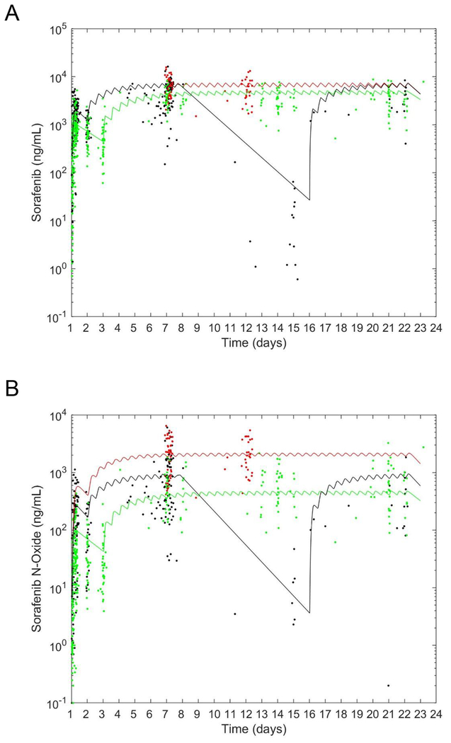
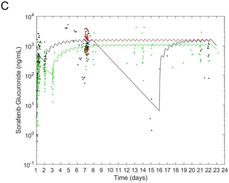
A total of 74 patients enrolled on either RELHEM (35 patients, 11 treated with the concurrent regimen and 24 with the sequential regimen) or ANGIO1 (39 patients) had evaluable pharmacokinetic studies in course 1. The PPK parameters for plasma concentrations of sorafenib (total number of plasma concentrations: n = 721), sorafenib N-oxide (n = 662), and sorafenib glucuronide (n = 474) for the base model (with only BSA-normalized parameters) are listed in Table 2, and their concentration-versus-time plots, along with the population estimated curve for each individual study (the concurrent and sequential arms of RELHEM and ANGIO1) are shown in Fig. 1. In RELHEM, sorafenib day 12 exposures were maintained relative to the day 7 exposure with the concurrent regimen. With the sequential regimen, the sorafenib model-predicted day 12 exposures (after 5 days of stopping sorafenib for Clo/AraC administration), as well as the observed day 15 exposures, were up to three orders of magnitude lower than the corresponding exposures with the concurrent regimen. The sorafenib exposure in ANGIO1 was lower than that in RELHEM.
Table 2.
Population pharmacokinetic parameters obtained from the base (body surface area normalized), and final covariate models. Covariate model: θ*exp(β*covariate) for continuous covariates and θ*β^covariate for categorical covariates. The categorical variables are defined as follows: CON = [0—sequential dosing in RELHEM and ANGIO1 or 1—concurrent dosing in RELHEM] and Study = [0–RELHEM or 1–ANGIO1].
| Parameter | Base estimate | RSE (CV %) | Final model estimate | RSE (CV %) | ||||
|---|---|---|---|---|---|---|---|---|
| Tlag (h) | 0.54 | 11.3 | 0.50 | 12.4 | ||||
| R0 (μg/h/m2) | 44751.7 | 20.8 | 42473.9 | 23.5 | ||||
| V1/f (L/m2) | 89.6 | 6.7 | 87.8 | 19.3 | ||||
| Clsorafenib/f (L/h/m2) | 2.14 | 9.3 | 2.46 | 14.3 | ||||
| β: Age—Clsorafenib | --- | --- | -0.23 | 52.8 | ||||
| β: Study—Clsorafenib | --- | --- | 0.72 | 24.9 | ||||
| K13 (h−1) | 0.00012 | 59.9 | 0.00017 | 70.1 | ||||
| V2/fn (L/m2) | 14.7 | 28.6 | 17.0 | 35.4 | ||||
| ClN-oxide/fn (L/h/m2) | 18.3 | 15.0 | 18.6 | 19.3 | ||||
| β: CON—ClN-oxide | --- | --- | 0.43 | 49.9 | ||||
| β: Bili—ClN-oxide | --- | --- | -0.35 | 73.1 | ||||
| V3/fg (L/m2) | 0.047 | 60.5 | 0.073 | 71.0 | ||||
| Clglucuronide/fg (L/h/m2) | 0.049 | 61.3 | 0.066 | 66.6 | ||||
| β: Bili—Clglucuronide | --- | --- | -0.28 | 88.6 | ||||
| σ prop | 0.098, 0.13, 0.043 | 9.5, 12.3, 19.3 | 0.10, 0.13, 0.044 | 12.6, 17.3, 42.5 | ||||
| −2 Log-likelihood | 21352.7 | 21293.1 | ||||||
| variance | RSE (%) | variance | RSE (%) | |||||
| IIV | IOV | IIV | IOV | IIV | IOV | IIV | IOV | |
| Tlag | 0.36 | --- | 34.3 | --- | 0.34 | --- | 42.7 | --- |
| R0 | 1.2 | --- | 24.1 | --- | 1.1 | --- | 29.1 | --- |
| V1/f | 0.087 | 0.16 | 46.9 | 50.7 | 0.072 | 0.17 | 87.6 | 90.7 |
| Clsorafenib/f | 0.15 | 0.18 | 45.2 | 24.6 | 0.059 | 0.048 | 83.5 | 30.8 |
| K13 | 0.21 | 0.18 | 59.0 | 99.3 | 0.13 | 0.39 | 70.5 | 223.0 |
| V2/fn | 0.75 | --- | 45.9 | --- | 0.94 | --- | 61.5 | --- |
| ClN-oxide/fn | 0.39 | 0.59 | 49.7 | 28.0 | 0.22 | 0.23 | 82.4 | 42.4 |
| V3/fg | --- | --- | --- | --- | --- | --- | --- | --- |
| Clglucuronide/fg | 0.28 | --- | 44.9 | --- | 0.23 | --- | 83.1 | --- |
Abbreviations: Tlag (h), the absorption lag time; R0 (μg/h/m2), the rate of the zero-order absorption; V1/f (L/m2), the apparent volume of sorafenib; Clsorafenib/f (L/h/m2), the sorafenib apparent clearance where f is the unidentifiable bioavailability; K13 (h−1), the sorafenib to sorafenib N-oxide rate constant; V2/fn (L/m2), the apparent volume of sorafenib N-oxide; ClN-oxide/fn (L/h/m2), the sorafenib N-oxide apparent clearance where fn is the unidentifiable formation fraction; V3/fg (L/m2), the apparent volume of sorafenib glucuronide; Clglucuronide/fg (L/h/m2), the sorafenib glucuronide apparent clearance where fg is the unidentifiable formation fraction; σ prop, proportional residual error for sorafenib, sorafenib n-oxide, and sorafenib glucuronide, respectively; IIV, inter-individual variability; IOV, inter-occasion variability; RSE, relative standard error; CV, coefficient of variation.
Figure 1. Population pharmacokinetic model-estimated sorafenib and metabolite concentration-time profiles on different schedules and studies.
Concentration-versus-time plots for (A) sorafenib, (B) sorafenib N-oxide, and (C) sorafenib glucuronide. Red curve and symbols: RELHEM with concurrent administration of sorafenib and clofarabine/cytarabine; black curve and symbols: RELHEM with sequential administration of sorafenib and clofarabine/cytarabine; green curve and symbols: ANGIO1.
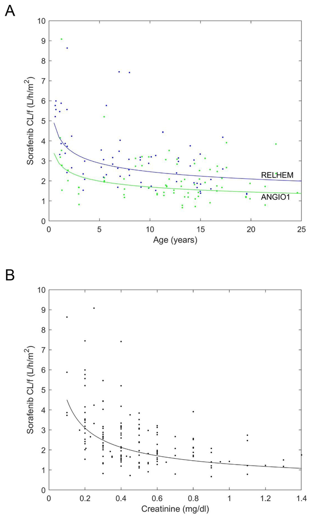
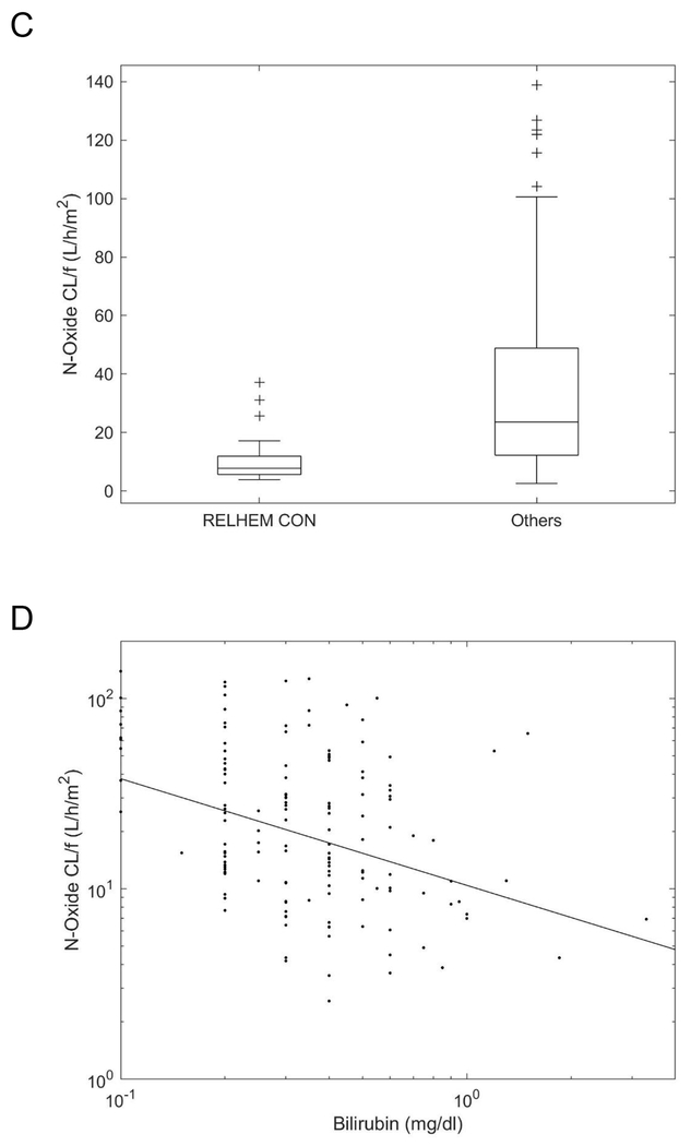
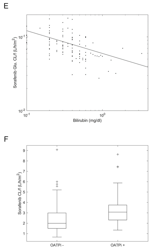
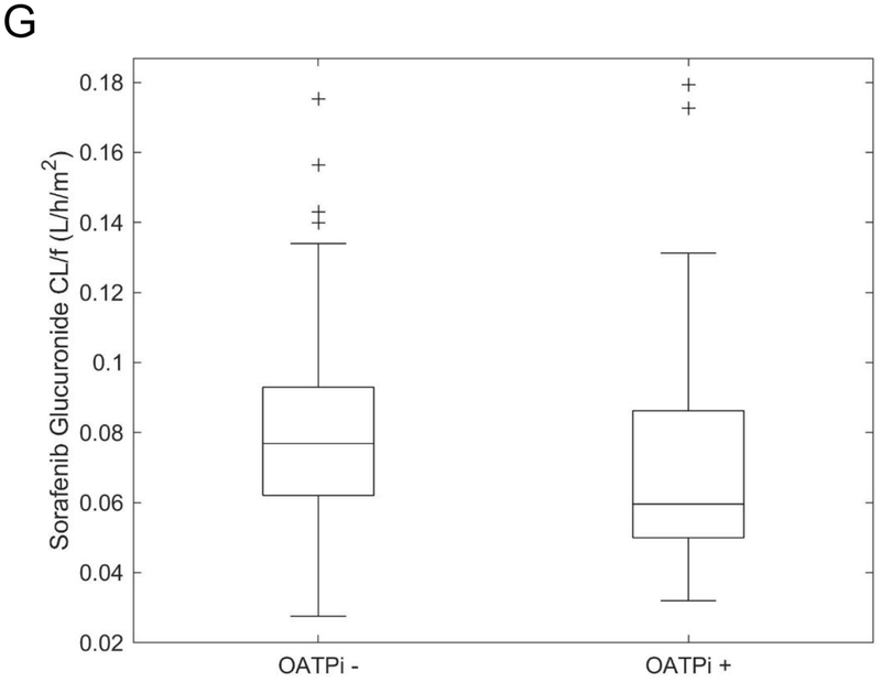
We evaluated each of the covariates and compared them to the base model. Among the covariates, age, study protocol (RELHEM versus ANGIO1), concurrent administration with Clo/AraC (versus others), bilirubin, creatinine, and OATP1B1 inhibitors, each significantly improved the model fit relative to the base model (Supplementary Table S3). OATP1B1 inhibitors were only given to RELHEM patients as infection prophylaxis (Supplementary Table S4). Sorafenib population apparent clearance decreased with increasing age from 3.8 L/h/m2 in a patient aged 1 year to 1.8 L/h/m2 in a patient aged 15 years (P = 1e−5), and the sorafenib population apparent clearance was 37% lower in ANGIO1 than in RELHEM (P = 9e−5) (Fig. 2A). In addition, a sorafenib population apparent clearance of 3.1 L/h/m2 with a creatinine level of 0.2 mg/dL decreased to 1.5 L/h/m2 with a creatinine level of 0.8 mg/dL (P = 7e−6) (Fig. 2B). Sorafenib N-oxide population apparent clearance was 65% lower in patients who received concurrent administration in RELHEM than in other patients (P = 7e−4) (Fig. 2C). Sorafenib N-oxide and glucuronide population apparent clearance were also lower in patients with higher bilirubin, declining from 25.7 and 0.098 L/h/m2 when the bilirubin level was 0.2 mg/dL to 10.4 and 0.064 L/h/m2 when the bilirubin level was 1.0 mg/dL, respectively (P = 1e−4) (Fig. 2D and 2E). Sorafenib population apparent clearance was 50% higher and the sorafenib glucuronide population apparent clearance was 22% lower in individuals who received OATP1B1 inhibitors (P = 3e−3) (Fig. 2F and 2G). The covariates, age, study protocol, creatinine, and OATP1B1 inhibitors accounted for 22%, 43%, 59%, and 13%, respectively, of the IIV in sorafenib apparent clearance; concurrent administration with Clo/AraC and bilirubin accounted for 49% and 12%, respectively, of the IIV in sorafenib N-oxide apparent clearance; and bilirubin and OATP1B1 inhibitors accounted for 24% and 13%, respectively, of the IIV in sorafenib glucuronide apparent clearance.
Figure 2. Association between covariates included in the population pharmacokinetic model and the apparent clearance of sorafenib or its metabolite.
(A) Sorafenib apparent clearance (CL/f) versus age in two studies. Blue curve and symbols: RELHEM (n = 35 patients); green curve and symbols: ANGIO1 (n = 39 patients). (B) Sorafenib apparent clearance versus creatinine levels (n = 74 patients). (C) Effects on sorafenib N-oxide apparent clearance of concurrent administration of sorafenib and clofarabine/cytarabine in RELHEM (RELHEM CON) (n = 11 patients) versus other regimens (sequential administration of sorafenib and clofarabine/cytarabine in RELHEM and ANGIO1) (n = 63 patients). (D) Sorafenib N-oxide apparent clearance versus bilirubin levels (n = 74 patients). (E) Sorafenib glucuronide apparent clearance versus bilirubin levels (n = 74 patients). (F) Sorafenib apparent clearance versus OATP1B1 inhibitors (OATPi) (OATPi+, n = 19 patients and OATPi-, n = 55 patients). (G) Sorafenib glucuronide apparent clearance versus OATP1B1 inhibitors (OATPi+, n = 19 patients and OATPi-, n = 55 patients).
We next considered combinations of covariates. Relative to the base model, the model that included age, study protocol (RELHEM vs. ANGIO1), and concurrent administration with Clo/AraC (vs. others) accounted for 67% of the IIV in sorafenib apparent clearance and 49% of the IIV in sorafenib N-oxide apparent clearance (Table 2 and Supplementary Table S3). The only laboratory chemistry result that remained significant relative to the above multivariate model was the bilirubin level. This accounted for an additional 8% and 26% of the IIV in sorafenib N-oxide and sorafenib glucuronide apparent clearance, respectively, relative to the previous model.
A summary of the base and final covariate model results and goodness-of-fit plots for sorafenib, sorafenib N-oxide, and sorafenib glucuronide are shown in Table 2 and Supplementary Figure S2, respectively. The apparent clearance of sorafenib N-oxide decreased over the period from day 1 to day 7 with daily administration (from a median 27.3 L/h/m2 to a median 12.3 L/h/m2, P = 2e−5) (Supplementary Figure S3A). The apparent clearance of sorafenib N-oxide correlated significantly with that of sorafenib (r = 0.7, P = 7e−13), whereas the apparent clearance of sorafenib glucuronide did not correlate with that of the parent compound (r = 0.06; P = 0.6) (Supplementary Figure S3B and S3C).
A summary of the post-hoc individual estimated exposure parameters for sorafenib, sorafenib N-oxide, and sorafenib glucuronide on days 1 and 7 is presented in Supplementary Table S5.
Exposure-toxicity association
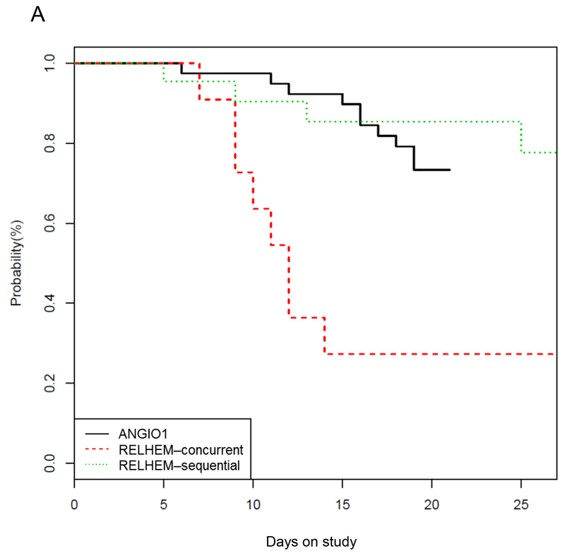
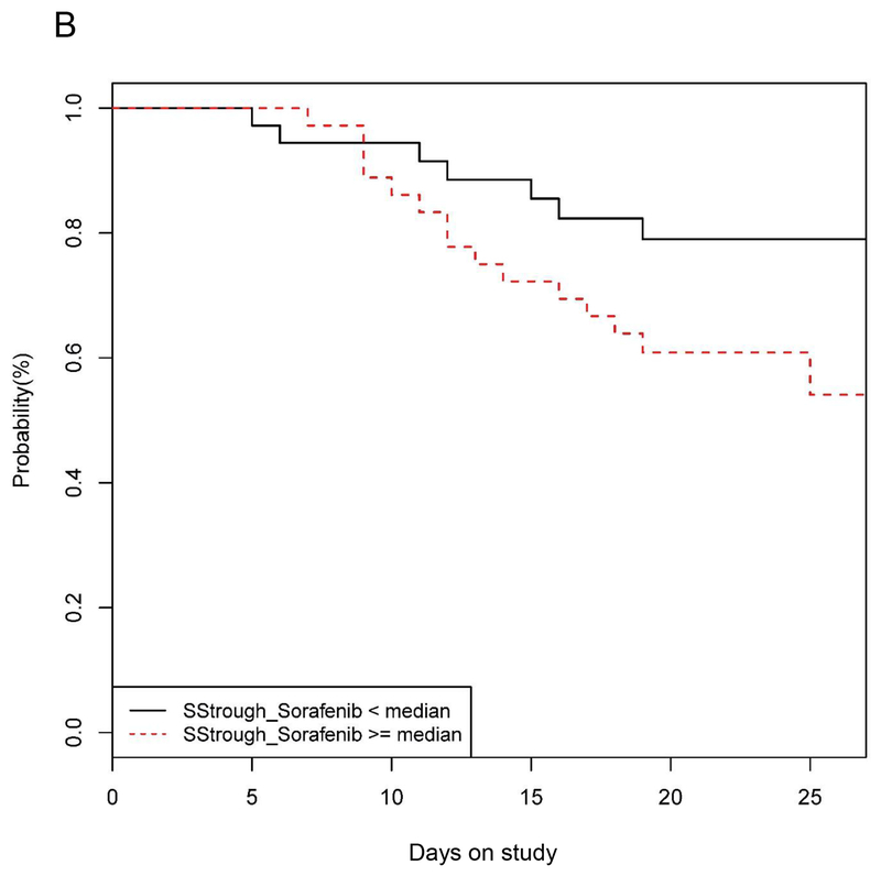
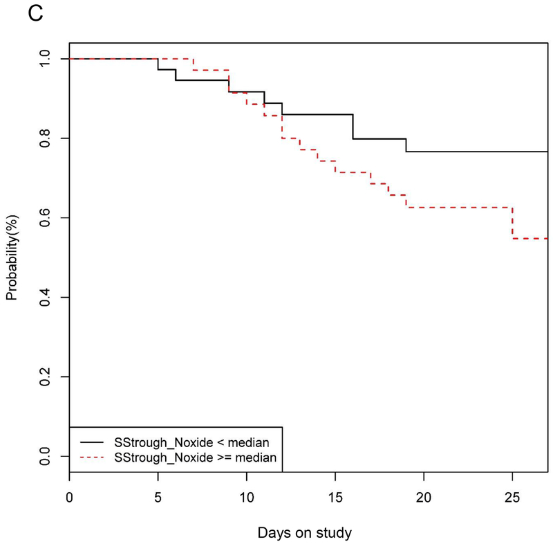
The determinants of sorafenib-induced HFSR were evaluated using Cox proportional hazards regression analysis. The rate of the first grade 2–3 HFSR was 4.72 times higher (95% CI = 1.81, 12.33; P = 0.0015) with concurrent administration of Clo/AraC compared with ANGIO1. However sequential administration of Clo/AraC versus ANGIO1 was not significantly associated with HFSR (hazard ratio = 0.72; 95% CI = 0.22, 2.35; P = 0.591) (Fig. 3A). Next, exposure to sorafenib or its metabolites were considered. Descriptively, patients experiencing grade 2–3 HFSR had higher sorafenib SS trough concentrations when compared to those experiencing grade 0–1 HFSR (median: 4.1 versus 3.3 mg/L respectively). A 1000-ng/mL increase in the sorafenib SS trough concentration was associated with a 1.45-fold increase in the HFSR rate (95% CI = 1.18, 1.78; P = 0.0004) (Fig. 3B), so the upper quartile concentration associates with an HFSR rate that is 2.16 times that of the lower quartile (95% CI = 1.41, 3.32). Patients experiencing grade 2–3 HFSR also had higher sorafenib N-oxide SS trough concentrations when compared to those experiencing grade 0–1 HFSR (median: 0.74 versus 0.47 mg/L respectively). In this case, a 100-ng/mL increase in the sorafenib N-oxide SS trough concentration was associated with a 1.04-fold increase in the HFSR rate (95% CI = 1.00, 1.09; P = 0.0275) (Fig. 3C), so that the upper quartile concentration associates with an HFSR rate that is 1.39 times that of the lower quartile (95% CI = 1.03, 1.86). In addition, each square-meter increase in BSA was associated with an increase in the rate of HFSR by a factor of 2.63 (95% CI = 1.18, 5.88; P = 0.0186), and after experiencing rash, patients experienced a 2.97-fold increase in the rate of HFSR (95% CI = 1.13, 7.79; P = 0.0268). Of the nine patients who had both rash and HFSR, rash was seen before HFSR in eight patients by a median of 1 day (range, 0–13 days).
Figure 3. Grade 2-3 hand-foot skin reaction (HFSR).
(A) Probability of absence of grade 2-3 HFSR according to the study protocol and the sequence of sorafenib and clofarabine/cytarabine administration. (B, C) The association between exposure to sorafenib (B) and sorafenib N-oxide (C) and probability of absence of grade 2–3 HFSR. Abbreviations: SStrough_Sorafenib and SStrough_Noxide, steady-state trough concentration of sorafenib and sorafenib N-oxide, respectively.
We next considered models with two predictors. By BIC, the four models with the greatest probability of being the best among those considered are the model with treatment regimen (concurrent or sequential administration with Clo/AraC versus ANGIO1) and BSA (47.9% probability); the model with BSA and sorafenib SS trough levels (10.8% probability); the model with treatment regimen and sorafenib SS trough levels (9.1% probability); and the model with prior occurrence of rash and sorafenib SS trough levels as predictors (8.8% probability) as predictors of HFSR (Supplementary Table S6). This analysis indicates that HFSR is associated with the concurrent administration with Clo/AraC, higher sorafenib SS trough levels, larger BSA, and previous occurrence of rash.
The above analysis indicated that rash may be an indicator of an increased risk for subsequent development of HFSR. Therefore, we performed Fine-Gray modeling analyses to evaluate factors that were associated with the development of grade 2–3 rash while treating development of HFSR before rash as a competing event. In this analysis, treatment regimen was significantly associated with the development of rash. Patients treated with concurrent administration of Clo/AraC developed rash at 21.4 times the rate of ANGIO1 patients (95% CI = 4.71, 97.24; P = 0.0001). Patients treated with sequential administration with Clo/AraC regimen developed rash at 4.48 times the rate of ANGIO1 patients (95% CI = 0.88, 22.91; P = 0.071). A 1000-ng/mL increase in the sorafenib SS trough concentration was associated with a 1.21-fold increase in the rash rate (95% CI = 0.98, 1.48; P = 0.074), and a 100-ng/mL increase in the sorafenib N-oxide SS trough concentration was associated with a 1.05-fold increase in the rash rate (95% CI = 1.01, 1.10; P = 0.018). In a BIC model selection analysis, treatment regimen appeared in all of the top four models (Supplementary Table S6).
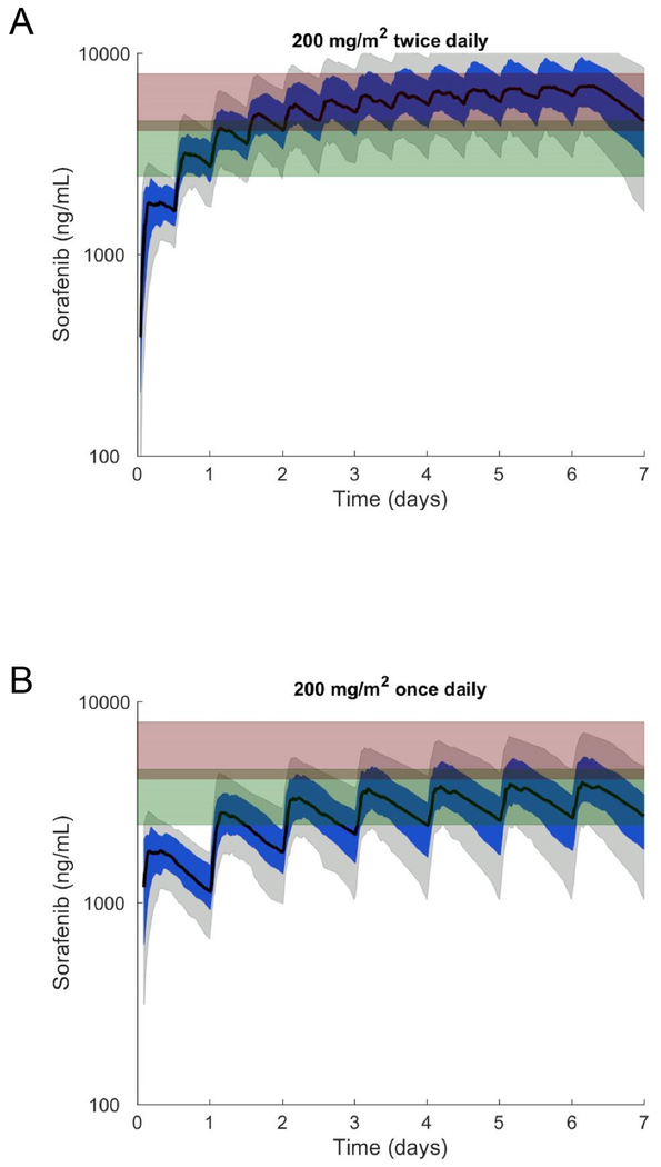
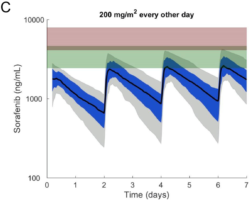
We performed pharmacokinetic simulations to evaluate alternative sorafenib administration schedules (once a day and every other day) that would minimize the number of patients whose sorafenib SS trough concentrations reached levels associated with HFSR (Fig. 4). Our simulations indicated that both the once-daily and every-other-day schedules of sorafenib 200 mg/m2 would minimize exposure to sorafenib SS trough concentrations associated with HFSR in most patients. Specifically, the median sorafenib SS trough concentration decreased from 5743 to 2564 or 934 ng/mL when the administration schedule changed from twice-daily to once-daily or every-other-day. This decrease in SS trough concentration corresponds to a 3.35 fold reduction in the HFSR rate when changing from twice-daily to once-daily administration (95% CI: 1.71, 6.55) and a 6.71 fold reduction when changing from twice-daily to every-other-day (95% CI: 2.26, 16.88). Of relevance to patients with AML and FLT3-ITD mutations, which are a target of sorafenib (5), we found that with once-daily or once-every-other-day administration, the estimated SS trough concentrations at the 10th percentile were well above the sorafenib concentration of 143 ng/mL (308 nM) that inhibited phospho-FLT3 of leukemia samples in a plasma inhibitory assay (30). We used this strategy to treat a patient with FLT3-ITD–positive AML with single-agent sorafenib, and remission was maintained for approximately 1 year (Supplementary Table S7).
Figure 4. Population pharmacokinetic model simulations of sorafenib plasma exposure on different administration schedules and day 7 trough concentrations associated with HFSR toxicity.
Simulations of sorafenib plasma concentrations after the administration of sorafenib at 200 mg/m2 (A) twice daily, (B) once daily, or (C) every other day. The black curve corresponds to the median concentration. The blue-shaded area corresponds to the 25th to 75th percentiles; the grey-shaded region corresponds to the 10th to 90th percentiles. The green-shaded rectangle corresponds to the 25th to 75th percentiles of the day 7 sorafenib trough concentrations for individuals with grade 0–1 HFSR; the red-shaded rectangle corresponds to the 25th to 75th percentiles of the day 7 sorafenib trough concentrations for individuals with grade 2–3 HFSR.
DISCUSSION
We have reported the PPK of sorafenib and its metabolites sorafenib N-oxide and sorafenib glucuronide, as well as the skin toxicity, in pediatric patients who were treated with a combination of sorafenib and Clo/AraC (for leukemia) or a combination of sorafenib and Bev/Cyclo (for solid tumor malignancies). Concurrent administration of sorafenib with Clo/AraC was associated with skin DLTs in three of six patients when sorafenib was administered at 200 mg/m2/day. Sequential administration of Clo/AraC administration and administration of lower sorafenib dose with Bev/Cyclo decreased the onset of HFSR. In the PPK analysis, older age, the study protocol (ANGIO1 versus RELHEM), and a higher creatinine level were associated with decreased sorafenib apparent clearance and administration of OATP1B1 inhibitors was associated with higher sorafenib apparent clearance. A higher bilirubin level, concurrent administration with Clo/AraC, and repeated drug administration for 7 days were associated with decreased sorafenib N-oxide apparent clearance. In addition, OATP1B1 inhibitors and increased bilirubin were associated with lower sorafenib glucuronide apparent clearance. In the exposure-toxicity analysis, grade 2–3 HFSR was significantly associated with concurrent administration of Clo/AraC, higher sorafenib SS trough levels, greater BSA, and previous occurrence of rash and grade 2–3 skin rash was associated with concurrent administration of Clo/AraC.
This paper reports the first PPK model for sorafenib in children. Notably, age was a significant covariate of sorafenib apparent clearance, with younger children exhibiting higher drug clearance than older children. Sorafenib is partly metabolized by CYP3A4 to the active metabolite sorafenib N-oxide and by UGT1A9 to sorafenib glucuronide (6). In young children, CYP450-catalyzed metabolism is increased (31), which could explain the increased sorafenib apparent clearance in younger patients in our PPK model. Greater BSA was associated with higher incidences of HFSR, which may reflect the association of reduced sorafenib clearance (higher exposure) with older age and increasing BSA. The differences in sorafenib apparent clearance between studies could be due to the different cancer types (solid tumor malignancies, which often involve organs, versus leukemia), the concomitant administration of OATP1B1 inhibitors in RELHEM patients, or differences in concomitant chemotherapy. Due to the small number of each solid tumor subtypes, we were unable to analyze based on the disease subtypes. However, this is the largest available population of pediatric patients with collected PK data. Although urinary excretion plays a minor role in sorafenib and metabolite elimination (accounting for <20% of the total), increased creatinine was associated with decreased apparent clearance of sorafenib. Recently, it has been shown that the clearance of drugs that are substrates for OATP uptake transporters, such as OATP1B1 and OATP1B3, which are located on the sinusoidal membrane of hepatocytes, decreases as kidney function declines (32). We previously demonstrated that sorafenib is a substrate for OATP1B1 and OATP1B3, which may also explain why creatinine is a covariate in the PPK model (33).
We have shown that sorafenib N-oxide also plays an important role in sorafenib-associated skin toxicities, as well as in anti-leukemia activities as an active metabolite (9,34). The apparent clearance of sorafenib N-oxide was decreased with higher bilirubin levels and repeated drug administration for 7 days, which could be associated with sorafenib glucuronide, with its high SS concentrations in the liver competing with sorafenib N-oxide for biliary excretion via the efflux transporters ABCB1 and/or ABCC2 (35). It is unclear why there was a difference in sorafenib N-oxide apparent clearance in patients receiving concurrent administration with Clo/AraC when compared to those in the other group.
In PPK analysis, several variables were associated with sorafenib glucuronide apparent clearance. Bilirubin accounted for a portion (24%) of the variability in the apparent clearance of sorafenib glucuronide. We also showed that concurrent administration of OATP1B1 inhibitors in RELHEM patients was associated with reduced sorafenib glucuronide apparent clearance (higher exposure) and a concomitant increase in sorafenib apparent clearance (lower exposure). This can be explained by inhibition of enterohepatic recirculation of sorafenib glucuronide (by inhibiting liver uptake), which accounts for approximately 50% of the circulating sorafenib parent compound (36). This is the first study to demonstrate a drug-drug interaction between OATP1B1 inhibitors and sorafenib.
The population estimates of sorafenib apparent clearance reported in this study are similar to those reported in several other population pharmacokinetic studies in adults. Specifically, our population estimated apparent clearance of sorafenib in a 10 year old (the median age of our population) was 2.46 and 1.77 L/h/m2 in RELHEM and ANGIO1 respectively. The population estimated apparent clearances of sorafenib reported by Widemann et al. (16) (median patient age, 14 years) and Hornecker et al. (37) (normalized to a BSA of 2.1) were 3.36 and 1.0 L/h/m2, respectively. In addition, the population estimated apparent clearance of sorafenib reported by Jain et al. (36) (normalized to a BSA of 2.1 and accounting for the fact that their estimate addressed enterohepatic recirculation) was 1.94 L/h/m2. Note that none of these studies measured metabolites.
HFSR is a common adverse event with sorafenib, occurring in approximately 60% of adults treated with this drug, and studies have aimed to identify risk factors for this toxicity, especially for the more severe grades that can decrease the quality of life of patients. Grade 2 or higher HFSR is reported to occur in 36% of patients and typically manifests within the first 2 weeks of treatment (38). The corresponding information on HFSR in pediatric populations is limited, and we observed an increased incidence of grade 2 or higher HFSR (67%) when sorafenib was administered concurrently with Clo/AraC, with onset occurring during the second week of treatment. The Clo/AraC regimen is frequently associated with skin toxicity, including palmar-plantar erythrodysesthesia (20,21), and probably contributed to the increased incidence of HFSR observed during concurrent treatment. In the sequential regimen, the drug holiday between days 8 and 14 led to exposures that were up to three orders of magnitude lower than the corresponding exposures with the concurrent regimen, which can be associated with lower incidences of HFSR. In the ANGIO1 trial, administration of lower doses of sorafenib and Bev/Cyclo, a chemotherapy regimen which has not been associated with skin toxicity, probably contributed to the decreased incidences of HFSR. Although it can be difficult to tease out the role of sorafenib exposure in skin toxicities in the presence of other chemotherapy, sorafenib trough level appeared in three of the top four models by the BIC model selection criteria. One of these top models used treatment regimen and sorafenib trough levels as predictors of HFSR. Therefore, sorafenib exposure, specifically day 7 SS trough concentration, is very likely to be a significant factor associated with HFSR.
In our study, patients with higher SS trough concentrations of sorafenib and sorafenib N-oxide experienced more grade 2 or higher HFSR. Even with the sequential administration of sorafenib in RELHEM or the lower doses of sorafenib in ANGIO1, approximately 20% of patients still experienced HFSR, supporting the importance of trough concentrations. These results are in line with those reported for two previous studies in adults with solid tumors who received single-agent sorafenib therapy, which found an association between a minimal-threshold sorafenib concentration of 5 mg/L or 5.8 mg/L and the incidence of grade 2 or higher HFSR (38,39). In another study, a nonlinear mixed-effect model was constructed to link sorafenib administration to the risk of HFSR in adults with solid tumors (40). The risk of HFSR increased with the number of doses administered per day (e.g., thrice daily > twice daily > once daily), and this factor was more influential than the total daily dose (range, 200 to 1600 mg/day). This finding suggests that administering higher daily doses of sorafenib on fewer occasions could minimize the risk of severe HFSR. However, this strategy would need to take into consideration the saturable absorption of sorafenib above single doses of 400 mg (37). Sorafenib is a potent inhibitor of FLT3-ITD–positive AML, and a sorafenib plasma concentration of 143 ng/mL was required to inhibit phospho-FLT3 in a plasma inhibitory assay (30). In the present study, the described population model was used to simulate sorafenib exposure with different administration schedules. These simulations suggest that sorafenib can be administered at a dose of 200 mg/m2 with a reduced frequency of once a day or every other day while maintaining trough concentrations above the threshold needed for the inhibition of FLT3-ITD and avoiding higher concentrations that are associated with HFSR. A similar modeling approach could be used to define alternative sorafenib dosing regimens for children with solid tumors. Further model refinements and the incorporation of physiologically based mechanisms can be used to define exposure-toxicity or exposure-response relationships and to develop alternative dosing strategies for sorafenib (41).
In conclusion, we evaluated the pharmacokinetics and skin toxicity profile of sorafenib administered in combination with Clo/AraC (either concurrently or sequentially) or Bev/Cyclo. We developed a PPK model for sorafenib and its metabolites in children that identified clinical covariates affecting their apparent clearances. We used the pharmacokinetic model to characterize associations between drug exposure and the dose-limiting toxicity HFSR. In addition, our pharmacokinetic model simulations suggested that an alternative sorafenib treatment regimen has the potential to decrease HFSR while maintaining efficacy, especially in patients with FLT3-ITD–positive AML. This regimen warrants further investigation.
Supplementary Material
Translational relevance.
As approximately 30% of pediatric cancer recurs, new therapeutic agents with different mechanisms of action are needed. This study examined the pharmacokinetics and skin toxicity profile in pediatric patients of the multikinase inhibitor sorafenib in combination with clofarabine/cytarabine (for relapsed/refractory leukemia) or bevacizumab/cyclophosphamide (for solid tumor malignancies). Concurrent administration of sorafenib with clofarabine/cytarabine was associated with severe skin toxicities, whereas sequential administration of sorafenib with clofarabine/cytarabine or a lower dose of sorafenib in combination with bevacizumab/cyclophosphamide was well tolerated. A population pharmacokinetic model identified covariates affecting the apparent clearance and steady-state exposure of sorafenib and its metabolites. Patients experiencing grade ≥2 hand-foot skin reaction, as compared to those with grade 0–1, had higher steady-state trough concentrations of sorafenib and sorafenib N-oxide. The described population pharmacokinetic model can be used to refine exposure-response relations and develop alternative dosing strategies for sorafenib to minimize skin toxicity.
ACKNOWLEDGEMENTS
We thank the clinical, research and pharmacokinetic nurses at St. Jude Children’s Research Hospital who participated in this study. The research reported in this paper was supported by the National Cancer Institute of the National Institutes of Health (R01CA138744 [to SDB] and P01CA21765), by ALSAC, and by Bayer/Onyx. We thank Keith A. Laycock, PhD, ELS, (St. Jude Children’s Research Hospital) for editorial assistance.
Acknowledgment of research support: This study was supported by National Institutes of Health (NIH) Cancer Center Support Grant P30 CA021765, by NIH R01 CA138744 (to SDB), by ALSAC, and by Bayer/Onyx.
Footnotes
Conflicts of interest: Dr. Inaba received research support from Bayer/Onyx.
REFERENCES
- 1.Zwaan CM, Kolb EA, Reinhardt D, Abrahamsson J, Adachi S, Aplenc R, et al. Collaborative efforts driving progress in pediatric acute myeloid leukemia. J Clin Oncol 2015;33(27):2949–62. doi 10.1200/JCO.2015.62.8289. [DOI] [PMC free article] [PubMed] [Google Scholar]
- 2.Rubnitz JE. Current management of childhood acute myeloid leukemia. Paediatr Drugs 2017;19(1):1–10. doi 10.1007/s40272-016-0200-6. [DOI] [PubMed] [Google Scholar]
- 3.Smith MA, Altekruse SF, Adamson PC, Reaman GH, Seibel NL. Declining childhood and adolescent cancer mortality. Cancer 2014;120(16):2497–506. doi 10.1002/cncr.28748. [DOI] [PMC free article] [PubMed] [Google Scholar]
- 4.Wilhelm S, Carter C, Lynch M, Lowinger T, Dumas J, Smith RA, et al. Discovery and development of sorafenib: a multikinase inhibitor for treating cancer. Nat Rev Drug Discov 2006;5(10):835–44. doi 10.1038/nrd2130. [DOI] [PubMed] [Google Scholar]
- 5.Zhang W, Konopleva M, Shi YX, McQueen T, Harris D, Ling X, et al. Mutant FLT3: a direct target of sorafenib in acute myelogenous leukemia. J Natl Cancer Inst 2008;100(3):184–98. doi 10.1093/jnci/djm328. [DOI] [PubMed] [Google Scholar]
- 6.Zimmerman EI, Roberts JL, Li L, Finkelstein D, Gibson A, Chaudhry AS, et al. Ontogeny and sorafenib metabolism. Clin Cancer Res 2012;18(20):5788–95. doi 10.1158/1078-0432.CCR-12-1967. [DOI] [PMC free article] [PubMed] [Google Scholar]
- 7.Borthakur G, Kantarjian H, Ravandi F, Zhang W, Konopleva M, Wright JJ, et al. Phase I study of sorafenib in patients with refractory or relapsed acute leukemias. Haematologica 2011;96(1):62–8. doi 10.3324/haematol.2010.030452. [DOI] [PMC free article] [PubMed] [Google Scholar]
- 8.Ravandi F, Cortes JE, Jones D, Faderl S, Garcia-Manero G, Konopleva MY, et al. Phase I/II study of combination therapy with sorafenib, idarubicin, and cytarabine in younger patients with acute myeloid leukemia. J Clin Oncol 2010;28(11):1856–62. doi 10.1200/JCO.2009.25.4888. [DOI] [PMC free article] [PubMed] [Google Scholar]
- 9.Inaba H, Rubnitz JE, Coustan-Smith E, Li L, Furmanski BD, Mascara GP, et al. Phase I pharmacokinetic and pharmacodynamic study of the multikinase inhibitor sorafenib in combination with clofarabine and cytarabine in pediatric relapsed/refractory leukemia. J Clin Oncol 2011;29(24):3293–300. doi 10.1200/JCO.2011.34.7427. [DOI] [PMC free article] [PubMed] [Google Scholar]
- 10.Röllig C, Serve H, Hüttmann A, Noppeney R, Müller-Tidow C, Krug U, et al. Addition of sorafenib versus placebo to standard therapy in patients aged 60 years or younger with newly diagnosed acute myeloid leukaemia (SORAML): a multicentre, phase 2, randomised controlled trial. Lancet Oncol 2015;16(16):1691–9. doi 10.1016/S1470-2045(15)00362-9. [DOI] [PubMed] [Google Scholar]
- 11.Antar A, Otrock ZK, El-Cheikh J, Kharfan-Dabaja MA, Battipaglia G, Mahfouz R, et al. Inhibition of FLT3 in AML: a focus on sorafenib. Bone Marrow Transplant 2017;52(3):344–51. doi 10.1038/bmt.2016.251. [DOI] [PubMed] [Google Scholar]
- 12.Llovet JM, Ricci S, Mazzaferro V, Hilgard P, Gane E, Blanc JF, et al. Sorafenib in advanced hepatocellular carcinoma. N Engl J Med 2008;359(4):378–90. doi 10.1056/NEJMoa0708857. [DOI] [PubMed] [Google Scholar]
- 13.Cheng AL, Kang YK, Chen Z, Tsao CJ, Qin S, Kim JS, et al. Efficacy and safety of sorafenib in patients in the Asia-Pacific region with advanced hepatocellular carcinoma: a phase III randomised, double-blind, placebo-controlled trial. Lancet Oncol 2009;10(1):25–34. doi 10.1016/S1470-2045(08)70285-7. [DOI] [PubMed] [Google Scholar]
- 14.Escudier B, Eisen T, Stadler WM, Szczylik C, Oudard S, Siebels M, et al. Sorafenib in advanced clear-cell renal-cell carcinoma. N Engl J Med 2007;356(2):125–34. doi 10.1056/NEJMoa060655. [DOI] [PubMed] [Google Scholar]
- 15.Escudier B, Worden F, Kudo M. Sorafenib: key lessons from over 10 years of experience. Expert Rev Anticancer Ther 2018:1–13. doi 10.1080/14737140.2019.1559058. [DOI] [PubMed] [Google Scholar]
- 16.Widemann BC, Kim A, Fox E, Baruchel S, Adamson PC, Ingle AM, et al. A phase I trial and pharmacokinetic study of sorafenib in children with refractory solid tumors or leukemias: a Children’s Oncology Group Phase I Consortium report. Clin Cancer Res 2012;18(21):6011–22. doi 10.1158/1078-0432.CCR-11-3284. [DOI] [PMC free article] [PubMed] [Google Scholar]
- 17.Navid F, Baker SD, McCarville MB, Stewart CF, Billups CA, Wu J, et al. Phase I and clinical pharmacology study of bevacizumab, sorafenib, and low-dose cyclophosphamide in children and young adults with refractory/recurrent solid tumors. Clin Cancer Res 2013;19(1):236–46. doi 10.1158/1078-0432.CCR-12-1897. [DOI] [PMC free article] [PubMed] [Google Scholar]
- 18.Castleberry RP, Crist WM, Holbrook T, Malluh A, Gaddy D. The cytosine arabinoside (Ara-C) syndrome. Med Pediatr Oncol 1981;9(3):257–64. [DOI] [PubMed] [Google Scholar]
- 19.Cetkovská P, Pizinger K, Cetkovský P. High-dose cytosine arabinoside–induced cutaneous reactions. J Eur Acad Dermatol Venereol 2002;16(5):481–5. [DOI] [PubMed] [Google Scholar]
- 20.Faderl S, Verstovsek S, Cortes J, Ravandi F, Beran M, Garcia-Manero G, et al. Clofarabine and cytarabine combination as induction therapy for acute myeloid leukemia (AML) in patients 50 years of age or older. Blood 2006;108(1):45–51. doi 10.1182/blood-2005-08-3294. [DOI] [PubMed] [Google Scholar]
- 21.Zhang B, Bolognia J, Marks P, Podoltsev N. Enhanced skin toxicity associated with the combination of clofarabine plus cytarabine for the treatment of acute leukemia. Cancer Chemother Pharmacol 2014;74(2):303–7. doi 10.1007/s00280-014-2504-y. [DOI] [PubMed] [Google Scholar]
- 22.Gomez P, Lacouture ME. Clinical presentation and management of hand-foot skin reaction associated with sorafenib in combination with cytotoxic chemotherapy: experience in breast cancer. Oncologist 2011;16(11):1508–19. doi 10.1634/theoncologist.2011-0115. [DOI] [PMC free article] [PubMed] [Google Scholar]
- 23.Anderson RT, Keating KN, Doll HA, Camacho F. The Hand-Foot Skin Reaction and Quality of Life Questionnaire: an assessment tool for oncology. Oncologist 2015;20(7):831–8 doi 10.1634/theoncologist.2014-0219. [DOI] [PMC free article] [PubMed] [Google Scholar]
- 24.Lacouture ME, Wu S, Robert C, Atkins MB, Kong HH, Guitart J, et al. Evolving strategies for the management of hand-foot skin reaction associated with the multitargeted kinase inhibitors sorafenib and sunitinib. Oncologist 2008;13(9):1001–11. doi 10.1634/theoncologist.2008-0131. [DOI] [PubMed] [Google Scholar]
- 25.Millisor VE, Roberts JK, Sun Y, Tang L, Daryani VM, Gregornik D, et al. Derivation of new equations to estimate glomerular filtration rate in pediatric oncology patients. Pediatr Nephrol 2017;32(9):1575–84. doi 10.1007/s00467-017-3693-5. [DOI] [PMC free article] [PubMed] [Google Scholar]
- 26.Kotsampasakou E, Brenner S, Jäger W, Ecker GF. Identification of novel inhibitors of organic anion transporting polypeptides 1B1 and 1B3 (OATP1B1 and OATP1B3) using a consensus vote of six classification models. Mol Pharm 2015;12(12):4395–404 doi 10.1021/acs.molpharmaceut.5b00583. [DOI] [PMC free article] [PubMed] [Google Scholar]
- 27.De Bruyn T, van Westen GJ, Ijzerman AP, Stieger B, de Witte P, Augustijns PF, et al. Structure-based identification of OATP1B1/3 inhibitors. Mol Pharm 2013;83(6):1257–67 doi 10.1124/mol.112.084152. [DOI] [PubMed] [Google Scholar]
- 28.Karlgren M, Vildhede A, Norinder U, Wisniewski JR, Kimoto E, Lai Y, et al. Classification of inhibitors of hepatic organic anion transporting polypeptides (OATPs): influence of protein expression on drug-drug interactions. J Med Chem 2012;55(10):4740–63 doi 10.1021/jm300212s. [DOI] [PMC free article] [PubMed] [Google Scholar]
- 29.Burnham KP, Anderson DR. Model selection and multimodel inference: a practical information-theoretic approach. 2nd edition New York: Springer; 2002. [Google Scholar]
- 30.Pratz KW, Cho E, Levis MJ, Karp JE, Gore SD, McDevitt M, et al. A pharmacodynamic study of sorafenib in patients with relapsed and refractory acute leukemias. Leukemia 2010;24(8):1437–44. doi 10.1038/leu.2010.132. [DOI] [PMC free article] [PubMed] [Google Scholar]
- 31.Stewart CF, Hampton EM. Effect of maturation on drug disposition in pediatric patients. Clin Pharm 1987;6(7):548–64. [PubMed] [Google Scholar]
- 32.Tan ML, Yoshida K, Zhao P, Zhang L, Nolin TD, Piquette-Miller M, et al. Effect of chronic kidney disease on nonrenal elimination pathways: a systematic assessment of CYP1A2, CYP2C8, CYP2C9, CYP2C19, and OATP. Clin Pharmacol Ther 2018;103(5):854–67. doi 10.1002/cpt.807. [DOI] [PMC free article] [PubMed] [Google Scholar]
- 33.Zimmerman EI, Hu S, Roberts JL, Gibson AA, Orwick SJ, Li L, et al. Contribution of OATP1B1 and OATP1B3 to the disposition of sorafenib and sorafenib-glucuronide. Clin Cancer Res 2013;19(6):1458–66. doi 10.1158/1078-0432.CCR-12-3306. [DOI] [PMC free article] [PubMed] [Google Scholar]
- 34.Zimmerman EI, Gibson AA, Hu S, Vasilyeva A, Orwick SJ, Du G, et al. Multikinase inhibitors induce cutaneous toxicity through OAT6-mediated uptake and MAP3K7-driven cell death. Cancer Res 2016;76(1):117–26. doi 10.1158/0008-5472.CAN-15-0694. [DOI] [PMC free article] [PubMed] [Google Scholar]
- 35.Vasilyeva A, Durmus S, Li L, Wagenaar E, Hu S, Gibson AA, et al. Hepatocellular shuttling and recirculation of sorafenib-glucuronide is dependent on Abcc2, Abcc3, and Oatp1a/1b. Cancer Res 2015;75(13):2729–36 doi 10.1158/0008-5472.CAN-15-0280. [DOI] [PMC free article] [PubMed] [Google Scholar]
- 36.Jain L, Woo S, Gardner ER, Dahut WL, Kohn EC, Kummar S, et al. Population pharmacokinetic analysis of sorafenib in patients with solid tumours. Br J Clin Pharmacol 2011;72(2):294–305. doi 10.1111/j.1365-2125.2011.03963.x. [DOI] [PMC free article] [PubMed] [Google Scholar]
- 37.Hornecker M, Blanchet B, Billemont B, Sassi H, Ropert S, Taieb F, et al. Saturable absorption of sorafenib in patients with solid tumors: a population model. Invest New Drugs 2012;30(5):1991–2000. doi 10.1007/s10637-011-9760-z. [DOI] [PubMed] [Google Scholar]
- 38.Karovic S, Shiuan EF, Zhang SQ, Cao H, Maitland ML. Patient-level adverse event patterns in a single-institution study of the multi-kinase inhibitor sorafenib. Clin Transl Sci 2016;9(5):260–66. doi 10.1111/cts.12408. [DOI] [PMC free article] [PubMed] [Google Scholar]
- 39.Fukudo M, Ito T, Mizuno T, Shinsako K, Hatano E, Uemoto S, et al. Exposure-toxicity relationship of sorafenib in Japanese patients with renal cell carcinoma and hepatocellular carcinoma. Clin Pharmacokinet 2014;53(2):185–96. doi 10.1007/s40262-013-0108-z. [DOI] [PubMed] [Google Scholar]
- 40.Hénin E, Blanchet B, Boudou-Rouquette P, Thomas-Schoemann A, Freyer G, Vidal M, et al. Fractionation of daily dose increases the predicted risk of severe sorafenib-induced hand-foot syndrome (HFS). Cancer Chemother Pharmacol 2014;73(2):287–97. doi 10.1007/s00280-013-2352-1. [DOI] [PubMed] [Google Scholar]
- 41.Edginton AN, Zimmerman EI, Vasilyeva A, Baker SD, Panetta JC. Sorafenib metabolism, transport, and enterohepatic recycling: physiologically based modeling and simulation in mice. Cancer Chemother Pharmacol 2016;77(5):1039–52. doi 10.1007/s00280-016-3018-6. [DOI] [PMC free article] [PubMed] [Google Scholar]
Associated Data
This section collects any data citations, data availability statements, or supplementary materials included in this article.


