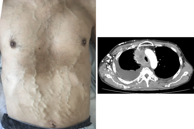
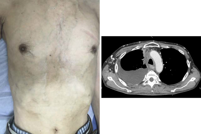
A man in his early 60s with mediastinal tumor was diagnosed with lung adenocarcinoma (cT4N2M1c, stage IVB). Despite various therapies, the mediastinal lesion continued to enlarge, and remarkable venous dilatations on the anterior thorax and abdomen, resembling caput medusae, were observed (Picture 1). Since a BRAFV600E mutation was detected, he was treated with dabrafenib and trametinib. The mediastinal tumor remarkably decreased, and the caput medusae-like venous dilatations were discernibly improved (Picture 2). Caput medusae are venous dilatations which develop as collateral circulation caused by an obstruction of either the portal venous system or vena cava. In a case of portal hypertension, the blood flow in the dilated vessels travels from the umbilical to the cranial direction. In this patient, the blood flow traveled in the opposite direction, as shown by the difference in the venous refilling time when one of the fingers was released after two fingers were used to empty the blood of the dilated veins, suggesting that his venous dilatation was due to obstruction of the superior vena cava (SVC), so-called SVC syndrome. In a case of caput medusae-like venous dilation secondary to lung cancer, SVC syndrome should be considered.
Picture 1.
Picture 2.
The authors state that they have no Conflict of Interest (COI).


