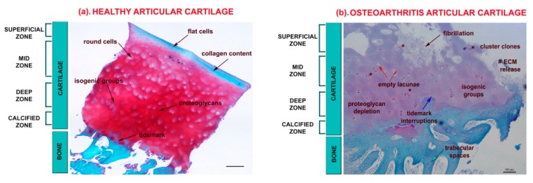
Figure 1.
Representative micrographs of articular cartilage tissues representing healthy (a) and OA (b) stained with Safranin-O/Fast Green staining taken from the laboratory archive. Representation of typical healthy and OA features in the superficial, mid, deep, and calcified zones of articular cartilage. Scale bar: 100 µm; red staining: proteoglycan content; pinkish staining: depletion of proteoglycan content; green staining: collagen content. (a) Healthy articular cartilage, black arrows report: (i) the presence of flat cells in the ECM rich of collagen fibres in the superficial zone; (ii) the presence of round cells in the ECM rich of proteoglycans in the mid-zone; (iii) the presence of isogenic groups in the ECM rich of proteoglycans in the deep zone; and (iv) the presence of tidemark in the calcified zone. (b) OA articular cartilage, black arrow reports the presence of fibrillation in the superficial zone; red arrows report the presence of empty lacunae; blue arrow: tidemark interruptions; green arrow: trabecular spaces; # reports ECM release; * cluster clones.

