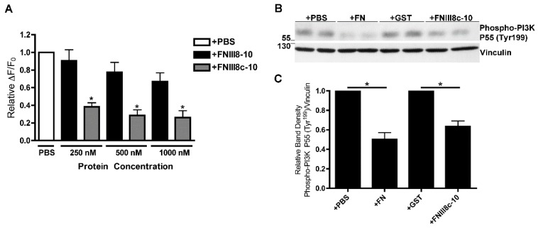
Figure 5.
Effects of FNIII8c-10 on PDGF-induced calcium release and phosphoinositol 3-kinase (PI3K) activity. (A) FN-null MEFs were seeded on collagen-coated wells (A, 1.1 × 10⁵ cells/cm2; B and C, 6 × 10⁴ cells/cm2) and allowed to adhere for 4 h. (A) Cells were treated the indicated protein or equal volume of PBS and incubated for 20 h. Cells were loaded with Fluo-4, stimulated with 30 ng/mL PDGF, and monitored for changes in fluorescence intensity. Data are presented as relative change in fluorescence intensity from baseline (ΔF/F0) and are expressed as mean ± SEM of 3 experiments performed in quadruplicate. Values were normalized to the “+PBS” condition, which was set to 1. * Significantly different versus +PBS by ANOVA (p < 0.05). (B,C) Cells were treated with PBS, 200 nM FN, 1 μM FNIII8c-10 or 1 μM GST and incubated for 20 h. Cells were then treated with 30 ng/mL PDGF for 5 min. Cell lysates were analyzed by immunoblotting. (B) Representative immunoblot represents 1 of 3 independent experiments performed in duplicate. (C) Phospho-PI3K and vinculin band densities were quantified by densitometry. The ratio of the average net intensity of phospho-PI3K P55 (Tyr199) bands to the average net intensity of vinculin bands was determined. Values were normalized to the control “+PBS” and “+GST” conditions, which were set to 1. Data are presented as the average ratio ± SEM of 3 experiments performed in duplicate. * Significantly different by ANOVA (p < 0.05).

