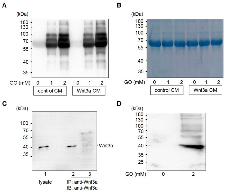
Figure 2.
Glycation of conditioned media (CM) with GO. (A) Control CM and Wnt3a CM were treated with different concentrations (0, 1, 2 mM) of GO for 5 h and analyzed by SDS-PAGE and Western blotting with anti-CML26 antibody. (B) After Western blot detection the same membrane was stained with Coomassie brilliant blue to show equal loading. The strong band corresponds to bovine serum albumin of the cell culture medium. (C) Western blot detection of Wnt3a from L-M(TK-)Wnt3A cell lysates (lane 1) or immunoprecipitated from serum-free Wnt3a CM (lane 2) or GO-treated Wnt3a CM (lane 3). (D) Treatment of 0.5 μg of purified Wnt3a protein with 2 mM GO induced Wnt3a carboxymethylation as detected with the anti-CML26 antibody on a Western blot. The untreated control was not modified.

