Abstract
The purpose of this study was to develop a minimum data set (MDS) registry for the prevention, diagnosis and treatment of chronic venous insufficiency (CVI) of the lower limbs. We designed the instrument in two phases, comprising a literature review and an e-Delphi study to validate the content. We obtained a total of 39 documents that we used to develop a registry with 125 items grouped in 7 categories, as follows: Patient examination, venous disease assessment methods, diagnostic tests to confirm the disease, ulcer assessment, treatments to manage the disease at all its stages, patient quality of life, and patient health education. The instrument content was validated by 25 experts, 88% of whom were primary healthcare and hospital nurses and 84% had more than 10 years’ experience in wound care. Using a two-round Delphi approach, we reduced the number of items in the MDS-CVI to 106 items. The categories remained unchanged. We developed an MDS for CVI with seven categories to assist healthcare professionals in the prevention, early detection, and treatment history of CVI. This tool will allow the creation of a registry in the primary care setting to monitor the venous health state of the population.
Keywords: diagnosis, information management, signs and symptoms, venous insufficiency, venous ulcer
1. Introduction
Chronic venous disease (CVD) of the lower limbs is a health problem with high prevalence and gradual progression. Developed countries are starting to manage this disease at early stages, in an attempt to prevent complications such as ulcers, when the human and economic burden is very heavy [1,2].
The evidence shows that lower limb venous disease can be staged by means of comprehensive history-taking that covers the classic signs and symptoms of venous disease, and correct Clinical, Etiological, Anatomical, and Pathophysiological (CEAP) classification [3,4]. The CEAP classification consensus document was published by the American Venous Forum in 1994 and was last updated in 2004. The aim of this instrument is to improve scientific communication when describing venous disease.
The CEAP clinical classification ranges from C0 (no visible or palpable signs of venous disease) to C6 (active venous ulcer). The system permits a patient’s status to be classified by the presence of signs such as reticular veins, oedema and trophic skin changes. These signs are accompanied by symptoms such as pain, heaviness, burning sensation, cramps, and pruritus [5]. The quality of life of individuals with CVD is drastically reduced as the disease advances [2].
Chronic venous insufficiency (CVI), defined as CEAP clinical classes C3–C6, affects 5% of the population, and an estimated 1–2% have a leg ulcer at some stage in their lives [6,7]. Active ulcers are responsible for the main financial impact of the disease process. The cost of caring for patients with CVI is estimated at 600–900 million euros in western Europe, accounting for 2% of healthcare expenditure. The estimated mean direct cost of each ulcer is €9000, representing 90% of the total CVI bill. This figure includes the cost of human resources (doctors and nurses), material for dressings, and hospital stays. Another less visible component is the indirect cost of CVI, which includes patients’ and relatives’ travel expenses, time off work, and even disability [5,8].
In the primary healthcare (PHC) setting, the clinical component (C) of CVD can be classified by means of patient questioning, thorough history taking, and a physical examination with the patient in a standing position, to observe dilated veins and skin abnormalities. The Doppler-assisted ankle-brachial index (ABI) must also always be calculated to make an accurate diagnosis and rule out peripheral arterial disease [6].
Venous disease prevention, diagnosis, and most treatment can take place in the primary care setting, but healthcare professionals must be appropriately trained and have the tools to provide this care. Patients may benefit from surgery at more advanced stages and will therefore need to be referred to the angiology or vascular surgery department [9].
Despite clear scientific evidence showing that the gold standard of CVI prevention and treatment is lower limb compression by means of bandaging, stockings, and other devices, in clinical practice, these measures are rarely implemented [3]. In fact, as many as 90% of patients with CVI receive no treatment whatsoever [10]. The literature describes several factors that might explain the low uptake of compression treatment, including a lack of awareness and skills among healthcare professionals [11,12].
A minimum data set (MDS) is a set of clearly defined items concerning a specific issue. MDSs have been shown to be effective in the prevention and early detection of different health problems, and to help guide their treatment [13,14]. A MDS permits interventions to be planned and followed up over time, and identifies which minimum quality indicators should be implemented [15]. The purpose of this study was to develop a MDS registry for CVI (MDS-CVI) of the lower limbs.
2. Methods
The instrument was designed in two phases, as follows: A literature review and an e-Delphi study with content validation by an expert panel.
2.1. Phase 1. Literature Review
We performed a literature review to define the MDS-CVI parameters. In December 2015, we carried out a literature search of keywords in MEDLINE (via PubMed), Cumulative Index to Nursing and Allied Health Literature (CINAHL), Scopus, and Cochrane Library Plus.
In PubMed and SCOPUS, we used the Medical Subject Headings (MeSH) terms ‘Diagnosis’, ‘Signs and Symptoms’, and ‘Venous Insufficiency’. In the CINAHL database, we used the MeSH terms ‘Diagnosis’ and ‘Venous insufficiency chronic’. The Boolean operator “AND” was used in all searches. In the Cochrane Library Plus database, we used the term “Venous Insufficiency”.
We used the Google search engine to find clinical practice guidelines and scientific society publications related to chronic wound care.
Inclusion criteria were language (English or Spanish), publication date (2011 or later), pathology (CVI of the lower limbs, venous ulcers), and treatment (of CVI of the lower limbs).
Two researchers analyzed the articles independently to identify concepts related to the prevention, diagnosis, or treatment of venous disease of the lower limbs. Then, they reached a consensus on the definitive items.
2.2. Phase 2. e-Delphi Study
We used an e-variant of the original Delphi study, which gathers experts’ opinions to reach a consensus on a complex issue. The e-Delphi format was used to obtain data through an online platform [16]. The purpose of the study was for wound care experts to assess the validity of the MDS-CVI content obtained through the literature review.
2.2.1. Sample
To create the expert panel, we contacted the six leading Spanish scientific societies for vascular diseases and wounds, as follows: Grupo Nacional para el Estudio y Asesoramiento en Úlceras por Presión y Heridas Crónicas (National Advisory Study Group for Pressure Ulcers and Chronic Wounds) (GNEAUPP), Asociación Nacional de Enfermería Dermatológica e Investigación del Deterioro de la Integridad Cutánea (National Association of Dermatology Nursing and Research into Deterioration of Skin Integrity) (ANEDIDIC), Sociedad Gallega de Heridas (Galician Society for Wounds) (SGH), Asociación Española de Enfermería Vascular y Heridas (Spanish Association for Vascular Nursing and Wounds) (AEEVH), Sociedad Española de Heridas (Spanish Society for Wounds) (SEHER), and the Sociedad Española de Angiología y Cirugía vascular (Spanish Society for Angiology and Vascular Surgery) (SEACV). These societies wrote to their members to describe the study objectives and methods, and provided an email address where members could request more information about the study with a view to participating in the panel.
2.2.2. Ethical Considerations
The study protocol was reviewed and approved by The Foundation University Institute for Primary Health Care Research Jordi Gol i Gurina (IDIAPJGol), under code P17/030.
All participants were required to sign a privacy agreement and study participation consent form before joining the expert panel.
2.2.3. Data Collection
The experts participated in two rounds by completing a questionnaire drawn up on the Google Forms platform.
2.2.4. e-Delphi Round 1
The first round, carried out in April 2017, contained the 125 items from the literature review, grouped into seven categories. The experts had to consider the suitability of the items for inclusion in the MDS-CVI and grade them on a scale of 1 to 5, where 1 was very unsuitable and 5 was very suitable.
The experts were informed that consensus would be established for items with a mean score of 4. A high consensus was defined as ≥72% of experts scoring ≥4 for an item, which is slightly higher than the 70% recommended by some authors [17]. Items that achieved this level of consensus were marked as definitive and excluded from the second round. Items with a mean score between 3.5 and <4 and a consensus of 50% to 72% were reviewed in the next round. Items with a mean score of <3.5 and a consensus of <50% were deleted. The experts were allowed to suggest new items and categories.
2.2.5. e-Delphi Round 2
In the second round, carried out in June 2017, the results from the first round were shared, new items proposed by the experts were added, and the method and criteria applied in the first round were repeated.
3. Results
3.1. Phase 1. Literature Review
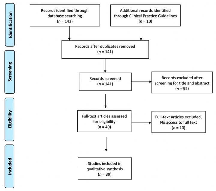
A total of 153 articles were obtained from the literature search (Figure 1). After removal of duplicate articles, those not meeting the inclusion criteria and those we were unable to access, 39 articles were included in the analysis.
Figure 1.
Flow diagram showing studies identified and selected.
With these 39 documents, we developed an MDS for the prevention, diagnosis, and treatment of CVI, with a total of 125 items grouped into seven categories, as follows:
-
(1)Patient examination [3,4,5,6,18,19,20,21,22,23,24,25,26,27,28,29,30,31,32,33,34,35,36,37,38,39,40,41,42,43,44,45,46,47,48,49,50,51,52,53,54,55] (Table 1), with two sub-categories, as follows:
-
(a)Risk factors, with 15 items covering personal circumstances that increase the likelihood of CVD. These items include age, sex, and family history of CVI.
-
(b)Leg conditions, with 22 items related to the signs and symptoms of venous disease of the lower limbs such as cramps, heaviness, and varicose veins.
-
(a)
-
(2)
Diagnostic studies [6,21,23,26,27,28,29,30,31,32,33,34,35,36,38,39,40,42,43,44,47,49,50,51,52,53] defining venous disease (Table 2), with eleven items describing existing diagnostic tests. These tests include continuous wave-Doppler and duplex ultrasound.
-
(3)
Scoring and classification systems [6,19,21,22,23,24,26,29,33,38,42,43,44,45,46,47,48,49,50,51] with three items. Venous disease scoring and classification systems consisted of the Villalta scale, the Venous Clinical Severity Score (VCSS) and the CEAP.
-
(4)
Ulcer examination [30,31,33,34,39,42] with seven items to describe ulcers, including photography, signs of infection, and pain.
-
(5)Different treatments at the various stages of venous disease (Table 3). This category has four sub-categories, as follows:
- (a)
- (b)
- (c)
- (d)
-
(6)
Patient quality of life [6,21,27,40,44,47,49,56] (Table 4), with five scales to assess patients’ quality of life at different stages of venous disease, such as the Chronic Venous Insufficiency Quality of Life Questionnaire (CIVIQ) for people with CVI and the Charing Cross Venous Ulcer Questionnaire for individuals with venous ulcers.
-
(7)
Health education [29,30,31,33,34,42,51] with 16 items, including recommendations to prevent complications and improve venous return, such as elevating the legs when resting, avoiding tight clothing, and taking regular exercise.
Table 1.
Items related to risk factors and patient’s leg conditions.
| Risk Factors | |||||
|---|---|---|---|---|---|
| Mean Round 1 | Exp ≥ 4 n (%) | Mean Round 2 | Exp ≥ 4 n (%) | Final Decision | |
| Usual medication [29,30]: Diuretics [36], Nonsteroidal anti-inflammatory drugs (NSAIDs) [36], Oral anticoagulants [38,40], Oral contraceptives [6,27,28,33,35] | 4.76 | 25 (100) | Kept | ||
| Mobility: Sedentary [31,34,42] | 4.76 | 24 (96) | Kept | ||
| Gender [6,21,22,24,26,27,28] | 4.76 | 23 (93) | Kept | ||
| Obesity (Body mass index ≥ 30) [6,19,21,22,23,26,28,29,31,33,35,42] | 4.72 | 25 (100) | Kept | ||
| Clinical history [30,34]: Diabetes Mellitus (DM) [26,35], Arterial Hypertension (HTA) [35,36] | 4.72 | 24 (96) | Kept | ||
| Family history of chronic venous insufficiency [6,22,23,24,28,31,34] | 4.72 | 24 (96) | Kept | ||
| Job [23,24,28,38,39,40,41] | 4.68 | 24 (96) | Kept | ||
| Age [3,6,18,19,20,21,22,23,24,25] | 4.6 | 23 (92) | Kept | ||
| Renal disease [26,35,36,40] | 4.52 | 22 (88) | Kept | ||
| Smoking status [21,26,28] | 4.52 | 21 (84) | Kept | ||
| Ankle mobility restrictions [29,30,33,34] | 4.36 | 22 (88) | Kept | ||
| Nutritional status [31,34] | 4.28 | 21 (84) | Kept | ||
| Bowel habit [28] | 4.20 | 20 (80) | Kept | ||
| Pregnancy (obstetric history) [22,23,26,27,28,34,38,40,43] | 4.12 | 20 (80) | Kept | ||
| Ethnicity [6,21,26,28] | 3.56 | 15 (52) | 3.44 | 15 (60) | Removed |
| History of leg ulcers | 4.88 | 25 (100) | Kept | ||
| Previous history of thrombosis | 4.84 | 25 (100) | Kept | ||
| Use of compression stockings | 4.68 | 23 (92) | Kept | ||
| Previous surgical background of the legs | 4.48 | 23 (92) | Kept | ||
| Year of diagnosis CVD/CVI | 4.24 | 20 (80) | Kept | ||
| Harmful alcohol consumption | 4.08 | 20 (80) | Kept | ||
| Leg conditions: Symptoms | |||||
| Heaviness [6,21,24,27,31,33,44,46,47,48,49] | 4.80 | 25 (100) | Kept | ||
| Itching [6,21,23,31,33,34,35,36,37,44,46,47,49] | 4.60 | 24 (96) | Kept | ||
| Pain [6,21,23,24,25,26,27,31,33,34,35,40,44,46,47,49,51,52] | 4.60 | 23 (92) | Kept | ||
| Cramps [6,23,30,31,33,35,44,47] | 4.52 | 24 (96) | Kept | ||
| Burning sensation [21,23,44,45,46] | 4.48 | 23 (92) | Kept | ||
| Paraesthesia [46] | 4.44 | 22 (88) | Kept | ||
| Discomfort legs [38,44,48] | 4.32 | 22 (88) | Kept | ||
| Tiredness [21,41,46,49] | 4.24 | 20 (80) | Kept | ||
| Leg conditions: Signs | |||||
| Active ulceration [19,21,26,27,28,33,35,42,44] | 4.96 | 25 (100) | Kept | ||
| Swelling (Oedema) [21,23,26,27,28,29,30,31,32,33,34,35,36,38,42,44,45,46,52,53,54] | 4.96 | 25 (100) | Kept | ||
| Varicose veins [19,23,28,31,34,38,39,40,42,44,45,46,47,48,50,52,55] | 4.92 | 25 (100) | Kept | ||
| Lipodermatosclerosis [28,32,34,35,39,44,46,51,53] | 4.88 | 25 (100) | Kept | ||
| Venous eczema [23,28,29,30,31,32,34,35,42,44,46] | 4.88 | 25 (100) | Kept | ||
| Atrophie blanche [28,31,33,34,39,42,46] | 4.84 | 25 (100) | Kept | ||
| Telangiectasias [24,26,28,35,38,44,46] | 4.80 | 25 (100) | Kept | ||
| Ocher dermatitis [33,42,44] | 4.80 | 24 (96) | Kept | ||
| Chronic skin changes [6,21,30,31,34,39,40,44,46,49,52] | 4.76 | 24 (96) | Kept | ||
| Corona phlebectatica [6,28] | 4.68 | 24 (96) | Kept | ||
| Varicophlebitis [34] | 4.68 | 24 (96) | Kept | ||
| Cellulitis [35] | 4.60 | 23 (92) | Kept | ||
| Reticular veins [24,28,44] | 4.60 | 23 (92) | Kept | ||
| Varicorrhage [21] | 4.56 | 22 (88) | Kept | ||
| Pitting edema | 4.76 | 24 (96) | Kept | ||
| Lymphedema | 4.04 | 20 (80) | Kept | ||
Table 2.
Items related to diagnosis, scoring classifications systems, and examination of the ulcer.
| Diagnostic Studies | |||||
|---|---|---|---|---|---|
| Mean Round 1 | Exp ≥ 4 n (%) | Mean Round 2 | Exp ≥ 4 n (%) | Final Decision | |
| Ankle brachial pressure index (ABPI) [6,29,30,31,33,34,35,39,42] | 4.56 | 22 (88) | Kept | ||
| Duplex ultrasound [6,21,23,27,28,31,33,34,38,39,40,44,47,49,50,51] | 4.44 | 22 (88) | Kept | ||
| D-dimer assay [35] | 3.44 | 14 (56) | 3.36 | 11 (44) | Removed |
| Trendelenburg test [28,31] | 3.6 | 18 (72) | 3.76 | 15 (60) | Removed |
| Perthes test [31] | 3.56 | 16 (64) | 3.76 | 16 (64) | Removed |
| Schwart test [33] | 3.56 | 16 (64) | 3.64 | 15 (60) | Removed |
| Continuous wave-doppler [6,21,26,30,31,32,33,35,36,40,43,44,47,50,53] | 3.36 | 16 (64) | Removed | ||
| Air-Plethismography [6,32,33,34,44] | 3.24 | 10 (40) | Removed | ||
| Venography [44,52,53] | 3.08 | 10 (40) | Removed | ||
| Pulse oximetry [34,39] | 3 | 10 (40) | Removed | ||
| Magnetic resonance [35,44,53] | 2.92 | 9 (36) | Removed | ||
| Samuels maneuver | 3.72 | 16 (64) | Removed | ||
| Scoring and classification systems | |||||
| CEAP classification of chronic venous disease [6,19,21,22,23,24,26,29,33,38,40,42,43,44,45,46,47,48,49,51] | 4.80 | 24 (96) | Kept | ||
| Venous Clinical Severity Score (VCSS) [6,21,40,44,47,50] | 4.60 | 22 (88) | Kept | ||
| Villalta score [6,44] | 3.92 | 18 (72) | 4.08 | 18 (72) | Kept |
| Examination of the ulcer | |||||
| Location [30,31,33,34,39,42] | 5 | 25 (100) | Kept | ||
| Appearance of ulcer bed [30,31,33,34,39,42] | 4.96 | 25 (100) | Kept | ||
| Characteristics of the ulcer [30,31,33,42] | 4.88 | 25 (100) | Kept | ||
| Edges [33,34,39,42] | 4.88 | 25 (100) | Kept | ||
| Pain [30,31,33,39] | 4.88 | 25 (100) | Kept | ||
| Amount and type of exudate [30,34,42] | 4.88 | 24 (96) | Kept | ||
| Signs of infection [34] | 4.88 | 24 (96) | Kept | ||
| Leg pulses | 4.64 | 23 (92) | Kept | ||
| RESVECH 2.0 score | 4.60 | 25 (100) | Kept | ||
Table 3.
Items related to treatments to manage all stages of venous disease.
| Compression Therapy | |||||
|---|---|---|---|---|---|
| Mean Round 1 | Exp ≥ 4 n (%) | Mean Round 2 | Exp ≥ 4 n (%) | Final Decision | |
| Graduated compression hosiery [19,21,26,27,30,32,33,34,35,38,39,41,42,44,46,49,50,51,53] | 4.84 | 25 (100) | Kept | ||
| Multi-layer compression bandage system [6,19,29,30,33,39,42] | 4.8 | 23 (92) | Kept | ||
| Long stretch compression bandages (LSB) [6,19,29,30,31,32,33,34,36,39,42,47,49] | 4.6 | 24 (96) | Kept | ||
| Short stretch compression bandages (SSB) [6,19,29,30,31,33,34,39,42,47] | 4.56 | 22 (88) | Kept | ||
| Adjustable Velcro bands [6] | 4.52 | 24 (96) | Kept | ||
| Unna boot [6,29,31,39] | 4.08 | 18 (72) | Kept | ||
| Pneumatic cuff compression [29,35,39,44] | 3.72 | 15 (60) | 3.52 | 15 (60) | Removed |
| Drug treatment | |||||
| Flavonoids/Phlebotonics [33,39,42,44,46] | 4.28 | 20 (80) | Kept | ||
| Sulodexide [6] | 4.28 | 20 (80) | Kept | ||
| Pentoxifylline [6,29,33,39,42,46] | 4.08 | 18 (72) | Kept | ||
| Antibiotic [21,39] | 3.96 | 15 (60) | 3.80 | 17 (68) | Removed |
| Acetylsalicylic acid [6,39] | 3.92 | 17 (68) | 4.08 | 18 (72) | Kept |
| Diuretic [35,52] | 3.88 | 16 (64) | 4.08 | 20 (80) | Kept |
| Oral anticoagulants [35,43,44,53] | 3.80 | 14 (56) | 4.08 | 19 (76) | Kept |
| Gabapentin [36] | 3.68 | 14 (56) | 3.40 | 12 (48) | Removed |
| Horse chestnut extract [35,44,46] | 3.40 | 11 (44) | Removed | ||
| Herbal substances | |||||
| Ruscus extract [44] | 3.40 | 11 (44) | Removed | ||
| Surgical treatment | |||||
| Foam sclerotherapy [6,19,23,28,44,49,50] | 4.16 | 19 (76) | Kept | ||
| Endovenous laser ablation (EVLA) [6,40,44,47,49] | 4.04 | 19 (76) | Kept | ||
| Percutaneous transluminal angioplasty and stenting [53,54] | 4.04 | 18 (72) | Kept | ||
| Radiofrequency ablation (RFA) [21,26,40,44,45,47,50] | 3.96 | 17 (68) | 3.92 | 17 (68) | Removed |
| Endovenous thermal ablation (EVTA) [6,21,23,44,50] | 3.92 | 17 (68) | 3.88 | 16 (64) | Removed |
| Ambulatory conservative haemodynamic management of varicose veins (CHIVA) [6,19,21,28,44,49,50,55] | 3.88 | 17 (68) | 3.92 | 16 (64) | Removed |
| Mechanochemical endovenous ablation (MOCA) [6,40,47,50] | 3.84 | 15 (60) | 3.84 | 17 (68) | Removed |
| Steam vein sclerosis (SVS) [43] | 3.72 | 14 (56) | 3.84 | 15 (60) | Removed |
| Cyanoacrylate embolization [21] | 3.68 | 14 (56) | 3.72 | 14 (56) | Removed |
| Venous ulcer treatment | |||||
| Cleansing [30,31,39] | 4.80 | 23 (92) | Kept | ||
| Moist environment dressing [6,29,30,31,33,39,44] | 4.76 | 24 (96) | Kept | ||
| Surrounding skin protection [33,39] | 4.76 | 23 (92) | Kept | ||
| Autolytic debridement [29,31,39] | 4.64 | 23 (92) | Kept | ||
| Sharp debridement [29,31,39] | 4.64 | 23 (92) | Kept | ||
| Biological debridement [29,31,39] | 4.52 | 23 (92) | Kept | ||
| Topical antimicrobials and antiseptics [29,30,33,39] | 4.48 | 20 (80) | Kept | ||
| Mechanical debridement [29,31,39] | 4.44 | 22 (88) | Kept | ||
| Vacuum assisted closure (VAC) [4,25,31,39] | 4.28 | 76 (19) | Kept | ||
| Osmotic debridement [29,31,39] | 4.12 | 18 (72) | Kept | ||
| Careful drying [30,31,39] | 4.08 | 17 (68) | Kept | ||
| Needle aspiration [31] | 4.04 | 72 (18) | Kept | ||
| Biopsy [34,39] | 3.92 | 17 (68) | 4.20 | 21 (84) | Kept |
| Skin graft [25,39,44] | 3.92 | 17 (68) | 4.08 | 18 (72) | Kept |
| Metalloproteinases [31] | 3.76 | 17 (68) | 3.80 | 17 (68) | Removed |
| Intermittent pneumatic compression [39] | 3.76 | 15 (60) | 3.44 | 14 (56) | Removed |
| Ultrasound therapy [39,44] | 3.56 | 14 (56) | 3.28 | 13 (52) | Removed |
| Hyperbaric oxygen therapy [39] | 3.44 | 48 (12) | Removed | ||
| Near-infrared light therapy [39] | 3.32 | 44 (11) | Removed | ||
| Electromagnetic therapy [39] | 3.32 | 48 (12) | Removed | ||
Table 4.
Items related to quality of life measurement and health education.
| Quality of Life Measurement | |||||
|---|---|---|---|---|---|
| Mean Round 1 | Exp ≥ 4 n (%) | Mean Round 2 | Exp ≥ 4 n (%) | Final Decision | |
| Chronic Venous Insufficiency Quality of Life Questionnaire (CIVIQ) [27] | 4.76 | 23 (92) | Kept | ||
| Charing Cross [56] | 4.64 | 22 (88) | Kept | ||
| EQ-5D [6,21,40,47] | 4.40 | 20 (80) | Kept | ||
| RAND-36 [6,40,44,47,49] | 3.96 | 18 (72) | 3.72 | 14 (56) | Removed |
| Aberdeen Varicose Vein Questionnaire (AVVQ) [21,40] | 3.88 | 18 (72) | 3.88 | 16 (64) | Removed |
| Health Education | |||||
| Exercise regularly [29,33,34,42] | 4.96 | 25 (100) | Kept | ||
| Keep mobile [33,42] | 4.92 | 25 (100) | Kept | ||
| Implement nutritional interventions/ weight loss [30,31,33,34,42] | 4.88 | 25 (100) | Kept | ||
| Use compression stockings [30,31,34,42] | 4.88 | 25 (100) | Kept | ||
| Elevate legs when resting [29,30,31,33,34,42,51] | 4.84 | 25 (100) | Kept | ||
| Avoid hot temperatures such as sitting too close to the fire [30,31,33,42] | 4.80 | 25 (100) | Kept | ||
| Keep legs raised at night [30,31,33] | 4.80 | 24 (96) | Kept | ||
| Wear well-fitted shoes [30,31,42] | 4.80 | 24 (96) | Kept | ||
| Avoid tight clothing [30,31,33,42] | 4.72 | 24 (96) | Kept | ||
| Shower as usual [30,33,42] | 4.68 | 22 (88) | Kept | ||
| Prevent constipation [31,33,42] | 4.60 | 24 (96) | Kept | ||
| Quit smoking [29] | 4.60 | 21 (84) | Kept | ||
| Leg massage [29] | 4.44 | 23 (92) | Kept | ||
| Use hyperoxygenated fatty acids [42] | 4.28 | 21 (84) | Kept | ||
| Take cold showers [30] | 4.28 | 20 (80) | Kept | ||
| Moisturize skin on legs [29,30,31,33,42] | 4.60 | 24 (96) | Kept | ||
3.2. Phase 2. e-Delphi Study
A total of 25 experts participated in both rounds, of whom 72% were men, 88% were nurses, and 12% were doctors specialized in vascular disease. Most worked in primary healthcare or hospital settings, and combined this work with university teaching (72%). A total of 84% had more than 10 years of experience in wound care.
In the first round, the experts added 11 items (see items without literature citation in the tables) and at the end of that round, 10 items were deleted, 25 were moved to the next round, and 90 were marked as definitive.
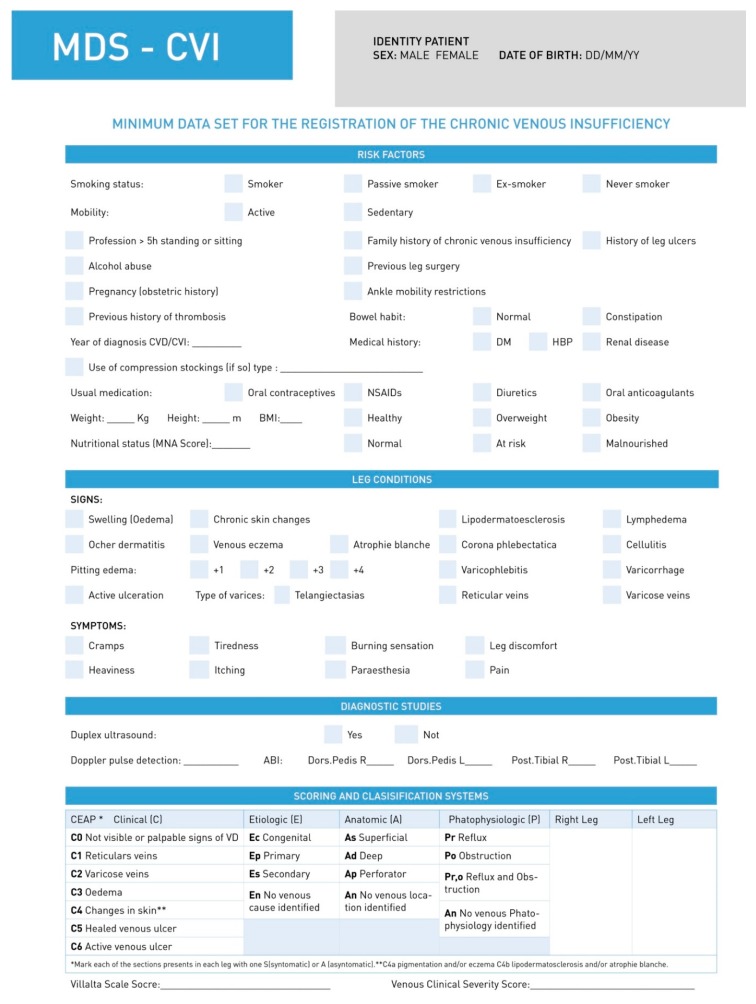
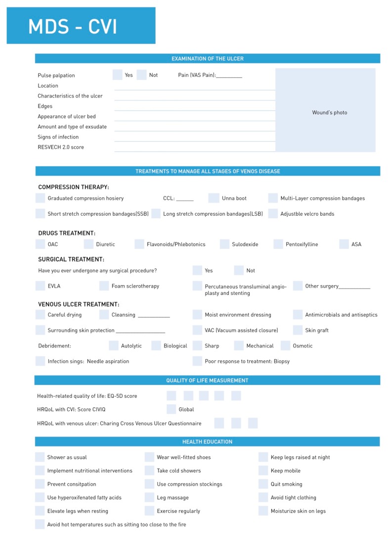
In the second round, the experts added no further items. At the end of the round, 20 items were deleted and 15 were accepted. The resulting MDS-CVI had a total of 106 items and 7 categories (Table 1, Table 2 and Table 3, Figure 2 and Figure 3).
Figure 2.
First page of the MSD-CVI.
Figure 3.
Second page of the MSD-CVI.
4. Discussion
The MDS-CVI is primarily a data-collection tool. However, when completing the registry, healthcare professionals are reminded of important actions that can be carried out in people with risk factors such as older age [3,6,18,19,20,21,22,23,24,25], female sex [6,21,22,24,26,28], and obesity [6,19,21,22,23,26,28,29,31,33,35,42], which increase their likelihood of having CVD [6]. Activities to promote health, prevent CVD, and diagnose it at earlier stages will help halt or delay disease progress. Healthcare professionals, and those working in primary health in particular, should aim to educate at-risk patients to lead a healthy lifestyle and use compression stockings.
Due to increased awareness of CVD, the tendency is generally for earlier diagnosis and treatment. However, in some countries, the disease is not detected until more advanced stages. There are gaps in healthcare professionals’ knowledge of venous leg ulcer physiology and its healing process [11], partly due to a lack of training at a degree level [12]. By applying and incorporating this MDS-CVI in patients’ health records, healthcare professionals will find it easier to monitor the disease course at every stage [6]. Above all, they should follow the recommendations to ensure correct diagnosis and treatment.
The CEAP classification system is a very easy method to classify venous disease and reach a reliable diagnosis of CVD/CVI in the population. The clinical part of the system can be obtained simply by observing a patient’s legs in the primary care setting. It is estimated that 80% of the population have the mildest level of symptoms (C1–C2, spider and varicose veins), while 5% have the most advanced stages (C3–C6) [6]. Implementation of this evidence-based MDS-CVI would result in more reliable data collection and facilitate monitoring of a specific population to observe disease progression, the treatments used, and their effectiveness [13,14]. With the existing level of evidence of the importance of therapeutic compression of the lower limbs, it is unacceptable that 90% of patients with CVI in Turkey [10] and 54% of patients with venous ulcers in Spain [57] are not given compression stockings. The MDS-CVI will also permit health managers to plan interventions according to the venous state of the population and identify which quality indicators should be applied [17].
People with CVI have a poor quality of life [58]. It is therefore important to determine how the venous disease affects each individual. Specific instruments are available to measure quality of life in these patients, such as the Aberdeen Varicose Vein Questionnaire (AVVQ) [21,40] or the Chronic Venous Insufficiency Quality of Life Questionnaire (CIVIC) [27] for patients with CVI, and the Charing Cross Venous Ulcer Questionnaire [56] for patients with venous ulcers. The instruments are valid for only certain languages and cultures [59] and they therefore need to be adapted to be effective.
Non-pharmacological measures are essential in the prevention and adjuvant therapy of CVD and healthcare professionals should therefore be aware of their existence and use them in their clinical practice. Recommendations such as weight loss [30,31,33,34,42] or taking regular exercise [29,33,34,42] will help venous return and delay symptom progression.
The MDS for CVI establishes minimum quality care criteria and can help to guide in the purchase of necessary services.
5. Limitations
One limitation of the review is that we were unable to access the full text of 10 articles that appeared in our literature search, although the addition of the 10 clinical practice guidelines helped overcome this limitation, at least in part.
In addition, all participants were from Spain, which may have given more or less importance to certain interventions and/or instruments than others. For example, the Aberdeen Varicose Vein Questionnaire was excluded from our study because no Spanish-language validation is available. On the contrary, the RESVECH 2.0 scale—an instrument that assesses chronic wound progression—was included but has no English-language validation [60]. Nevertheless, the literature review and the details of the items that were added and excluded by the experts make it easy to view the items that were assessed, and they can be easily adapted according to the needs of each health system.
Another limitation of the study is that most participants were nurses, and this may explain the elimination of some items from the e-Delphi data set related to non-nursing procedures, such as radiofrequency ablation.
6. Conclusions
We have developed a MDS for CVI with seven categories and 106 items to assist healthcare professionals in the prevention, early detection, and treatment history of CVI. This MDS-CVI also enables the creation of a population-based registry in the primary care setting to monitor the venous health state of the population, the pathological evolution over time, characteristics of the population, attention provided, and the distribution of health resources destined or necessary for the complete care of the person suffering from CVI.
Acknowledgments
We appreciate the translation into English and manuscript editing provided by Emma Goldsmith (https://www.goldsmithtranslations.com/).
Author Contributions
Conceptualization, E.H.-R. and A.R.-C.; Methodology, E.H.-R.; Software, A.R.-C.; Validation, E.H.-R.; Formal Analysis, E.H.-R. and A.R.-C.; Investigation, E.H.-R.; Resources, E.H.-R. and A.R.-C.; Data Curation, A.R.-C.; Writing—Original Draft Preparation, E.H.-R. and A.R.-C.; Writing—Review & Editing, E.H.-R. and A.R.-C.; Visualization, E.H.-R.; Supervision, E.H.-R.; Project Administration, E.H.-R.; Funding Acquisition, E.H.-R. and A.R.-C.
Funding
This work was supported by Girona University (MPCUdG2016/066), as part of the Professional Nursing Development project and Health Department of the Generalitat de Catalunya with the PERIS Project (SLT002/16/00199).
Conflicts of Interest
The authors declare no conflict of interest. The sponsors had no role in the design, execution, interpretation, or writing of the study.
References
- 1.Escudero Rodríguez J.R., Fernández Quesada F., Bellmunt Montoya S. Prevalencia y características clínicas de la enfermedad venosa crónica en pacientes atendidos en Atención Primaria en España: Resultados del estudio internacional Vein Consult Program. Cir. Esp. 2014;92:539–546. doi: 10.1016/j.ciresp.2013.09.013. [DOI] [PubMed] [Google Scholar]
- 2.Green J., Jester R., McKinley R., Pooler A. The impact of chronic venous leg ulcers: A systemic review. J. Wound Care. 2014;23:601–612. doi: 10.12968/jowc.2014.23.12.601. [DOI] [PubMed] [Google Scholar]
- 3.Amsler F., Blättler W. Compression therapy for occupational leg symptoms and chronic venous disorders: A meta-analysis of randomised controlled trials. Eur. J. Vasc. Endovasc. Surg. 2008;35:366–372. doi: 10.1016/j.ejvs.2007.09.021. [DOI] [PubMed] [Google Scholar]
- 4.Mosti G., De Maeseneer M., Cavezzi A., Parsi K., Morrison N., Nelzén O., Rabe E., Partsch H., Caggiati A., Simka M., et al. Society for Vascular Sugery and American Venous Forum Guidelines on the management of venous leg ulcers: The point of view of the International Union of Phlebology. Int. Angiol. 2015;34:202–218. [PubMed] [Google Scholar]
- 5.Eberhardt R.T., Raffetto J.D. Chronic Venous Insufficiency. Circulation. 2014;130:333–346. doi: 10.1161/CIRCULATIONAHA.113.006898. [DOI] [PubMed] [Google Scholar]
- 6.Wittens C., Davies A.H., Bækgaard N., Broholm R., Cavezzi A., Chastanet S., De Wolf M., Eggen C., Giannoukas A., Gohel M., et al. Editor’s Choice—Management of Chronic Venous Disease: Clinical Practice Guidelines of the European Society for Vascular Surgery (ESVS) Eur. J. Vasc. Endovasc. Surg. 2015;49:678–737. doi: 10.1016/j.ejvs.2015.02.007. [DOI] [PubMed] [Google Scholar]
- 7.Wrona M., Jöckel K.H., Pannier F., Bock E., Hoffmann B., Rabe E. Association of Venous Disorders with Leg Symptoms: Results from the Bonn Vein Study 1. Eur. J. Vasc. Endovasc. Surg. 2015;50:360–367. doi: 10.1016/j.ejvs.2015.05.013. [DOI] [PubMed] [Google Scholar]
- 8.Gohel M. Which treatments are cost-effective in the management of varicose veins? Phlebology. 2013;28(Suppl. 1):153–157. doi: 10.1177/0268355513477003. [DOI] [PubMed] [Google Scholar]
- 9.Bellmunt Montoya S., Díaz Sánchez S., Sánchez Nevárez I., Fuentes Camps E., Fernández Quesada F., Piquer Farrés N. Criteria for referral between levels of care of patients with peripheral vascular disease. SEMFYC-SEACV consensus document. Aten Primaria. 2012;44:e1–e555. doi: 10.1016/j.aprim.2012.05.002. [DOI] [PMC free article] [PubMed] [Google Scholar]
- 10.Akbulut B., Uçar H.I., Oç M., Ikizler M., Yorgancioglu C., Dernek S., Böke E. Characteristics of venous insufficiency in western Turkey: VEYT-I study. Phlebology. 2012;27:374–377. doi: 10.1258/phleb.2011.011100. [DOI] [PubMed] [Google Scholar]
- 11.Ylönen M., Stolt M. Leino-Kilpi, H.; Suhonen, R. Nurses’ knowledge about venous leg ulcer care: A literature review. Int. Nurs. Rev. 2014;61:194–202. doi: 10.1111/inr.12088. [DOI] [PubMed] [Google Scholar]
- 12.Romero-Collado A., Raurell-Torreda M., Zabaleta-del-Olmo E., Homs-Romero E., Bertran-Noguer C. Course content related to chronic wounds in nursing degree programs in Spain. J. Nurs. Scholarsh. 2015;47:51–61. doi: 10.1111/jnu.12106. [DOI] [PubMed] [Google Scholar]
- 13.Alipour J., Ahmadi M., Mohammadi A. The need for development a national minimum data set of the information management system for burns in Iran. Burns. 2016;42:710. doi: 10.1016/j.burns.2015.05.016. [DOI] [PubMed] [Google Scholar]
- 14.Jebraeily M., Ghazisaeidi M., Safdari R., Makhdoomi K., Rahimi B. Hemodialysis Adequacy Monitoring Information System: Minimum Data Set and Capabilities Required. Acta Inform. Med. 2015;23:239–242. doi: 10.5455/aim.2015.23.239-242. [DOI] [PMC free article] [PubMed] [Google Scholar]
- 15.Hjaltadóttir I., Ekwall A.K., Nyberg P., Hallberg I.R. Quality of care in Icelandic nursing homes measured with Minimum Data Set quality indicators: Retrospective analysis of nursing home data over 7 years. Int. J. Nurs. Stud. 2012;49:1342–1353. doi: 10.1016/j.ijnurstu.2012.06.004. [DOI] [PubMed] [Google Scholar]
- 16.Donohoe H., Stellefson M., Tennant B. Advantages and limitations of the e-Delphi technique: Implications for health education researchers. Am. J. Health Educ. 2012;43:38–46. doi: 10.1080/19325037.2012.10599216. [DOI] [Google Scholar]
- 17.Feo R., Conroy T., Jangland E., Muntlin Athlin Å., Brovall M., Parr J., Blomberg K., Kitson A. Towards a standardised definition for fundamental care: A modified Delphi study. J. Clin. Nurs. 2018;27:2285–2299. doi: 10.1111/jocn.14247. [DOI] [PubMed] [Google Scholar]
- 18.Alvarez Fernández L.J., Lozano F., Marinel·lo-Roura J., Masegosa-Medina J.A. Encuesta epidemiológica sobre insuficiencia venosa crónica en España: Estudio DETECT-IVC 2006. Angiología. 2008;60:27–36. doi: 10.1016/S0003-3170(08)01003-1. [DOI] [Google Scholar]
- 19.Reich-Schupke S., Murmann F., Altmeyer P., Stuücker M. Compression therapy in elderly and overweight patients. Vasa. 2012;41:125–131. doi: 10.1024/0301-1526/a000175. [DOI] [PubMed] [Google Scholar]
- 20.Weller C.D., Buchbinder R., Johnston R.V. Interventions for helping people adhere to compression treatments for venous leg ulceration. Cochrane Database Syst. Rev. 2013:CD008378. doi: 10.1002/14651858.CD008378.pub2. [DOI] [PubMed] [Google Scholar]
- 21.Morrison N., Gibson K., McEnroe S., Goldman M., King T., Weiss R., Cher D., Jones A. Randomized trial comparing cyanocrylate embolization and radiofrequency ablation for incompetent great saphenous vein (VeClose) J. Vasc. Surg. 2015;61:985–994. doi: 10.1016/j.jvs.2014.11.071. [DOI] [PubMed] [Google Scholar]
- 22.Musil D., Kaletova M., Herman J. Age, body mass index and severity of primary chronic venous disease. Biomed. Pap. Med. Fac. Univ. Palacky Olomouc Czech Repub. 2011;155:367–371. doi: 10.5507/bp.2011.054. [DOI] [PubMed] [Google Scholar]
- 23.Van den Boezem P.B., Klem T.M., le Cocq d’Armandville E., Wittens C.H. The management of superficial venous incompetence. BMJ. 2011;343:d4489. doi: 10.1136/bmj.d4489. [DOI] [PubMed] [Google Scholar]
- 24.Pitsch F. VEIN CONSULT Program: Interim results from the first 70,000 secreened patients in 13 countries. Phlebolymphology. 2012;19:132–137. [Google Scholar]
- 25.Dumville J.C., Land L., Evans D., Peinemann F. Negative pressure wound therapy for treating leg ulcers. Cochrane Database Syst. Rev. 2015:CD011354. doi: 10.1002/14651858.CD011354.pub2. [DOI] [PMC free article] [PubMed] [Google Scholar]
- 26.Carruthers T.N., Farber A., Rybin D., Doros G., Eslami M.H. Interventions on the superficial venous system for chronic venous insufficiency by surgeons in the modern era: An analysis of ACS-NSQIP. Vasc. Endovasc. Surg. 2014;48:482–490. doi: 10.1177/1538574414561226. [DOI] [PubMed] [Google Scholar]
- 27.Allegra C., Antignani P.L., Will K., Allaert F. Acceptance. compliance and effects of compression stockings on venous functional symptoms and quality of life of Italian pregnant women. Int. Angiol. 2014;33:357–364. [PubMed] [Google Scholar]
- 28.Robertson L.A., Evans C.J., Lee A.J., Allan P.L., Ruckley C.V., Fowkes F.G. Incidence and risk factors for venous reflux in the general population: Edinburgh Vein Study. Eur. J. Vasc. Endovasc. Surg. 2014;48:208–214. doi: 10.1016/j.ejvs.2014.05.017. [DOI] [PubMed] [Google Scholar]
- 29.Association for the Advancement of Wound Care (AAWC) Venous Ulcer Guideline. Association for the Advancement of Wound Care (AAWC); Malvern, PA, USA: 2010. [PubMed] [Google Scholar]
- 30.Muñoz Rodríguez A., Escanciano Pérez I., Ballesteros Úbeda M.V., Polimón Olibarrieta I., Díaz Ramírez C., González Sánchez J., Aparicio Martín A., Sánchez Morantes A., Búa Ocaña S., López Hernández R., et al. Manual de Protocolos y Procedimientos en el Cuidado de las Heridas Crónicas. Hospital Universitario de Móstoles; Madrid, Spain: 2011. [Google Scholar]
- 31.Caicedo González R., Castañeda Robles C., Cossío Gómez F., Delgado Uría A., Fernández Sáiz B., Gómez España M.V. Manual de Prevención y Cuidados Locales en Heridas Crónicas. Servicio Cántabro de Salud; Cantabria, Spain: 2011. Edita: Servicio Cántabro de Salud. [Google Scholar]
- 32.Suehiro K., Morikage N., Murakami M., Yamashita O., Ueda K., Samura M., Hamano K. Study of leg edema in immobile patients. Circ. J. 2014;78:1733–1739. doi: 10.1253/circj.CJ-13-1599. [DOI] [PubMed] [Google Scholar]
- 33.Asociación Española de Enfermería Vascular y Heridas . Guía de Práctica Clínica: Consenso Sobre Úlceras Vasculares y Pie Diabético. 2nd ed. AEEVH; Sevilla, Spain: 2014. [Google Scholar]
- 34.Australian Wound Management Association & New Zealand Wound Care Society . Australian and New Zealand Clinical Practice Guideline for Prevention and Management of Venous Leg Ulcers. Cambridge Publishing; Melbourne, VIC, Australia: 2011. [Google Scholar]
- 35.Trayes K., Studdiford J., Pickle S., Tully A. Edema: Diagnosis and Management. Am. Fam. Phys. 2013;88:102–110. [PubMed] [Google Scholar]
- 36.Thaler H.W., Pienaar S., Wirnsberger G. Roller-Wirnsberger, R.E. Bilateral leg edema in an older woman. Z. Gerontol. Geriatr. 2015;48:49–51. doi: 10.1007/s00391-013-0557-x. [DOI] [PubMed] [Google Scholar]
- 37.Benigni J.P., Bihari I., Rabe E., Uhl J.F., Partsch H., Cornu-Thenard A., Jawien A. UIP—Union Internationale de Phlébologie. Venous symptoms in C0 and C1 patients: UIP consensus document. Int. Angiol. 2013;32:261–265. [PubMed] [Google Scholar]
- 38.Blazek C., Amsler F., Blaettler W., Keo H.H., Baumgartner I., Willenberg T. Compression hosiery for occupational leg symptoms and leg volume: A randomized crossover trial in a cohort of hairdressers. Phlebology. 2013;28:239–247. doi: 10.1258/phleb.2011.011108. [DOI] [PubMed] [Google Scholar]
- 39.Scottish Intercollegiate Guidelines Network . A National Clinical Guideline. Scottish Intercollegiate Guidelines Network; Edinburgh, UK: 2010. Management of chronic venous leg ulcers. [Google Scholar]
- 40.Boersma D., van Eekeren R.R., Kelder H.J., Werson D.A., Holewijn S., Schreve M.A., Reijnen M.M., de Vries J.P. Mechanochemical endovenous ablation versus radiofrequency ablation in the treatment of primary small saphenous vein insufficiency (MESSI trial): Study protocol for a randomized controlled trial. Trials. 2014;29:421. doi: 10.1186/1745-6215-15-421. [DOI] [PMC free article] [PubMed] [Google Scholar]
- 41.Robertson L., Yeoh S.E., Kolbach D.N. Non-pharmacological interventions for preventing venous insufficiency in a standing worker population. Cochrane Database Syst. Rev. 2013:CD006345. doi: 10.1002/14651858.CD006345.pub3. [DOI] [PMC free article] [PubMed] [Google Scholar]
- 42.Álvaro Rangil T., Berenguer Pérez M., Cegri Lombardo F., García Arcos E., Manuel Martí B., Marquilles Bonet C. Guia D’úlceres Venoses. AIFICC; Barcelona, Spain: 2014. [Google Scholar]
- 43.Milleret R., Huot L., Nicolini P., Creton D., Roux A.S., Decullier E., Chapuis F.R., Camelot G. Great saphenous vein ablation with steam injection: Results of a multicentre study. Eur. J. Vasc. Endovasc. Surg. 2013;45:391–396. doi: 10.1016/j.ejvs.2013.01.027. [DOI] [PubMed] [Google Scholar]
- 44.Miquel Abad C., Rial Horcajo R., Ballesteros Ortega M.D., García Madrid C. Guías de Práctica Clínica en Enfermedad Venosa Crónica. IDMedica; Torrejón de Ardoz, Spain: 2015. [Google Scholar]
- 45.Korkmaz K., Yener A.Ü., Gedık H.S., Budak A.B., Yener Ö., Genç S.B., Lafçi A. Tumescentless endovenous radiofrequency ablation with local hypothermia and compression technique. Cardiovasc. J. Afr. 2013;24:313–317. doi: 10.5830/CVJA-2013-053. [DOI] [PMC free article] [PubMed] [Google Scholar]
- 46.Perrin M., Ramelet A.A. Pharmacological treatment of primary chronic venous disease: Rationale. results and unanswered questions. Eur. J. Vasc. Endovasc. Surg. 2011;41:117–125. doi: 10.1016/j.ejvs.2010.09.025. [DOI] [PubMed] [Google Scholar]
- 47.Van Eekeren R.R., Boersma D., Konijn V., de Vries J.P., Reijnen M.M. Postoperative pain and early quality of life after radiofrequency ablation and mechanochemical endovenous ablation of incompetent great saphenous veins. J. Vasc. Surg. 2013;57:445–450. doi: 10.1016/j.jvs.2012.07.049. [DOI] [PubMed] [Google Scholar]
- 48.Amsler F., Rabe E., Blätter W. Leg Symptoms of Somatic. Psychic. and Unexplained Origin in the Population-based Bonn Vein Study. Eur. J. Vasc. Endovasc. Surg. 2013;46:255–262. doi: 10.1016/j.ejvs.2013.04.026. [DOI] [PubMed] [Google Scholar]
- 49.Bakker N.A., Schieven L.W., Bruins R.M., van den Berg M., Hissink R.J. Compression stockings after endovenous laser ablation of the great saphenous vein: A prospective randomized controlled trial. Eur. J. Vasc. Endovasc. Surg. 2013;46:588–592. doi: 10.1016/j.ejvs.2013.08.001. [DOI] [PubMed] [Google Scholar]
- 50.Lawson J., Gauw S. van Vlijmen, C.; Pronk, P.; Gaastra, M.; Mooij, M.; Wittens, C.H. Sapheon: The solution? Phlebology. 2013;28(Suppl. 1):2–9. doi: 10.1177/0268355513475970. [DOI] [PubMed] [Google Scholar]
- 51.Kelechi T.J., Mueller M., Zapka J.G., King D.E. The effect of a cryotherapy gel wrap on the microcirculation of skin affected by chronic venous disorders. J. Adv. Nurs. 2011;67:2337–2349. doi: 10.1111/j.1365-2648.2011.05680.x. [DOI] [PMC free article] [PubMed] [Google Scholar]
- 52.Hsieh M.C., Chang P.Y., Hsu W.H., Yang S.H., Chan W.P. Role of three-dimensional rotational venography in evaluation of the left iliac vein in patients with chronic lower limb edema. Int. J. Cardiovasc. Imaging. 2011;27:923–929. doi: 10.1007/s10554-010-9745-6. [DOI] [PubMed] [Google Scholar]
- 53.Anaya-Ayala J.E., Adams M.K., Telich-Tarriba J.E., Dresser K.L., Ismail N., Peden E.K. Complex left profunda femoris vein to renal vein bypass for the management of progressive chronic iliofemoral occlusion. Ann. Vasc. Surg. 2013;27:112.e5–112.e8. doi: 10.1016/j.avsg.2012.05.018. [DOI] [PubMed] [Google Scholar]
- 54.Adams M.K., Anaya-Ayala J.E., Ismail N., Peden E.K. Surgical femorocaval bypass for recalcitrant iliofemoral venous occlusion to endovascular treatment. Vasc. Endovasc. Surg. 2012;46:578–581. doi: 10.1177/1538574412454584. [DOI] [PubMed] [Google Scholar]
- 55.Bellmunt-Montoya S., Escribano J.M., Dilme J., Martinez-Zapata M.J. CHIVA method for the treatment of chronic venous insufficiency. Cochrane Database Syst. Rev. 2015:CD009648. doi: 10.1002/14651858.CD009648.pub3. [DOI] [PMC free article] [PubMed] [Google Scholar]
- 56.Robertson L., Lee A.J., Evans C.J., Boghossian S., Allan P.L., Ruckley C.V., Fowkes F.G. Incidence of chronic venous disease in the Edinburgh Vein Study. J. Vasc. Surg. Venous Lym. Dis. 2013;1:59–67. doi: 10.1016/j.jvsv.2012.05.006. [DOI] [PubMed] [Google Scholar]
- 57.Guinot-Bachero J., Balaguer-López E., Loma-Osorio R., Rivera Álvarez A., Ros-Mora M.C., González-Jiménez F., Garralón-Pérez A., Herrera-Herzog E., Gombau Baldrich Y., García-Molina P. Heridas en consultas de enfermería de Atención Primaria. Rev. ROL Enferm. 2018;41:126–133. [Google Scholar]
- 58.Soydan E., Yılmaz E., Baydur H. Effect of socio-demographic characteristics and clinical findings on the quality of life of patients with chronic venous insufficiency. Vascular. 2017;25:382–389. doi: 10.1177/1708538116685945. [DOI] [PubMed] [Google Scholar]
- 59.Marinel lo Roura J., Verdú Soriano J. Documento de Consenso 2018. 2nd ed. Ergon; Madrid, Spain: 2018. Conferencia nacional de consenso sobre las úlceras de la extremidad inferior (CONUEI) [Google Scholar]
- 60.Restrepo-Medrano J.C., Verdu Soriano J. Development of a wound healing index for chronic wounds. GEROKOMOS. 2011;22:176–183. doi: 10.4321/S1134-928X2011000400005. [DOI] [Google Scholar]


