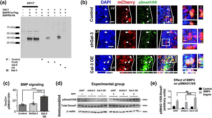
Figure 4.

Gal‐3 increases BMP signaling. (a) Co‐immunoprecipitation of Gal‐3 and BMPR1α and BMPR2. (b) Representative confocal images 3DPE. DAPI+ nuclei, mCherry+ electroporated cells, and pSmad 1/5/8+ cells. Arrows indicate pSmad1/5/8+ and arrowheads pSmad1/5/8‐labeled cells. Boxed areas are enlarged as orthogonal views. Scale bars: 20 μm for main panels, 5 μm for insets. (c) Quantification of B. One‐way ANOVA, Tukey's test for multiple comparisons. n = 5–6. (d) Western blot 24 hr after nucleofection with shNT (control), shGal‐3, or Gal‐3 OE, cells were treated with control or 5 ng/ml BMP4 for 24 hr. Immunoblot for pSmad 1/5/8, total Smad1 and β‐Actin. (e) Quantification of pSmad1/5/8 band intensities from (A1‐2) normalized to total Smad 1. 2‐way ANOVA
