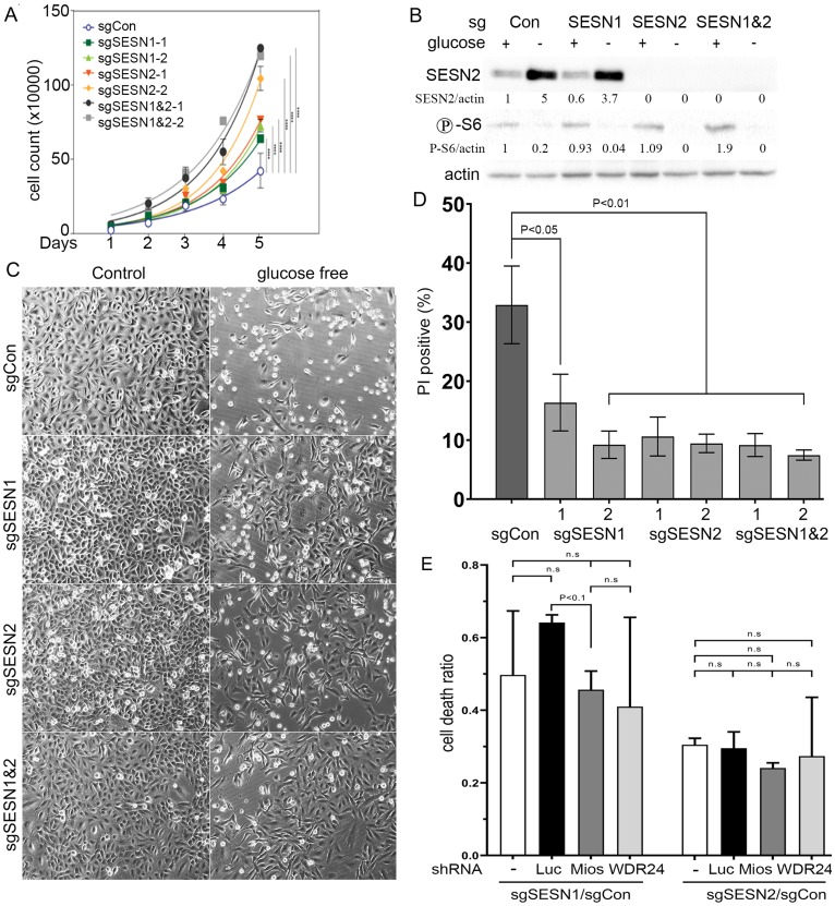
Figure 5. Inactivation of SESN1 and/or SESN2 in lung adenocarcinoma A549 cells stimulates cell proliferation rate and increases resistance to glucose starvation.
(A) Cells of indicated genotypes were plated onto 6-well plates (50,000 cells/well) and counted every 24 h for 5 days. (B) Western blot analysis of SESN2 expression and S6 phosphorylation of cells of the indicated genotypes incubated 18 h in glucose-free medium. (C) A549 cells with inactivation of SESN1 and/or SESN2 are resistant to cell death in glucose-free medium. Cell death was analyzed by phase-contrast microscopy. (D) Analysis of PI-positive cells incubated 24 h in glucose-free medium by flow cytometry. Data are presented as mean ± SEM. P values were calculated relative to control using one-way ANOVA followed by correction for multiple comparisons using Dunnett correction (α = 0.05) in three independent experiments. (E) SESN1 and SESN2 support cell death in a GATOR2-independent manner. Mios and WDR24 were silenced in control, SESN1- and SESN2- deficient cells and the levels of cell death were evaluated by flow cytometry analysis of PI inclusion. The data are presented as a ratio of the cell death in sgSESN1/sgControl(Con) or sgSESN2/sgCon cells in the presence of control shRNA (shLuc), shMios, or shWDR24. In the experiments (B–E) cells were incubated with glucose-free DMEM supplemented with L-glutamine, penicillin/streptomycin, and 10% dialyzed fetal bovine serum.

