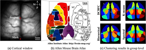
Fig. 4.
Comparison with other existing methods for in vivo Vglut2-GCaMP6s mouse data. Two markers (bregma and one arbitrary marker) were made on the sagittal suture (a) and registered with the Allen atlas (b). (c) The clustering results of DCBFC, k-means, hierarchical, and spectral clustering at group level (). All pixels are color-coded based on the functional module to which they belong. Some functional modules may contain more than one brain region.

