Abstract
The stringent response is a general bacterial stress response that allows bacteria to adapt and survive adverse conditions. This reprogramming of cell physiology is caused by the accumulation of the alarmone (p)ppGpp which, in Escherichia coli, depends on the (p)ppGpp synthetase RelA and the bifunctional (p)ppGpp synthetase/hydrolase SpoT. Although conditions that control SpoT-dependent (p)ppGpp accumulation have been described, the molecular mechanisms regulating the switching from (p)ppGpp degradation to synthesis remain poorly understood. Here, we show that the protein YtfK promotes SpoT-dependent accumulation of (p)ppGpp in E. coli and is required for activation of the stringent response during phosphate and fatty acid starvation. Our results indicate that YtfK can interact with SpoT. We propose that YtfK activates the stringent response by tilting the catalytic balance of SpoT toward (p)ppGpp synthesis.
Subject terms: Bacterial physiology, Bacteriology, Microbial genetics
The enzyme SpoT is important for accumulation of the alarmone (p)ppGpp, which triggers the stringent response in E. coli. Here, Germain et al. show that the protein YtfK promotes SpoT-dependent accumulation of (p)ppGpp and is required for activation of the stringent response during phosphate and fatty acid starvation.
Introduction
Fast and robust adaptive responses are critical for life in harsh environments. Therefore bacteria, including major pathogens, have evolved molecular mechanisms that allow them to respond and to adapt to their surrounding environment. In particular, the ubiquitous stringent response is a general bacterial stress response induced by diverse nutritional and environmental stresses that switches the metabolism balance from growth and division to stress response and survival. This global resetting of bacterial cell physiology relies on the accumulation of the alarmones ppGpp (guanosine 5′-diphosphate, 3′-diphosphate) and pppGpp (guanosine 5′-triphosphate, 3′-diphosphate) [collectively named (p)ppGpp]1.
In Gram-negative bacteria, (p)ppGpp reprograms transcription via binding to RNA polymerase, thereby altering its promoter selectivity2–5. Indeed, (p)ppGpp influences the transcription rates of around 500 genes in E. coli6,7. In addition to transcriptional control, (p)ppGpp directly inhibits several enzymes, including DNA primase, translation factors, polyphosphate kinase, and lysine decarboxylase8.
The RelA–SpoT Homologue (RSH) family of bifunctional proteins constitutes the key players in synthesizing and degrading (p)ppGpp9. Therefore, the balance between both activities represents a critical point of regulation to control the intracellular level of (p)ppGpp. Long RSH proteins have two functional regions: the enzymatic N-terminal half (NTD), encompassing the synthetase (SYNTH) and the hydrolase (HD) domains, and the C-terminal regulatory half (CTD)10,11. In most gamma and beta-proteobacteria, including E. coli, the stringent response is controlled by two RSHs called RelA and SpoT. SpoT has both (p)ppGpp hydrolytic and synthetic activities similar to the Rel enzymes, the long bifunctional RSH enzyme present in the vast majority of bacteria12–14. However, despite the strong homology between RelA and SpoT, RelA maintains a pseudo-hydrolase domain that is structurally conserved but nevertheless enzymatically inactive15–17. Thus, RelA is a monofunctional (p)ppGpp synthetase. Amino acid starvation that leads to loading of RelA•tRNA complexes at the ribosomal A-site activates RelA’s (p)ppGpp synthetic activity in vivo and in vitro18,19. In contrast, SpoT functions as a hub protein that integrates various environmental signals, including fatty acid (FA)20, carbon12, iron21, and phosphate starvation22. Importantly, the hydrolysis function of SpoT is crucial for balancing cellular (p)ppGpp concentration in the presence of RelA and disruption of spoT in wild-type E. coli strain is lethal due to the toxic accumulation of (p)ppGpp12. Hence SpoT-dependent regulation of intracellular (p)ppGpp level can reflect changes in either synthetase or hydrolase activity. How the switching between the two enzymatic activities is regulated remains largely unknown despite intensive investigations. However, control of SpoT activities through protein–protein interaction plays an important role. Indeed, the acyl carrier protein (ACP) binds SpoT and triggers (p)ppGpp synthesis in response to FA starvation23. More recently, the anti-σ70 factor Rsd of E. coli was shown to interact directly with SpoT to regulate stringent response during carbon source downshift by stimulating (p)ppGpp hydrolysis24.
Here, we present a genetic assay for the identification of protein candidates that can modulate SpoT-dependent (p)ppGpp synthesis in E. coli. Overproduction of one of these proteins, YtfK, promotes accumulation of (p)ppGpp in vivo. YtfK is required to maintain elevated (p)ppGpp levels in response to phosphate and FA starvation, and enhances survival and promotes formation of antibiotic tolerant cells under nutritional stress conditions. YtfK appears to interact with SpoT. We propose that YtfK acts by tilting the catalytic balance of SpoT toward (p)ppGpp synthesis rather than hydrolysis.
Results
YtfK promotes SpoT-dependent accumulation of (p)ppGpp
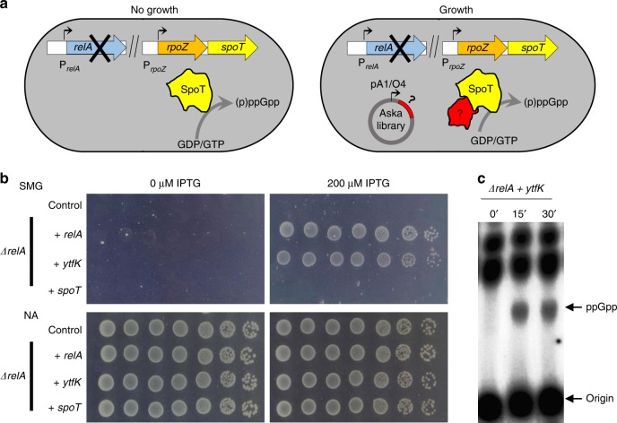
We designed a simple genetic assay allowing us to identify protein partners in E. coli that potentially modulate SpoT-dependent accumulation of (p)ppGpp. We exploited the fact that growth in the presence of 1 mM of serine, methionine, and glycine (SMG medium), which induces isoleucine starvation, requires elevated levels of (p)ppGpp25. Indeed, expression of isoleucine biosynthetic operon is stimulated by (p)ppGpp and, as a result, a ΔrelA mutant is unable to grow on SMG plates (Fig. 1a)25. To identify potential activators of SpoT-dependent (p)ppGpp synthesis, we selected for genes that, in multiple copies, would suppress the growth defect of an MG1655 E. coli K-12 ΔrelA strain on SMG agar plates. We pooled a collection of plasmids obtained from the ASKA library containing almost all E. coli K-12 genes26, each cloned into the high-copy-number vector pCA24N downstream of the isopropyl β-D-1-thiogalactopyranoside (IPTG)-inducible PT5-lac promoter, and identified several putative candidate genes as described in the Methods section. Among them, ytfK suppressed the growth defect of a ΔrelA mutant on SMG medium. This result was further confirmed after re-cloning the coding region of ytfK (without any tag) in a more suitable physiological plasmid harboring a tightly IPTG-inducible PT5-lac promoter (pEG25) (Fig. 1b; Supplementary Fig. 1a, b). Interestingly, ytfK encodes a small protein of 8 kDa and is expressed during phosphate starvation27, a condition known to trigger SpoT-dependent accumulation of (p)ppGpp22.
Fig. 1. YtfK promotes SpoT (p)ppGpp synthesis.
a Genetic setup based on the multicopy suppression (ASKA library) of the nongrowing phenotype of ΔrelA mutant on SMG plates. SMG plate induces isoleucine starvation and requires high (p)ppGpp level to de-repress isoleucine biosynthesis operon (see the Methods section). Question mark denotes putative candidate that promotes SpoT-dependent (p)ppGpp accumulation. b ytfK overexpression suppresses the nongrowing phenotype of the ΔrelA mutant on SMG plates. WT and ΔrelA mutant were transformed with pEG25 harboring either relA, ytfK, or spoT under an IPTG-inducible promoter. Cells were serial diluted and spotted both on nutrient agar (NA) and SMG plates with or without IPTG. This experiment was repeated five times with identical results. Additional controls and concentration of IPTG are provided in Supplementary Fig. 1. c In vivo (p)ppGpp accumulation following ectopic expression of ytfK. ΔrelA mutant carrying ytfK on pEG25 was grown exponentially in phosphate MOPS minimal medium (see the Methods section). Samples were collected before and after ytfK induction (1 mM IPTG) prior to nucleotide extraction and separation by TLC. Representative autoradiograph of the TLC plates is shown. This experiment was repeated three times with equivalent pattern of (p)ppGpp accumulation. Source data are provided as a Source Data file.
To assess the role of YtfK in the stringent response, we monitored the (p)ppGpp level after ectopic ytfK expression in a ΔrelA strain. As shown in Fig. 1c, the (p)ppGpp level increased dramatically 15 min after ytfK induction. Importantly, overexpression of ytfK failed to suppress amino acid auxotrophic phenotype characteristic of ppGpp0 strain (ΔrelA spoT mutant) while relA expression complements the growth defect, showing that YtfK is not a small alarmone synthetase (Supplementary Fig. 1a). Taken together our results show that YtfK triggers SpoT-dependent accumulation of (p)ppGpp in E. coli.
The SpoT-YtfK ratio controls the switch of SpoT activities
Interestingly, we did not select the spoT allele in the multicopy suppressor assay. To determine whether SpoT level is involved in the regulation of (p)ppGpp synthetase and hydrolase activities, we monitored the growth of a ΔrelA mutant carrying plasmid pEG25-spoT on SMG plates. As shown in Fig. 1b, induction of spoT failed to suppress the growth defect of a ΔrelA mutant on SMG medium. This result supports the notion that ectopic production of SpoT is not associated with an increase level of (p)ppGpp. Importantly, we observed that induction of spoT above 50 µM of IPTG rendered WT strain unable to grow on SMG plates (Supplementary Fig. 1a). Moreover, strong induction of spoT triggered amino acid auxotrophic phenotype in ΔrelA mutant (Supplementary Fig. 1a). These results strongly support that high level of SpoT reduces basal level of (p)ppGpp, consistent with previous observations13. Finally, we observed that induction of spoT complements the amino acid auxotrophic phenotype of the ppGpp0 strain only at concentration below 250 µM IPTG. Taken together, these data suggest that artificial control of SpoT level determines the switching from (p)ppGpp synthesis to degradation.
Our observation that ectopic production of YtfK triggers SpoT-dependent (p)ppGpp accumulation in absence of external stresses (Fig. 1c) raises the possibility that the regulation of SpoT activity by YtfK is likely to be acting by a change in the SpoT/YtfK ratio.
To address this possibility, we thoroughly monitored the growth phenotype of the WT strain on SMG plates as function of SpoT and YtfK levels. As observed in Fig. 2 (and in Supplementary Fig. 2), a gradual induction of SpoT rendered WT cells unable to grow on SMG plate. Remarkably, this growth defect is shifted toward higher concentration of SpoT when YtfK is increased (Fig. 2; Supplementary Fig. 2). Therefore, the SpoT-YtfK ratio controls the switching from (p)ppGpp degradation to synthesis. Taken together, our results show that SpoT synthetase activity is subjected to YtfK limitation in vivo.
Fig. 2. The YtfK/SpoT ratio determines growth on SMG plates.
WT strain was co-transformed with pEG25 harboring spoT gene under an IPTG-inducible promoter and with pBbS2K harboring ytfK gene under an anhydrotetracyclin (aTc) promoter. Cells were serially diluted and spotted both on SMG and NA medium with gradual concentration of IPTG (to induce spoT) and aTc (to induce ytfK). Additional controls with empty plasmids are provided in Supplementary Fig. 2. Experiments have been repeated four times with similar results.
YtfK is required in response to phosphate and FA starvation
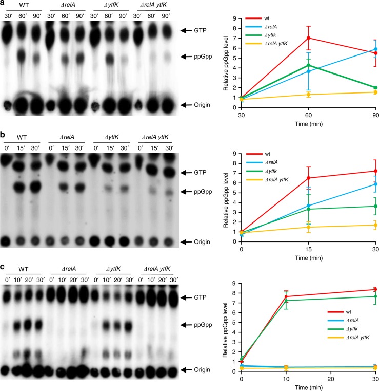
Previous work suggested that the transcriptional activator of the Pho regulon (PhoB) or one of the Pho products is involved in the control of (p)ppGpp accumulation during phosphate (Pi) starvation28. Since ytfK is a member of the Pho regulon27,29 and is therefore induced during Pi starvation (Supplementary Fig. 3a), we naturally tested the hypothesis that YtfK could play a physiological role in (p)ppGpp accumulation under this condition. As shown in Fig. 3a, Pi starvation triggers fast accumulation of (p)ppGpp in wild-type (WT) E. coli strain. A similar accumulation is observed in a ΔrelA strain, consistent with the previous observation that E. coli cells starved for Pi accumulates (p)ppGpp in a SpoT-dependent manner22,30. Interestingly, deletion of ytfK reduces the accumulation of (p)ppGpp in the WT strain. Remarkably, this accumulation is abolished in the ΔrelA ytfK strain. Taken together, our results show that YtfK is required to maintain normal (p)ppGpp level during Pi starvation (Fig. 3a).
Fig. 3. ytfK is required for normal accumulation of (p)ppGpp during phosphate and fatty acid starvation.
Exponentially growing cells of MG1655 (WT) and isogenic deletion strains, ΔrelA, ΔytfK, and ΔrelA ytfK were challenged for different starvations. Samples were taken at the indicated time, after being washed and resuspended in low-phosphate medium to induce phosphate starvation (a), after addition of 250 µg/mL cerulenin to induce fatty acid starvation (b), and after addition of 500 µg/mL L-Valine to induce amino acid starvation (c) (see the Methods section). Nucleotides were extracted and separated by TLC. Representative autoradiograph of the TLC is shown on the left, and quantification is presented on the right panel. Error bars indicate the standard deviations of averages of four independent experiments for phosphate starvation, six independent experiments for fatty acid starvation, and three independent experiments for amino acid starvation. Source data are provided as a Source Data file.
The ability of SpoT to sense many sources of nutrient stress other than amino acid starvation has long been puzzling. However, an important mechanism allowing SpoT to sense FA synthesis limitation involving the acyl carrier protein (ACP) has been previously described23,31,32. We therefore tested the intriguing possibility that YtfK may also be required for (p)ppGpp synthesis under FA starvation. As shown in Fig. 3b, we observed a rapid and similar production of (p)ppGpp in cells of WT and the ΔrelA strain following FA starvation (induced by the addition of cerulenin, a specific inhibitor of FA synthesis), consistent with previous observations that FA starvation triggers SpoT-dependent (p)ppGpp synthesis in presence of amino acids20,23. Importantly, deletion of ytfK impaired the accumulation of (p)ppGpp in response to FA starvation. Moreover, we observed that YtfK rapidly accumulated during FA starvation (Supplementary Fig. 3b). Taken together, these results point to an important role of YtfK in (p)ppGpp accumulation under this condition.
To further characterize the role of YtfK in (p)ppGpp physiology, we assessed its importance during amino acid starvation, a signal well known to trigger RelA-dependent (p)ppGpp synthesis18. To test this, isoleucine starvation was provoked by the addition of valine33. Under these conditions, we observed a rapid accumulation of (p)ppGpp in the WT strain. As expected, (p)ppGpp does not accumulate in the ΔrelA strain. Interestingly, we do not detect any differences in (p)ppGpp accumulation in the cells of a ΔytfK strain (Fig. 3c). Moreover, YtfK protein does not seem to accumulate after amino acid starvation (Supplementary Fig. 3c). These observations suggest that YtfK is not required to trigger RelA (p)ppGpp synthesis during amino acid starvation (Fig. 3c). Taken together, our results show that YtfK is essential for full accumulation of (p)ppGpp during stresses known to trigger SpoT-dependent synthesis of (p)ppGpp.
YtfK enhances cell survival
Cerulenin is commonly considered as a bacteriostatic antibiotic. However, a recent study revealed that cerulenin becomes bactericidal concomitantly with a loss of cell envelope integrity in a strain unable to accumulate (p)ppGpp34. We therefore investigated the role of YtfK in cerulenin tolerance. As previously observed, the plating efficiency of WT cells was unaffected after 4 h of cerulenin treatment, while the plating efficiency of ppGpp0 strain drops by more than 100,000-fold (Fig. 4a)34. Deletion of relA or ytfK does not significantly affect cerulenin tolerance (Fig. 4a). However, the plating efficiency of ΔrelA ytfK double-mutants drops by more than 10,000-fold after 4 h of cerulenin treatment, and this phenotype is trans-complemented by pEG220-ytfK. Interestingly, we also observed that while cerulenin efficiently inhibits cell growth, this inhibition is sharper in the WT and ΔrelA strains than what observed in the ytfK mutants and in the ppGpp0 strain. These results suggest that (p)ppGpp level contributes to growth arrest during FA starvation (Figs. 3b, 4b; Supplementary Fig. 4a). Therefore, our results show that YtfK contributes to cerulenin tolerance, and further underscores the role of (p)ppGpp as a guardian of cell viability in the absence of FA synthesis34.
Fig. 4. YtfK contributes to antibiotic tolerance.
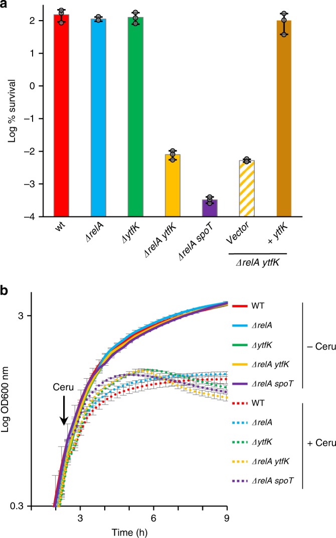
a YtfK is required for cerulenin survival in absence of relA. Growing cells of MG1655 (WT) and isogenic deletion strains ΔrelA, ΔytfK, ΔrelA ytfK, ΔrelA spoT, and ΔrelA ytfK harboring the pEG220 and the ΔrelA ytfK harboring the pEG220-ytfK (with native promoter) were exposed to 500 µg/mL of cerulenin for 4 h (see the Methods section). Percentage of survival (log scale) after 4 h is shown. Error bars indicate the SDs of averages of three independent experiments. b Growth curve of WT and isogenic deletion strains ΔrelA, ΔytfK, ΔrelA ytfK and ΔrelA spoT, in presence (dashed line) or absence (solid line) of cerulenin. Overnight culture were 1000 times diluted in LB, and growth was monitored at 600 nm using a microplate reader. At the indicated time, 250 µg/ml of cerulenin was added. Error bars indicate the SDs of averages of three independent experiments. Source data are provided as a Source Data file.
Finally and consistent with the proposed role of (p)ppGpp as factor contributing to antibiotic tolerance34–39, we observed that cerulenin pretreatment renders WT and ΔrelA mutant cells 10,000-fold more tolerant to ampicillin, but fails to substantially protect the ΔytfK mutant cells (Supplementary Fig. 4b).
YtfK can interact with SpoT and modulate its activity
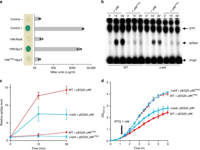
Several protein partners of the E. coli SpoT have been identified, and regulation of SpoT activities through protein–protein interactions have been reported23,24,40. We therefore tested whether YtfK is able to interact with SpoT in vivo using a bacterial two-hybrid (BTH) assay41. To this end, the complementary T18 and T25 domains of Bordetella pertussis adenylate cyclase were fused to N-terminus coding sequences of YtfK and SpoT and transformed in the cya-deficient E. coli strain BTH101. When this strain is co-transformed with both plasmids harboring T18 and T25 fusions, SpoT displayed a strong interaction in vivo with YtfK in both vector combinations. (Fig. 5a; Supplementary Fig. 5a).
Fig. 5. YtfK can interact with SpoT and trigger (p)ppGpp synthesis.
a YtfK interacts with SpoT in BTH assays. Briefly, overnight culture of BTH101 strain harboring pUT18c ytfK (or pUT18c ytfKP42L) and pKT25-spoT (or pKT25 relA) were spotted on X-Gal agar base plates (see the Methods section). The blue color indicates a positive interaction. The results of the β-galactosidase assays using the same strains are shown on the horizontal graphs on the right. Error bars indicate the SDs of averages of three independent experiments. b In vivo (p)ppGpp accumulation following ectopic expression of both ytfK and ytfKP42L. WT strain or ΔrelA mutant carrying ytfK or ytfKP42L on pEG25 were grown exponentially in phosphate MOPS minimal medium (see the Methods section). Samples were collected before and after ytfK induction (1 mM IPTG) prior to nucleotides extraction and were then separated by TLC. Representative autoradiograph of the TLC plates is shown and quantification is provided in (c). Error bars indicate the standard deviations of averages of three independent experiments. d The intracellular level of (p)ppGpp induced by ytfK overexpression controls the growth rate. Growth curve of WT (red) strain or ΔrelA mutant (blue) carrying ytfK (solid lane) or ytfKP42L (dashed lane) on pEG25 in the LB medium. Overnight culture was diluted 100 times, and growth was monitored at 600 nm. At the indicated time, 1 mM of IPTG was added to induce ytfK expression. Error bars indicate the standard deviations of averages of three independent experiments. Source data are provided as a Source Data file.
Even though SpoT and RelA share clear sequence homologies and similarities, no interaction is detected between YtfK and full-length RelA by bacterial two-hybrid assay (Fig. 5a; Supplementary Fig. 5b, c). To further gain insight into the YtfK-SpoT interaction and its role in (p)ppGpp synthesis, we randomly mutagenized ytfK. One mutant that had proline 42 substituted with leucine (P42L), exhibited impaired interaction with SpoT (Fig. 5a). Both T18-YtfK and T18-YtfKP42L recombinant proteins were correctly expressed as shown by western blot with Anti-CyaA (Supplementary Fig. 5d). Overexpression of ytfK but not ytfKP42L suppressed the growth defect of ΔrelA mutant on SMG plates (Supplementary Fig. 6a). This result predicts that the lack of interaction with SpoT abrogates the stimulatory effect of YtfK on (p)ppGpp accumulation. Indeed, it is the case. As judged by TLC analysis, ytfKP42L failed to promote (p)ppGpp synthesis when overexpressed in either WT or in ΔrelA strains in contrast to the massive accumulation of (p)ppGpp observed when the WT copy of ytfK is induced (Fig. 5b, c).
Consistent with the proposed role of SpoT and (p)ppGpp in controlling the bacterial growth rate42,43, we observed that induction of ytfK with 1 mM of IPTG strongly reduces the growth rate in both WT and ΔrelA strains, but not in the ppGpp0 strain (Fig. 5d; Supplementary Fig. 6b). Interestingly, the tougher growth rate control observed in WT strain is correlated to a higher (p)ppGpp level (Fig. 5c, d). Moreover, even if YtfK and YtfKP42L are produced at similar levels (Supplementary Fig. 6c, d), induction of ytfKP42L does not measurably affect the growth rate (Fig. 5d). Taken together, our results support the notion that a specific interaction between SpoT and YtfK controls SpoT activity, both during exponential growth (Fig. 5) and during nutrient stresses (Fig. 3).
YtfK can interact with the catalytic domains of SpoT
Using the BTH assay, we determined which domain(s) of SpoT are involved in the interaction with YtfK. The RSH protein family has very similar domain structures with the N-terminal half of the protein comprising the synthetase (SYNTH) and hydrolase activities (HD) and the C-terminal part, involved in the regulation of these activities, encompassing a Thr-tRNA synthetase, GTPase, and SpoT domain (TGS domain); a helical domain; a domain containing conserved cysteines (CC domain); and an Aspartokinase, Chorismate mutase, and TyrA domain (ACT domain) (Fig. 6)44. To test which of these domains are required for YtfK interaction, we constructed several truncated SpoT proteins fused to the T25 domain of B. pertussis adenylate cyclase (Fig. 6).
Fig. 6. YtfK can interact with the catalytic domain of SpoT in vivo.
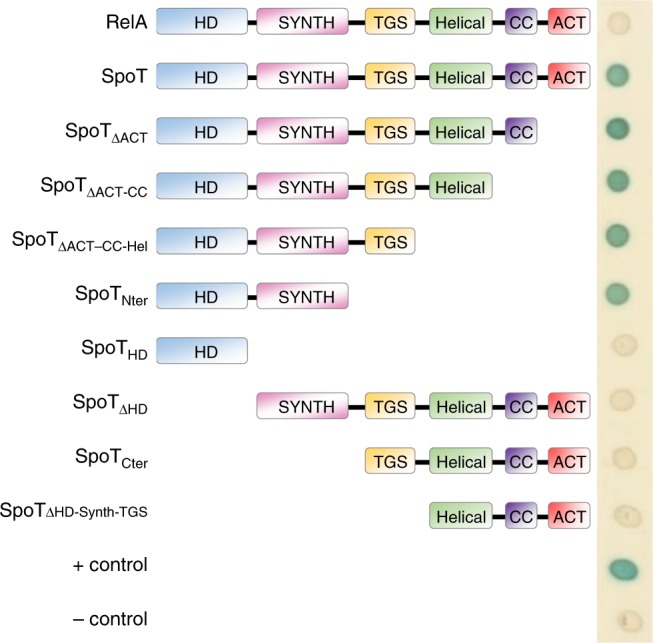
Bacterial two-hybrid assay with YtfK and several truncated version of SpoT. BTH101 strain harboring pUT18c ytfK and truncated pKT25 spoT were spotted on X-Gal agar base plates (see the Methods section). The blue color indicates a positive interaction. This experiment was repeated four times with identical results.
The analysis revealed that YtfK interacts with the N-terminal half of SpoT comprising the catalytic domains (Fig. 6). Moreover, none of the five domains encompassing the C-terminal regulatory part of SpoT are required for this interaction. However, truncated SpoT proteins fusion lacking either the synthetase domain or the hydrolase domain fail to interact in vivo with YtfK (Fig. 6; Supplementary Fig. 7a, b). Thus, both catalytic domains seem to be involved in the interaction with YtfK. Interestingly, the interaction with the N-terminal enzymatic half of SpoT seems specific, because YtfK does not interact with the homologous region of RelA by BTH assay (Supplementary Fig. 7c).
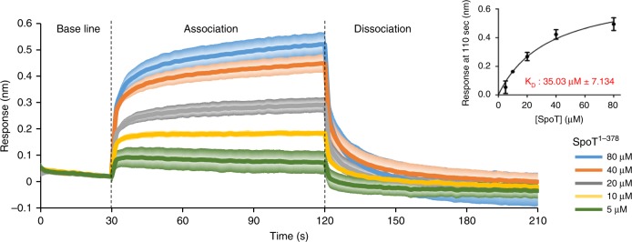
To confirm the SpoT-YtfK in vivo interaction revealed by BTH assay, we used BioLayer interferometry (BLI), an in vitro protein–protein interaction approach. SpoT catalytic domains (SpoT1–378) and YtfK proteins were produced and purified by two consecutive affinity and size-exclusion chromatography experiments (Supplementary Fig. 8). YtfK was biotinylated and immobilized on streptavidin biosensors as the ligand. Interaction experiments were performed with purified SpoT1–378 as the analyte. Upon addition of SpoT, a concentration-dependent association is recorded and decreases during the washing step of the sensor (dissociation step) (Fig. 7). These results indicate that YtfK interacts in vitro with the catalytic domains of SpoT with an estimated dissociation constant (KD) of 35 µM (Fig. 7). Moreover, YtfK does not interact with the homologous N-terminal enzymatic half of RelA in vitro, supporting the specificity of interaction between YtfK and SpoT (Supplementary Fig. 9). Taken together, our results suggest that the catalytic domains of SpoT are necessary and sufficient to interact with YtfK.
Fig. 7. YtfK can interact with the catalytic domain of SpoT in vitro.
BioLayer interferometry assay. The graph shows the subtracted reference binding responses during the association and dissociation of SpoT on YtfK. For kinetic titration analysis: increasing concentrations of recombinant SpoT1–378 (5–80 μM) were bounded to biotinylated YtfK (2.5 µM). Error bars (presented in degraded colors) indicate the standard deviations of averages of three experiments. The inset graph shows the specific BlI response (nm) 10 s before the end of association as a function of SpoT1–378 concentration (5–80 µM). Each data point (mean +/− SD) is the result from triplicates studies used to determine dissociation constant KD. Source data are provided as a Source Data file.
Discussion
In most gamma and beta-proteobacteria, to which E. coli belongs, the stringent response is orchestrated by two proteins RelA and SpoT encoding, respectively, for the (p)ppGpp synthase I and II activity. The metabolic cycle of (p)ppGpp is also further driven by the hydrolase activity encoded by SpoT, which is crucial for balancing the intracellular level of (p)ppGpp. Therefore, the molecular mechanisms regulating the switch between SpoT synthetase and hydrolase activities constitute a fine-tuning point of control for stress response and adaptation in E. coli. In this report, we discovered an important molecular mechanism regulating (p)ppGpp accumulation in E. coli. This regulation may involve physical interaction between the catalytic domains of SpoT and the protein YtfK. YtfK is a poorly characterized small basic protein specific to gamma-proteobacteria that seems limited to orders of Enterobacteriales, Vibrionales, Aeromonadales, Pasteurellales, and Alteromonadales45. We show that YtfK is important to maintain normal (p)ppGpp levels in response to both FA and phosphate starvation (Fig. 3). This regulation seems particularly important for cell survival. Indeed, we observed that ytfK actively contributes to cell viability under FA starvation (Fig. 4a). Moreover, it has been recently observed that deletion of ytfK also affects cell viability under phosphate starvation and H2O2 stress46. The stringent response is also recurrently linked to antibiotic tolerance and virulence. In this respect and consistent with the role of YtfK in controlling SpoT and (p)ppGpp levels, YtfK also contributes to stress-induced antibiotic tolerance (Supplementary Fig. 4b).
RelA and SpoT evolved from a common ancestor9, however, despite the same domains organization and strong sequence homologies (30.5/64% identity/similarity), we were not able to detect physical interaction between YtfK and RelA (Fig. 5a; Supplementary Fig. 9), suggesting a specific physiological role of YtfK in triggering SpoT activities. The lack of functional interaction between RelA and YtfK is further supported by the observation that YtfK does not affect the level of (p)ppGpp under amino acid starvation, a signal well known to trigger (p)ppGpp synthesis by RelA18 (Fig. 3c). However, we observed that YtfK triggers a stronger accumulation of (p)ppGpp when overexpressed in WT compared with the ΔrelA strain (Fig. 5b). Moreover, ectopic expression of ytfK reduced the growth rate of WT even more tightly to that observed in ΔrelA strain (Fig. 5d). Therefore, RelA also participates to the accumulation of (p)ppGpp promoted by YtfK. Even if we cannot rule out a direct activation of RelA by YtfK under these conditions, we suggest that this effect is rather indirect by residual activation of RelA. Indeed, it is well described that the hydrolysis activity of SpoT is required for balancing (p)ppGpp level in the presence of RelA, and disruption of the spoT gene in E. coli is therefore lethal12. Moreover, it is currently admitted that RSHs avoid to simultaneously synthesize and degrade (p)ppGpp primarily to prevent futile cycle10. Thus, we propose that YtfK pushes the catalytic balance of SpoT toward (p)ppGpp synthesis rather than hydrolysis therefore enabling residual activation of RelA to maximize the alarmone production. Therefore, the regulation of SpoT activities by YtfK points to an additional layer of regulation to the current stringent response model.
SpoT functions as a central protein that integrates various nutritional stress signals12,20–22. How different types of nutrient starvation can use the same or independent signal transduction pathways to trigger SpoT-dependent (p)ppGpp accumulation remains still unclear. It has been shown that the ACP transduces the status of FA metabolism to SpoT, thereby triggering (p)ppGpp accumulation in response to FA starvation23,31,32. Moreover, FA metabolism could also be the transducing signal that triggers SpoT during carbon starvation. Indeed, carbon depletion would lead to FA starvation through a decrease of the acetyl-CoA pool that is supplied by glycolysis47. Our finding that YtfK, in addition to the well-described role of ACP, is required for (p)ppGpp accumulation under FA starvation renders more sophisticated the current model for the switch linking SpoT to fatty acid metabolism23,31,32. Interestingly, YtfK protein level plays an important role for adjusting intracellular (p)ppGpp level. Indeed, ectopic expression of ytfK is sufficient to trigger SpoT-dependent (p)ppGpp accumulation in absence of nutritional stresses (Figs. 1c, 2). Moreover, ytfK is rapidly expressed under Pi and FA starvation, and is required for full (p)ppGpp accumulation under these conditions (Fig. 3a, b; Supplementary Fig. 3a, b) strongly arguing that YtfK protein level is indeed important to trigger the stringent response under nutrient stress conditions. However, how the control of YtfK protein level becomes critical for adjusting (p)ppGpp level in response to other stresses known to trigger SpoT-dependent (p)ppGpp accumulation remains unknown. Interestingly, ytfK is also induced and involved in H2O2 tolerance by stimulating expression of at least the detoxifying enzyme encoded by katG46. Whether this stimulation is direct or indirect through activation of SpoT remains to be uncovered.
The CTD region of RSH enzymes are thought to play pivotal roles in sensing nutrient starvation and to controlling the reciprocal regulation of the two enzymatic states10,48,49. Consistent with a regulatory role of the CTD, the TGS domain is required for ACP and Rsd binding to SpoT of E. coli. Moreover, the ACT domain is also needed for interaction with the phosphorylated enzyme IIANtr to promote (p)ppGpp accumulation in Caulobacter crescentus in response to glutamine deprivation50,51. In contrast, we observed that the NTD region of SpoT is necessary and sufficient for YtfK binding (Figs. 6, 7), suggesting a distinct mode of regulation. The crystal structure of the HD and SYNTH domains of the Rel/Spo homologue from Streptococcus equisimilis has revealed two conformations of the enzyme corresponding to reciprocal activity states: (p)ppGpp-hydrolase-OFF/synthetase-ON and (p)ppGpp-hydrolase-ON/synthetase-OFF11. A docking experiment using modeled structures predicts that YtfK might bind SpoT at the small synthetase/hydrolase interdomain interface (Supplementary Fig. 10). The molecular basis of the apparent regulation of SpoT by YtfK can only be speculated upon. However, based on our preliminary in vitro results (Fig. 7), we propose that YtfK might directly interact with SpoT to trigger conformational changes in the catalytic domains. Alternatively, additional indirect effects of YtfK on SpoT activity are also possible, and further structural and functional analysis are currently under investigation to test whether direct binding of YtfK at the interface between the opposing site of SpoT affects the antagonistic regulation of (p)ppGpp synthesis and degradation.
Methods
Bacterial strains and plasmids
Bacterial strains and plasmids are listed in Supplementary Table 1 and Supplementary Data 1, respectively. DNA oligonucleotides are listed in Supplementary Data 2.
MG1655 YtfK::mNeonGreen construction
The chromosomal YtfK::mNeonGreen fusion was generated by lambda red recombination adapted from Blank et al.52. The DNA fragment was amplified by PCR using primers pEJM230/231 with plasmid pWRG100 as a template52. The resulting PCR product was electroporated into MG1655 cells after 1 h of lambda recombinase expression from pKD46 plasmid53. After 1 h of phenotypic expression, the cells were plated on nutrient agar (NA) containing 25 μg/mL chloramphenicol. Selected colonies contained a ytfK::sce-I::cm cassette which has been then newly transduced in MG1655 cells harboring the pWRG99 plasmid52. The chloramphenicol cassette was then removed by counter selection using lambda red recombination to insert a PCR product complementary to the flanking regions of the sce-I::cm cassette on the chromosome. The PCR product was generated with primers pEJM244/245 on pJF119EH encoding mNeonGreen as a template (Generous gift from Axel Magalon’s lab). PCR product was then electroporated into MG1655 ytfK::sce-I::cm expressing lambda recombinase from pWRG9952, and after 1 h of phenotypic expression serially diluted and plated on NA containing 100 μg/mL ampicillin and 1 μg/mL anhydrotetracycline. pWRG99 also encoded for the meganuclease I-Sce-I under the control of an anhydrotetracycline (aTc) inducible promoter. The proper integration of the mNeonGreen was confirmed by diagnostic PCR and then sequenced.
Plasmid construction
pEG25 relA and spoT: Due to their intrinsic activities, RelA and SpoT overproduction become toxic. To circumvent this problem and therefore be able to express relA in ΔrelA spoT strain, we have engineered a robust system to tightly regulate intracellular level of RelA. First, we have constructed the pEG25 plasmid which contains the Pt5 promoter from the pCA24N. This promoter contains three Lac operators, and is therefore less leaky (see below). Most importantly, we played with constants that contribute with the process of translation initiation. Indeed by engineering the Shine-Dalgarno sequence, the initiation codon and the spacer between them we were able to tightly regulate the intracellular level of RelA (SD4 TTG) and SpoT (SD8 TTG). The chosen Shine-Dalgarno sequences are described in the primers list.
pEG25: pMG25 derivative where the PLac promoter has been replaced by Pt5 from the pCA24N26. Pt5 promoter has been amplified by PCR using primers EG186/EG187, digested with XhoI and EcoRI, and ligated in the plasmid digested with the same enzymes.
pEG25 derivatives: Genes inserted have been PCR amplified from the chromosomal DNA and with primers listed in the Supplementary Data 2, digested with EcoRI and BamHI and ligated in the pEG25 digested with the same enzymes.
pBbS2K: The original plasmid pBbS2K(RFP) has been digested with BglII/BamHI, and then self ligated. The resulting plasmid has been then used as a negative control.
pBbS2K(RFP) derivatives plasmids: Genes inserted have been PCR amplified from the chromosomal DNA with primers listed in the Supplementary Data 2, digested with EcoRI/BamHI, and ligated in the pBbS2K(RFP) plasmid digested with the same enzymes.
pEG220: pNDM220 (Amp) has been engineered and modified to a kanamycin plasmid. Kanamycin cassette has been amplified from pKD4 using pEG287/288 and transformed to DY331 strain with the lambda red recombineering system on the chromosome. Antibiotic cassette exchange has been checked by PCR and then sequenced.
pUT18C-derivative plasmids: Genes inserted have been PCR amplified from the chromosomal DNA (or mutated plasmid) with primers listed in the Supplementary Data 2, digested with XbaI and KpnI, and ligated in pUT18C digested with the same enzymes.
pKT25-derivative plasmids: Genes inserted have been PCR amplified from the chromosomal DNA (or from mutated plasmid) with primers listed in Supplementary Data 2, digested with XbaI and KpnI, and ligated in pKT25 digested with the same enzymes.
pET28a + -TRX: TRX sequence, including thioredoxin-6his-tev, has been amplified from pLic07 with primers listed in Supplementary Data 2, digested with NcoI and BamHI, and ligated in pET28a + digested with the same enzymes.
pET28a + -ytfK: ytfK gene has been amplified from E. coli chromosomal DNA with primers listed in Supplementary Data 2, digested with BamHI and HindIII, and ligated in pET28a + -TRX digested with the same enzymes.
Media and antibiotics
Luria—Bertani broth from OXOID (LP0021B; LP0042B) was prepared as previously described54. MOPS minimal medium was prepared as previously described55, and was supplemented with 2 mM phosphate (or 0.4 mM phosphate for uniform P32 labeling), 0.2% glucose, and 40 µg/ml (each) of the 20 amino acids. Terrific broth (TB) :1.2% peptone, 2.4% yeast extract, 72 mM K2HPO4, 17 mM KH2PO4, and 0.4% glycerol.
Rich solid medium used in this study is NA from Oxoid (CM0003B). M9 minimal medium: M9 salt (60 mM Na2HPO4; 22 mM KH2PO4; 8 mM NaCl; 20 mM NH4Cl) 1 mM MgSO4; 100 µM CaCl2; thiamine 1 µg/mL; 0.2% glucose. SMG solid minimal medium: M9 medium supplemented with 40 µg/mL of L-serine; 40 µg/mL L-methionine; 40 µg/mL glycine; and 15 g/L of agar.
When required, media was supplemented with antibiotic (50 µg/ml ampicillin; 50 µg/ml chloramphenicol; 25 µg/ml kanamycin). Expression of protein from plasmids carrying PLac promoter was induced by addition of IPTG.
Suppression of the nongrowing phenotype of ΔrelA on SMG
A pooling mixture of the 6His-tagged genes (minus GFP) ASKA plasmid library26 has been transformed into a ΔrelA deletion strain. Cell were then spread on SMG plates supplemented with chloramphenicol and different concentration of IPTG (0, 100, and 200 µM) to induce the expression of ASKA plasmid-encoded genes and incubated for 36 h at 37 °C. Plasmids that produced colonies under this nonpermissive condition were sequenced, and we identified ytfK, metL, sdaB, ymgB, yaaA, thil, murI, and relA as candidate genes that enable growth in a ΔrelA strain on high concentration of serine, methionine, and glycine (SMG)25.
Bacterial two-hybrid assay (BTH)
We used BTH assay to test protein–protein interaction in vivo. This method is based on the reconstitution of the adenylate cyclase from Bordetella pertussis41. Proteins of interest were fused to T18 and T25 fragments of adenylate cyclase using pUT18c and pKT25. After co transformation in BTH101 strain, the plates were incubated overnight at 30 °C. Colonies were re-streaked overnight at 30 °C. Then, 1 mL of LB supplemented with ampicillin and kanamycin was inoculated in the morning and incubated at 30 °C with shaking for 8 h. In total, 5 µl of undiluted cultures were then spotted on NA agar plates supplemented with X-Gal 40 µg/mL with different concentrations of IPTG. For β-galactosidase experiments, the same inoculum has been used, and β-galactosidase activity was determined as described by Miller with the use of the TECAN microplate reader to follow OD600 and OD420 over time.
Screening for loss of interaction by ytfK random mutagenesis
pUT18c-ytfK plasmid has been mutagenized randomly through propagation in the mutator XL1-Red strain from Agilent Technologies. XL1-Red strain are deficient in three of the primary DNA repair pathways (mutS, mutD, and mutT). BTH101 harboring pKT25-spoT was then transformed with the resulting pUT18c-ytfK mutated plasmid library and screened on X-Gal plates. White colonies were selected and sequenced.
In vivo (p)ppGpp measurement
(p)ppGpp accumulation was measured in cells grown and uniformly labeled with 32P in MOPS minimal medium supplemented as described below. In total, 100 µl samples were taken at the indicated times, and 40 µl of 21 M formic acid was added to stop the reaction. Samples were then left on ice for 20 min, and spun down at 14,000 g for 20 min at 4 °C. In all, 5 µl of each sample were loaded on PEI Cellulose TLC plates (purchased from MercK-Millipore). Plates were revealed by PhosphoImaging (GE Healthcare) and analyzed using ImageQuant software (GE Healthcare). Quantification was performed using FIJI software. ppGpp levels were normalized to that of the WT strain at time zero for each starvation condition.
For the phosphate starvation, overnight cultures were grown in MOPS 2 mM phosphate supplemented with 40 µg/mL of the 20 amino acids and 0.2% glucose. Overnight cultures were then diluted 1/100-fold in the same medium and incubated at 37 °C with shaking up to OD600: 0.4–0.5. Cells were harvested and washed once in MOPS without phosphate and then resuspended in MOPS with 50 µM phosphate and labeled with 150 µCi of 32P.
For fatty acid starvation, overnight cultures were grown in MOPS 2 mM phosphate supplemented with 40 µg/mL of the 20 amino acids and glucose 0.2%. Overnight cultures were then diluted 1/100-fold in the same medium with 0.4 mM phosphate instead of 2 mM and incubated at 37 °C with shaking up to OD600: 0.4–0.5. Cells were then diluted down to OD600: 0.05 in the same medium, labeled with 150 µCi of 32P and grew up to OD600: 0.25. Fatty acid starvation is then induced by addition of 250 µg/mL of cerulenin.
For amino acid starvation, cells were grown in MOPS 2 mM phosphate supplemented with 0.2% glucose without amino acids. Overnight cultures were then diluted 1/100-fold in the same medium with 0.4 mM phosphate instead of 2 mM and incubated at 37 °C with shaking up to OD600: 0.4–0.5. Cells were then diluted down to OD600: 0.05 in the same medium, labeled with 150 µCi of 32P and grew up to OD600: 0.25. Amino acid starvation is then induced by addition of 500 µg/mL of L-Valine.
For yfK overexpression, WT strain and ΔrelA mutant harboring pEG25 ytfK or pEG25 ytfK-6his and pEG25 ytfKP42L or pEG25 ytfKP42L-6his were grown in MOPS 2 mM phosphate supplemented with 40 µg/mL of the 20 amino acids and glucose 0.2%. Overnight cultures were diluted 1/100-fold in MOPS 0.4 mM phosphate and incubated at 37 °C with shaking up to OD600: 0.4–0.5. Cells were then diluted down to OD600: 0.05 in the same medium, labeled with 150 µCi of 32P and grew up to OD600: 0.25. ytfK expression was then induced by the addition of 1 mM of IPTG.
Cerulenin antibiotic tolerance
Strains of WT, ΔrelA, ΔytfK, ΔrelA ytfK, ΔrelA spoT, ΔrelA ytfK harboring the pEG220 and the ΔrelA ytfK harboring the pEG220-ytfK were tested for the effect of 500 µg/mL of cerulenin. Overnight cultures were diluted 1000-fold in 10 ml of fresh LB medium, and incubated ~2.5 h at 37 °C with shaking (typically reaching 1–2 × 10⁸ cells/mL). For complementation assay, the medium was supplemented by 25 µg/ml of kanamycin. Then aliquots of 1 mL were transferred into a 15 mL falcon tube containing 10 µL of cerulenin 50 mg/mL and incubated 4 h at 37 °C with shaking. For determination of CFUs, 200 µl aliquots were removed, the cells harvested, resuspended in fresh medium, serially diluted, and plated on solid NA medium. Surviving fraction was calculated by dividing the number of CFU/ml in the culture after 4 h of incubation with the cerulenin by the number of CFU/ml in the culture before adding the cerulenin.
Ampicillin tolerance after cerulenin pre-treatments
WT, ΔrelA, ΔytfK, ΔrelA ytfK, and ΔytfK harboring the pEG220 and the ΔytfK harboring the pEG220-ytfK were challenged against ampicillin treatment (100 µg/ml) after being pre-treated with 250 µg/mL of cerulenin to induce (p)ppGpp synthesis in a SpoT-dependent manner. Overnight cultures were diluted 1000-fold in 10 ml of fresh LB medium and incubated for about 2.5 h at 37 °C with shaking (typically reaching 1–2 × 10⁸ cells/mL). For complementation assay, medium was supplemented with 25 µg/ml of kanamycin. Then 3 mL aliquots were transferred into a 50 -mL falcon tube containing 15 µL of cerulenin 50 mg/mL and incubated during 30 min at 37 °C with shaking and then treated for 4 h with ampicillin (100 µg/ml). For determination of CFUs at the indicated time points, 800 µl aliquots were removed, the cells harvested, resuspended in fresh medium, serially diluted, and plated on solid NA medium. Surviving fraction was calculated by dividing the number of CFU/ml in the culture after 4 h of incubation with ampicillin by the number of CFU/ml in the culture before adding ampicillin.
Protein production
For SpoT1–378 and RelA1–396 (catalytic domains) production, BL21(DE3) competent cells were transformed with the pMG25-SpoT1–378 -6His or pEG25-RelA1–396 -6His and plated on NA plates containing 100 mg/L ampicillin. Several colonies were picked up and inoculated into 100 ml of LB containing 100 mg/L ampicillin. The cultures were grown with shaking at 30 °C overnight. Overnight cultures were dispersed as inoculum of 20 mL aliquots into 1L of TB media with of 100 mg/L of ampicillin and shaken at 30 °C until OD600 reached 0.5, then cells were induced with 0.5 mM IPTG and incubated at 30 °C for 4 h.
For YtfK production, BL21 (DE3) competent cells were transformed by the pET28a-ytfK plasmid and plated on NA plates with 50 mg/L kanamycin. Several colonies were picked up and inoculated into 100 ml LB containing 50 mg/L kanamycin. The cultures were grown with shaking at 30 °C overnight. Overnight cultures were dispersed as inoculum of 20 ml aliquots into 1L LB media in the presence of 50 mg/L of kanamycin and shaken at 30 °C until OD600 0.4 unit, then cells were induced with 0.5 mM IPTG and incubated at 20 °C for 4 h.
Finally, cells were centrifuged at 9000 × g for 20 min at 4 °C. Dry cell pellet was stored at −80 °C.
Protein purification
For YtfK purification, cells were harvested from 4L culture, suspended in lysis buffer (Tris-HCl 50 mM pH 8.0, NaCl 300 mM, EDTA 1 mM, lysozyme 0.5 mg/mL, phenylmethylsulfonyl fluoride (PMSF)), DNase 20 μg/mL, and MgCl2 20 mM. The mixture was incubated for 1 h at 4 °C with gentle shacking and then subjected to two cycles of French-press lysis steps. The soluble fraction was obtained by centrifugation for 30 min at 200,000 × g. Recombinant proteins were purified by ion metal affinity chromatography using a 5-mL Nickel (HiTrapHP) Column on an ÄKTA pure 25 (GE healthcare) pre-equilibrated in Tris-HCl 50 mM pH 8.0, NaCl 300 mM, 10 mM imidazole (buffer A). After several washes in buffer A, 6 × His-tagged proteins were eluted in buffer A supplemented with 250 mM imidazole and immediately desalted using Hiprep 26/10 Desalting column pre-equilibrated with buffer A. The resulting desalted proteins were mixed with 0.2 mg/mL of TEV protease and incubated for 2 h at RT and then loaded onto a HisTrapTM column pre-equilibrated in buffer A, which selectively retains the TEV, TRX, and the uncleaved proteins and contaminants. Untagged YtfK was collected in the flow through, concentrated on a Centricon (Millipore; cutoff of 3 kDa), and passed through a HiLoad 16/600 Superdex 200 column pre-equilibrated with 50 mM Tris-HCl pH 8.0, 500 mM NaCl, 500 mM KCl, 2 mM β-mercaptoethanol, glycerol 2%. The purity of YtfK preparations was assessed by SDS–PAGE (Supplementary Fig. 8a, b) and spectrophotometrically [OD260/OD280] ratio below 0.52.
For SpoT1–378 and RelA1–396 purification, cells were harvested from 4L culture, then cell pellets were resuspended in lysis buffer composed of 50 mM Tris-HCl pH 8.0, 500 mM NaCl, 10 mM imidazole, 2 mM β-mercaptoethanol, 0.5% CHAPS, glycerol 2%, 1 mM EDTA, 0.5 mg/mL lysozyme, 1 mM phenylmethylsulfonyl fluoride, 20 µg/mL DNase, and 20 mM MgCl2. The mixture was incubated for 1 h at 4 °C with gentle shaking. Further French-Press lysis step was performed to ensure the complete cell lysis. Pellet and soluble fractions were separated by centrifugation for 30 min at 20,000 × g. The soluble fraction containing SpoT1–378 or RelA1–396 proteins were loaded at room temperature onto a 5-ml nickel column (HiTrapHP) using an ÄKTA pure 25 apparatus (GE healthcare) pre-equilibrated with equilibrium buffer 50 mM Tris-HCl pH 8.0, 500 mM NaCl, 10 mM imidazole, 2 mM β-mercaptoethanol, glycerol 2%, and the immobilized proteins were eluted in elution buffer 50 mM Tris-HCl pH 8.0, 500 mM NaCl, 500 mM imidazole, 2 mM β-mercaptoethanol, and glycerol 2%. Eluted proteins were immediately subjected to size-exclusion chromatography (SEC) purification using a HiLoad 26/600 Superdex 200 pg column pre-equilibrated with 50 mM Tris-HCl pH 8.0, 500 mM NaCl, 500 mM KCl, 2 mM β-mercaptoethanol, and glycerol 2%.
The purity of SpoT and RelA preparations was assessed by SDS–PAGE (Supplementary Fig. 8c–e), and spectrophotometrically [OD260/OD280] ratio below 0.61 for SpoT and below 0.57 for RelA.
Biolayer interferometry (BLI)
YtfK was biotinylated using the EZ-Link NHS-PEG4-Biotin kit (Perbio Science, France) with 1:1 ratio of YtfK and Biotin at 4 °C, respectively. The reaction was stopped, by removing the excess of the biotin using a Zeba Spin Desalting column (Perbio Science, France). BLI studies were performed at 25 °C using the Blitz apparatus (ForteBio, USA) with shaking at 2200 rpm with the following steps; 30 s baseline, 90 s association, and 90 s dissociation. Streptavidin biosensor tips (ForteBio, USA) were first hydrated with 0.2 ml of 50 mM Tris-HCl pH 8.0, 500 mM NaCl, 500 mM KCl, 2 mM β-mercaptoethanol, glycerol 2% for 10 min, and then loaded with 2.5 µM of biotinylated YtfK in the same buffer. To study the binding of YtfK to SpoT1–378 or RelA1–396, increasing concentrations of SpoT1–378 (5 to 80 µM) or RelA (40 to 120 µM) in interaction buffer (50 mM Tris-HCl pH 8.0, 500 mM NaCl, 500 mM KCl, 2 mM β-mercaptoethanol, glycerol 2%, and BSA 1 mg/ml) were used, and the association and dissociation phases were monitored, respectively, with constant agitation at 2200 rpm. To avoid the nonspecific binding of SpoT1–378 or RelA1–396 to the Streptavidin biosensors, the biosensors were incubated with 10 µg/mL biocytin in interaction buffer (50 mM Tris-HCl pH 8.0, 500 mM NaCl, 500 mM KCl, 2 mM β-mercaptoethanol, glycerol 2% BSA 1 mg/ml) for 90 s. In all experiments, the BLItz ProTM software performed a reference subtraction of the SpoT1–378 (or RelA1–396) protein response on the uncoated biosensors for each tested concentrations. The dissociation constants (KD), i.e., affinity of YtfK for SpoT, were estimated using the GraphPad Prism 5.0 software on the basis of the maximum responses in nm at 110 s, directly related to the concentration of SpoT1–378. The KD was estimated by plotting on the x-axis, the different concentration of SpoT1–378 and the different responses of SpoT1–378 (10 s before the end of the association) on the y-axis. For KD calculation, a nonlinear regression fit for xy analysis was used with a one site (specific binding) as a model which corresponds to the equation y = Bmax × x/(KD + x).
Western blot analysis
The western blot using Penta-His-HRP-conjugated against 6His-tagged proteins has been done following the recommendation of the company (Qiagen ref: 34460). Cells were grown exponentially at 37 °C in LB (OD600 0.5), then cells were induced with 200 µM of IPTG for 1 h. Sample was prepared, and equal amount of total proteins were loaded per lane on SDS–PAGE. Immunoblot analysis was performed with the antibody diluted 1/10,000 and visualized by using luminata crescendo HRP substrate (Millipore ref: WBLUR0100) and ImageQuant Las4000 (GE Healthcare).
The western blot experiment using anti-cyaA antibody against adenylate cyclase protein encoded by pUT18c plasmid has been performed using PBS + 0.1% Tween20 (PBST) for all steps of the immunoblot. The membrane was incubated in blocking buffer for 1 h (PBST + 5% milk). The first anti-cyaA antibody has been diluted 1/5.000 (Santa Cruz, ref: sc-13582) in PBST milk and incubated for 1 h at room temperaure. The secondary antibody (Sigma, ref: A4416) has been diluted 1/10.000 in PBST milk and incubated for 1 h at room temperature. Immunoblot was visualized by using luminata crescendo HRP substrate (Millipore, ref: WBLUR0100) and ImageQuant Las4000 (GE Healthcare). For sampling, MG1655 was transformed with the all different truncated versions of SpoT encoded by the pUT18c. Cells grew during 3H00 (OD: 0.5) at 30 °C in LB. Protein productions were induced by addition of 1 mM IPTG for 4H00. Sample was prepared and equal amount of total proteins were loaded per lane on SDS–PAGE.
Protein modeling and molecular docking simulation
The available atomic coordinates of RelA (PDB ID: 5KPV) from E. coli was used as a template to generate a model of E. coli SpoT (30.4%/64.3% identity/similarity) using SWISS-MODEL (a web-based integrated service for protein structure homology modeling)56. Similarly, the model of E. coli YtfK was generated using the NMR structure of the YtfK homologue from Shewanella oneidensis (PDB ID: 2JRO; 30.3%/65.8% identity/similarity with E. coli YtfK).
For the modeling of YtfK-SpoT1–378 binding interface, we used the HADDOCK2.2 webserver interface. To define Haddock run restraints, we used the surface exposed residues of both YtfK and SpoT as active residues that could be directly involved in the interaction. For SpoT and YtfK, we selected the surface exposed residues manually using PyMol (Supplementary Table 2). Three independent docking simulations were performed. For each run, ten clusters were generated, and classified based on their HADDOCK score. Five YtfK/SpoT models picked from the best five clusters are shown in Supplementary Fig. 10, and the parameters of the respective clusters generated by Haddock are shown in Supplementary Table 3.
Fluorescence microscopy and analysis
Overnight culture of WT and ytfK-L-mNeonGreen strains in MOPS minimal medium were diluted 1000 times in the same medium and incubated at 37 °C until OD600 nm reached 0.25. Cells were then starved using the same protocol used to measure (p)ppGpp level in vivo. The microscopy images were taken using an inverted epifluorescence microscope Nikon Eclipse TiE PFS (100 × oil objective NA 1.45 Phase Contrast) and a Hamamatsu Digital camera C11440 ORCA-Flash4.0 LT. Images were collected with NS elements software. Samples (3 µl of cells) were spotted on a 24 × 50 mm coverslip, and covered directly with a thin pad of minimal medium supplemented with 1.5% agarose. Twenty fields were taken for each point using the perfect focus system (PFS) and a motorized stage. The fluorescence images have been acquired with diascopy (DIA) and a GFP filter (482/35 nm excitation, 536/40 nm emission). The exposure time were 30 ms and 300 ms, respectively. The images were analyzed in TIFF format with the plugin MicrobeJ57 available for downloading in FIJI software.
A threshold was determined for each experiment to detect bacteria. Then, the backgrounds were subtracted with a rolling ball radius of 30 pixels, and the fluorescence of each conditions was measured with MicrobeJ. All the images have the same contrast and have been acquired and analyzed in the same way.
Reporting summary
Further information on research design is available in the Nature Research Reporting Summary linked to this article.
Supplementary information
Description of Additional Supplementary Files
Acknowledgements
This work was supported by the European Research Council starting grant (ERC StG) under the European Union’s Horizon 2020 and innovation program grant agreement no. 714934 “Stringency” to E.M. We thanks Axel Magalon for the generous gift of the pJF119EH encoding for the mNeonGreen. We thank Mike Cashel, Aurelia Battesti, Anne Galinier, and the members of the Maisonneuve group for stimulating discussions.
Source data
Author contributions
E.M. and E.G. conceptualized the study. E.M., E.G., P.G., D.B., B.D. and M.D. performed the experiments. E.M., E.G., P.G., D.B., B.D. and M.D. analyzed the data. E.M. and E.G. wrote the original draft of the paper. D.B., reviewed and edited the paper. E.M. acquired funding.
Data availability
All relevant data are available in this article and its Supplementary Information files. The source data underlying Figs. 1c, 3a–c, 4a, b, 5b–d and 7, and Supplementary Figs. 4b, 5d, 6b–d, 7b, and 9 are provided as a Source Data file. Any additional data relevant to this paper are available from the corresponding authors upon request.
Competing interests
The authors declare no competing interests.
Footnotes
Peer review information Nature Communications thanks Christiane Wolz and the other, anonymous, reviewer(s) for their contribution to the peer review of this work. Peer reviewer reports are available.
Publisher’s note Springer Nature remains neutral with regard to jurisdictional claims in published maps and institutional affiliations.
Contributor Information
Elsa Germain, Email: egermain@imm.cnrs.fr.
Etienne Maisonneuve, Email: emaisonneuve@imm.cnrs.fr.
Supplementary information
Supplementary information is available for this paper at 10.1038/s41467-019-13764-4.
References
- 1.Potrykus K, Cashel M. (p)ppGpp: still magical? Annu. Rev. Microbiol. 2008 doi: 10.1146/annurev.micro.62.081307.162903. [DOI] [PubMed] [Google Scholar]
- 2.Zuo Y, Wang Y, Steitz TA. The mechanism of E. coli RNA polymerase regulation by ppGpp is suggested by the structure of their complex. Mol. Cell. 2013 doi: 10.1016/j.molcel.2013.03.020. [DOI] [PMC free article] [PubMed] [Google Scholar]
- 3.Ross W, Vrentas CE, Sanchez-Vazquez P, Gaal T, Gourse RL. The magic spot: a ppGpp binding site on E. coli RNA polymerase responsible for regulation of transcription initiation. Mol. Cell. 2013 doi: 10.1016/j.molcel.2013.03.021. [DOI] [PMC free article] [PubMed] [Google Scholar]
- 4.Sanchez-Vazquez P, Dewey CN, Kitten N, Ross W, Gourse RL. Genome-wide effects on Escherichia coli transcription from ppGpp binding to its two sites on RNA polymerase. Proc. Natl Acad. Sci. USA. 2019 doi: 10.1073/pnas.1819682116. [DOI] [PMC free article] [PubMed] [Google Scholar]
- 5.Ross W, et al. PpGpp binding to a site at the RNAP-DksA interface accounts for its dramatic effects on transcription initiation during the stringent response. Mol. Cell. 2016 doi: 10.1016/j.molcel.2016.04.029. [DOI] [PMC free article] [PubMed] [Google Scholar]
- 6.Traxler MF, et al. The global, ppGpp-mediated stringent response to amino acid starvation in Escherichia coli. Mol. Microbiol. 2008 doi: 10.1111/j.1365-2958.2008.06229.x. [DOI] [PMC free article] [PubMed] [Google Scholar]
- 7.Durfee T, Hansen A-M, Zhi H, Blattner FR, Jin DJ. Transcription profiling of the stringent response in Escherichia coli. J. Bacteriol. 2008 doi: 10.1128/JB.01092-07. [DOI] [PMC free article] [PubMed] [Google Scholar]
- 8.Kanjee U, Ogata K, Houry WA. Direct binding targets of the stringent response alarmone (p)ppGpp. Mol. Microbiol. 2012 doi: 10.1111/j.1365-2958.2012.08177.x. [DOI] [PubMed] [Google Scholar]
- 9.Atkinson GC, Tenson T, Hauryliuk V. The RelA/SpoT homolog (RSH) superfamily: distribution and functional evolution of ppgpp synthetases and hydrolases across the tree of life. PLoS One. 2011 doi: 10.1371/journal.pone.0023479. [DOI] [PMC free article] [PubMed] [Google Scholar]
- 10.Mechold U, Murphy H, Brown L, Cashel M. Intramolecular regulation of the opposing (p)ppGpp catalytic activities of RelSeq, the Rel/Spo enzyme from streptococcus equisimilis. J. Bacteriol. 2002 doi: 10.1128/JB.184.11.2878-2888.2002. [DOI] [PMC free article] [PubMed] [Google Scholar]
- 11.Hogg T, Mechold U, Malke H, Cashel M, Hilgenfeld R. Conformational antagonism between opposing active sites in a bifunctional RelA/SpoT homolog modulates (p)ppGpp metabolism during the stringent response. Cell. 2004 doi: 10.1016/S0092-8674(04)00260-0. [DOI] [PubMed] [Google Scholar]
- 12.Xiao, H. et al. Residual guanosine 3′,5′-bispyrophosphate synthetic activity of relA null mutants can be eliminated by spoT null mutations. J. Biol. Chem. 266, 5980–5990 (1991). [PubMed]
- 13.Murray KD, Bremer H. Control of spoT-dependent ppGpp synthesis and degradation in Escherichia coli. J. Mol. Biol. 1996 doi: 10.1006/jmbi.1996.0300. [DOI] [PubMed] [Google Scholar]
- 14.An, G., Justesen, J., Watson, R. J. & Friesen, J. D. Cloning the spoT gene of Escherichia coli: identification of the spoT gene product. J. Bacteriol. 137, 1100–1110 (1979). [DOI] [PMC free article] [PubMed]
- 15.Brown A, Fernández IS, Gordiyenko Y, Ramakrishnan V. Ribosome-dependent activation of stringent control. Nature. 2016 doi: 10.1038/nature17675. [DOI] [PMC free article] [PubMed] [Google Scholar]
- 16.Arenz S, et al. The stringent factor RelA adopts an open conformation on the ribosome to stimulate ppGpp synthesis. Nucleic Acids Res. 2016 doi: 10.1093/nar/gkw470. [DOI] [PMC free article] [PubMed] [Google Scholar]
- 17.Loveland AB, et al. Ribosome.RelA structures reveal the mechanism of stringent response activation. eLife. 2016 doi: 10.7554/eLife.17029. [DOI] [PMC free article] [PubMed] [Google Scholar]
- 18.Haseltine W. A., Block R. Synthesis of Guanosine Tetra- and Pentaphosphate Requires the Presence of a Codon-Specific, Uncharged Transfer Ribonucleic Acid in the Acceptor Site of Ribosomes. Proceedings of the National Academy of Sciences. 1973;70(5):1564–1568. doi: 10.1073/pnas.70.5.1564. [DOI] [PMC free article] [PubMed] [Google Scholar]
- 19.Winther Kristoffer Skovbo, Roghanian Mohammad, Gerdes Kenn. Activation of the Stringent Response by Loading of RelA-tRNA Complexes at the Ribosomal A-Site. Molecular Cell. 2018;70(1):95-105.e4. doi: 10.1016/j.molcel.2018.02.033. [DOI] [PubMed] [Google Scholar]
- 20.Seyfzadeh M, Keener J, Nomura M. spoT-dependent accumulation of guanosine tetraphosphate in response to fatty acid starvation in Escherichia coli. Proc. Natl Acad. Sci. USA. 1993 doi: 10.1073/pnas.90.23.11004. [DOI] [PMC free article] [PubMed] [Google Scholar]
- 21.Vinella D, Albrecht C, Cashel M, D’Ari R. Iron limitation induces SpoT-dependent accumulation of ppGpp in Escherichia coli. Mol. Microbiol. 2005 doi: 10.1111/j.1365-2958.2005.04601.x. [DOI] [PubMed] [Google Scholar]
- 22.Spira B, Silberstein N, Yagil E. Guanosine 3′,5′-bispyrophosphate (ppGpp) synthesis in cells of Escherichia coli starved for P(i) J. Bacteriol. 1995;177:4053–4058. doi: 10.1128/jb.177.14.4053-4058.1995. [DOI] [PMC free article] [PubMed] [Google Scholar]
- 23.Battesti A, Bouveret E. Acyl carrier protein/SpoT interaction, the switch linking SpoT-dependent stress response to fatty acid metabolism. Mol. Microbiol. 2006;62:1048–1063. doi: 10.1111/j.1365-2958.2006.05442.x. [DOI] [PubMed] [Google Scholar]
- 24.Lee J-W, Park Y-H, Seok Y-J. Rsd balances (p)ppGpp level by stimulating the hydrolase activity of SpoT during carbon source downshift in Escherichia coli. Proc. Natl Acad. Sci. USA. 2018 doi: 10.1073/pnas.1722514115. [DOI] [PMC free article] [PubMed] [Google Scholar]
- 25.Uzan M, Danchin A. Correlation between the serine sensitivity and the derepressibility of the ilv genes in Escherichia coli relA-mutants. MGG Mol. Gen. Genet. 1978;165:21–30. doi: 10.1007/BF00270372. [DOI] [PubMed] [Google Scholar]
- 26.Kitagawa M, et al. Complete set of ORF clones of Escherichia coli ASKA library (a complete set of E. coli K-12 ORF archive): unique resources for biological research. DNA Res. 2005;12:291–299. doi: 10.1093/dnares/dsi012. [DOI] [PubMed] [Google Scholar]
- 27.Baek JH, Lee SY. Novel gene members in the Pho regulon of Escherichia coli. FEMS Microbiol. Lett. 2006;264:104–109. doi: 10.1111/j.1574-6968.2006.00440.x. [DOI] [PubMed] [Google Scholar]
- 28.Spira B, Yagil E. The relation between ppGpp and the PHO regulon in Escherichia coli. Mol. Gen. Genet. 1998;257:469–477. doi: 10.1007/s004380050671. [DOI] [PubMed] [Google Scholar]
- 29.Yoshida Y, et al. Identification of PhoB binding sites of the yibD and ytfK promoter regions in Escherichia coli. J. Microbiol. 2011;49:285–289. doi: 10.1007/s12275-011-0360-6. [DOI] [PubMed] [Google Scholar]
- 30.Bougdour A, Gottesman S. ppGpp regulation of RpoS degradation via anti-adaptor protein IraP. Proc. Natl Acad. Sci. USA. 2007 doi: 10.1073/pnas.0705561104. [DOI] [PMC free article] [PubMed] [Google Scholar]
- 31.Battesti A, Bouveret E. Bacteria possessing two RelA/SpoT-like proteins have evolved a specific stringent response involving The acyl carrier protein-SpoT interaction. J. Bacteriol. 2009;191:616–624. doi: 10.1128/JB.01195-08. [DOI] [PMC free article] [PubMed] [Google Scholar]
- 32.Angelini, S., My, L. & Bouveret, E. Disrupting the acyl carrier protein/SpoT interaction in vivo: identification of ACP residues involved in the interaction and consequence on growth. PLoS ONE7, e36111 (2012). [DOI] [PMC free article] [PubMed]
- 33.Leavitt, R. I. & Umbarger, H. E. Isoleucine and valine metabolism in Escherichia coli. XI. Valine inhibition of the growth of Escherichia coli strain K-12. J. Bacteriol. 83, 624–630 (1962). [DOI] [PMC free article] [PubMed]
- 34.Vadia S, et al. Fatty acid availability sets cell envelope capacity and dictates microbial cell size. Curr. Biol. 2017;27:1757–1767.e5. doi: 10.1016/j.cub.2017.05.076. [DOI] [PMC free article] [PubMed] [Google Scholar]
- 35.Nguyen D, et al. Active starvation responses mediate antibiotic tolerance in biofilms and nutrient-limited bacteria. Science. 2011 doi: 10.1126/science.1211037. [DOI] [PMC free article] [PubMed] [Google Scholar]
- 36.Helaine S, et al. Internalization of salmonella by macrophages induces formation of nonreplicating persisters. Science. 2014;343:204–208. doi: 10.1126/science.1244705. [DOI] [PMC free article] [PubMed] [Google Scholar]
- 37.Amato SM, Orman MA, Brynildsen MP. Metabolic control of persister formation in Escherichia coli. Mol. Cell. 2013 doi: 10.1016/j.molcel.2013.04.002. [DOI] [PubMed] [Google Scholar]
- 38.Amato SM, Brynildsen MP. Persister heterogeneity arising from a single metabolic stress. Curr. Biol. 2015 doi: 10.1016/j.cub.2015.06.034. [DOI] [PubMed] [Google Scholar]
- 39.Rodionov DG, Ishiguro EE. Dependence of peptidoglycan metabolism on phospholipid synthesis during growth of Escherichia coli. Microbiology. 1996 doi: 10.1099/13500872-142-10-2871. [DOI] [PubMed] [Google Scholar]
- 40.Raskin DM, Judson N, Mekalanos JJ. Regulation of the stringent response is the essential function of the conserved bacterial G protein CgtA in Vibrio cholerae. Proc. Natl Acad. Sci. USA. 2007;104:4636–4641. doi: 10.1073/pnas.0611650104. [DOI] [PMC free article] [PubMed] [Google Scholar]
- 41.Karimova G, Pidoux J, Ullmann A, Ladant D. A bacterial two-hybrid system based on a reconstituted signal transduction pathway. Proc. Natl Acad. Sci. USA. 1998;95:5752–5756. doi: 10.1073/pnas.95.10.5752. [DOI] [PMC free article] [PubMed] [Google Scholar]
- 42.Ryals, J., Little, R. & Bremer, H. Control of rRNA and tRNA syntheses in Escherichia coli by guanosine tetraphosphate. J. Bacteriol. 151, 1261–1268 (1982). [DOI] [PMC free article] [PubMed]
- 43.Sarubbi E, Rudd KE, Cashel M. Basal ppGpp level adjustment shown by new spoT mutants affect steady state growth rates and rrnA ribosomal promoter regulation in Escherichia coli. MGG Mol. Gen. Genet. 1988;213:214–222. doi: 10.1007/BF00339584. [DOI] [PubMed] [Google Scholar]
- 44.Hauryliuk V, Atkinson GC, Murakami KS, Tenson T, Gerdes K. Recent functional insights into the role of (p)ppGpp in bacterial physiology. Nat. Rev. Microbiol. 2015;13:298–309. doi: 10.1038/nrmicro3448. [DOI] [PMC free article] [PubMed] [Google Scholar]
- 45.Gao B, Mohan R, Gupta RS. Phylogenomics and protein signatures elucidating the evolutionary relationships among the Gammaproteobacteria. Int. J. Syst. Evol. Microbiol. 2009 doi: 10.1099/ijs.0.002741-0. [DOI] [PubMed] [Google Scholar]
- 46.Iwadate Y, Kato JI. Involvement of the ytfK gene from the PHoB regulon in stationary-phase H2O2 stress tolerance in Escherichia Coli. Microbiology. 2017 doi: 10.1099/mic.0.000534. [DOI] [PubMed] [Google Scholar]
- 47.DiRusso CC, Nyström T. The fats of Escherichia coli during infancy and old age: regulation by global regulators, alarmones and lipid intermediates. Mol. Microbiol. 1998 doi: 10.1046/j.1365-2958.1998.00645.x. [DOI] [PubMed] [Google Scholar]
- 48.Jain V, Saleem-Batcha R, China A, Chatterji D. Molecular dissection of the mycobacterial stringent response protein Rel. Protein Sci. 2006 doi: 10.1110/ps.062117006. [DOI] [PMC free article] [PubMed] [Google Scholar]
- 49.Schreiber, G. et al. Overexpression of the relA gene in Escherichia coli. J. Biol. Chem. 266, 3760–3767 (1991). [PubMed]
- 50.Ronneau S, Petit K, De Bolle X, Hallez R. Phosphotransferase-dependent accumulation of (p)ppGpp in response to glutamine deprivation in Caulobacter crescentus. Nat. Commun. 2016 doi: 10.1038/ncomms11423. [DOI] [PMC free article] [PubMed] [Google Scholar]
- 51.Ronneau S, et al. Regulation of (p)ppGpp hydrolysis by a conserved archetypal regulatory domain. Nucleic Acids Res. 2019 doi: 10.1093/nar/gky1201. [DOI] [PMC free article] [PubMed] [Google Scholar]
- 52.Blank K, Hensel M, Gerlach RG. Rapid and highly efficient method for scarless mutagenesis within the salmonella enterica chromosome. PLoS One. 2011 doi: 10.1371/journal.pone.0015763. [DOI] [PMC free article] [PubMed] [Google Scholar]
- 53.Datsenko KA, Wanner BL. One-step inactivation of chromosomal genes in Escherichia coli K-12 using PCR products. Proc. Natl Acad. Sci. USA. 2000 doi: 10.1073/pnas.120163297. [DOI] [PMC free article] [PubMed] [Google Scholar]
- 54.Clark DJ, Maaløe O. DNA replication and the division cycle in Escherichia coli. J. Mol. Biol. 1967 doi: 10.1016/S0022-2836(67)80070-6. [DOI] [Google Scholar]
- 55.Neidhardt, F. C., Bloch, P. L. & Smith, D. F. Culture medium for enterobacteria. J. Bacteriol. 119, 736–747 (1974). [DOI] [PMC free article] [PubMed]
- 56.Waterhouse A, et al. SWISS-MODEL: homology modelling of protein structures and complexes. Nucleic Acids Res. 2018 doi: 10.1093/nar/gky427. [DOI] [PMC free article] [PubMed] [Google Scholar]
- 57.Ducret A, Quardokus EM, Brun YV. MicrobeJ, a tool for high throughput bacterial cell detection and quantitative analysis. Nat. Microbiol. 2016 doi: 10.1038/nmicrobiol.2016.77. [DOI] [PMC free article] [PubMed] [Google Scholar]
Associated Data
This section collects any data citations, data availability statements, or supplementary materials included in this article.
Supplementary Materials
Description of Additional Supplementary Files
Data Availability Statement
All relevant data are available in this article and its Supplementary Information files. The source data underlying Figs. 1c, 3a–c, 4a, b, 5b–d and 7, and Supplementary Figs. 4b, 5d, 6b–d, 7b, and 9 are provided as a Source Data file. Any additional data relevant to this paper are available from the corresponding authors upon request.


