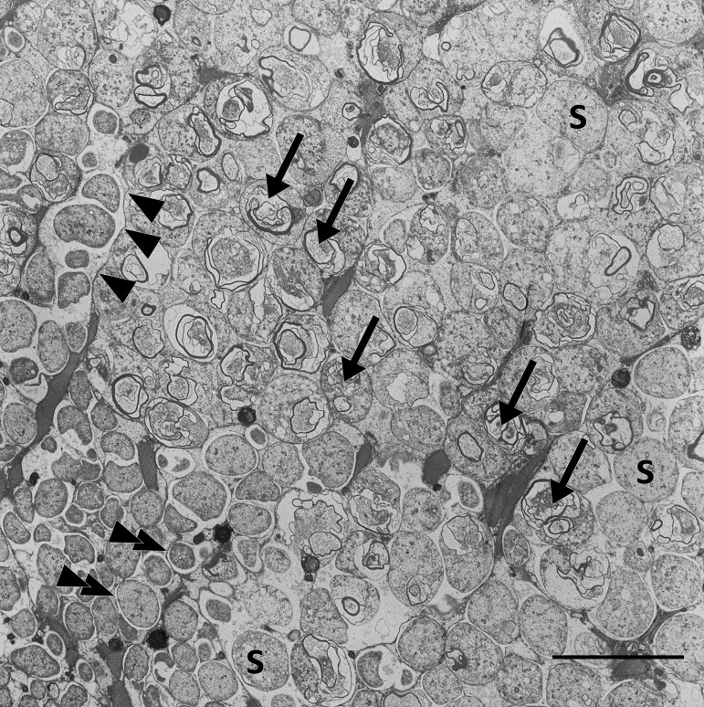
FIG 3.
Transmission electron micrograph of a Riftia trophosome tissue section. Within the lobular trophosome tissue, this section shows the median and peripheral zones of an individual lobule with host bacteriocytes containing intracellular coccoid symbionts (S) located in dedicated vesicles (arrowheads, bacteriocyte membrane; double arrowheads, vesicle membrane). While the lower left area of the image shows mostly intact symbiont cells, arrows in the central area point to symbiont cells in the state of digestion by the host, where cell degradation is indicated by the presence of lamellar bodies. Image brightness and contrast were adjusted for visual clarity. Scale bar, 10 μm.

