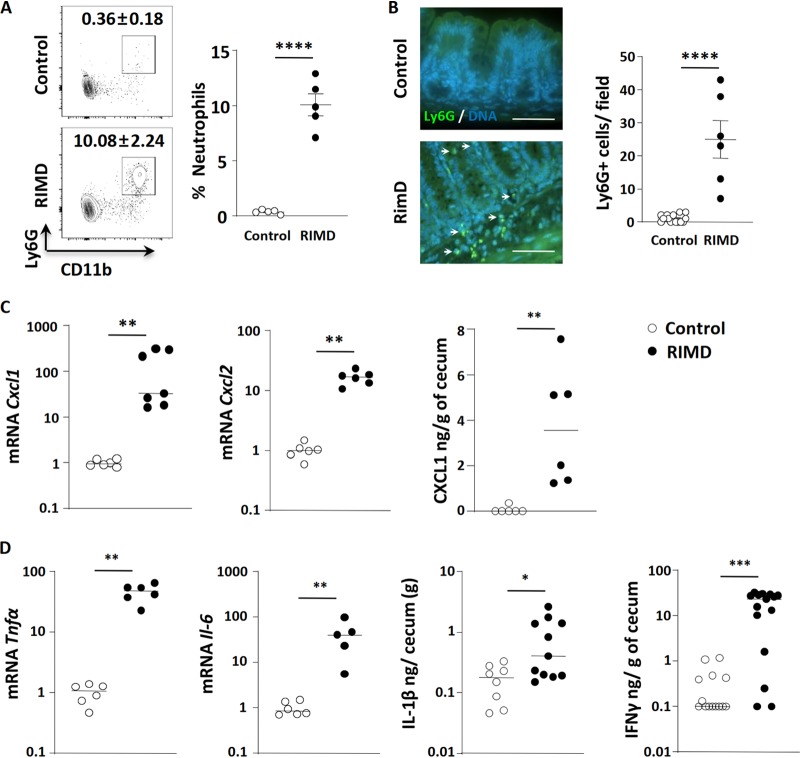
FIG 2.
V. parahaemolyticus RIMD2210633 infection triggers increased intestinal inflammation. V. parahaemolyticus-infected germfree C57BL/6 mice were euthanized at 21 h p.i. for analysis. (A) Flow cytometry analysis showed significant CD11b+ Ly6G+ neutrophil infiltration into the lower gastrointestinal tract (cecum and colon). (B) Immunostaining showed Ly6G+ neutrophil (green; shown by white arrows) infiltration into the cecal mucosa and submucosa during V. parahaemolyticus infection. DNA in cell nuclei were stained in blue. Bars, 50 μm. (C) Transcription of neutrophil-attracting chemokine genes Cxcl1 and Cxcl2 increased in infected cecal tissues, as did the protein level of CXCL1 as determined by ELISA from cecal homogenates. (D) Increased expression/production of proinflammatory cytokines (Tnfα and Il-6 by qPCR and IL-1β and IFN-γ by ELISA) in the infected cecum. In the graphs, bars show the mean ± SEM (A and B) or the median (C and D), and each symbol represents an individual mouse from 2 to 3 independent experiments. *, P < 0.05; **, P < 0.01; ***, P < 0.001; ****, P < 0.0001, by two-tailed Student’s t test (A and B) or Mann-Whitney test (C and D).

