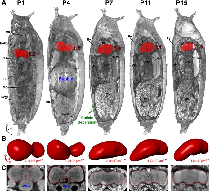
Fig. 2.
Developmental anatomy of Drosophila brains during pupation viewed by µ-CT. (A) 3D representation of pupae at stage P1, early P4, P7, P11 and P15. Part of the pupal cuticle and underlying soft tissue were digitally removed to reveal internal structures (axis denotes origin; A, anterior; D, dorsal; R, right). Brain is highlighted in red; values denote brain size relative to P1. Note the prominent abdominal air bubble at early P4 and cuticle separation between the pupal and adult epidermis at the pharate adult stage P7. BWM, body wall muscles; Cd, cardia; DLMs, dorsal longitudinal muscles; E-AD, eye-antennal discs; Ey, eye; FB, fat body; Lm, lamina; MG, midgut; MP, mouthparts; VNC, ventral nerve cord; Wg, wing. (B) Isolated brains from A, shown in same orientation, rendered as a 3D surface. Volume measurements are given below. (C) 2D xy anterior view of the brains in A. A, anterior; D, dorsal; L, left; R, right. Scale bars: 100 µm. Stained with 0.1 N iodine and scanned in slow mode at an image pixel size of 1.4 µm.

